Primary Adenocarcinoma of the Minor Duodenal Papilla
- Affiliations
-
- 1Department of Internal Medicine 2, Fukushima Medical University School of Medicine, Fukushima, Japan. irisawa@fmu.ac.jp
- 2Department of Surgery 2, Fukushima Medical University School of Medicine, Fukushima, Japan.
- 3Department of Pathology 1, Fukushima Medical University School of Medicine, Fukushima, Japan.
- KMID: 1084510
- DOI: http://doi.org/10.3349/ymj.2008.49.2.333
Abstract
- A 70-year-old man was admitted to our institution due to aggravation of blood-sugar level control and because an abdominal CT showed dilatation of the main pancreatic duct. Upper gastrointestinal endoscopy revealed a flat elevated tumor with central ulceration in the second portion of the duodenum. Subsequent duodenoscopy for a more detailed examination showed that the tumor had originated in the minor duodenal papilla. A biopsy specimen showed moderately differentiated adenocarcinoma. Endoscopic retrograde pancreatography via the major duodenal papilla revealed a slightly dilated main pancreatic duct and obstruction of the accessory pancreatic duct. Endoscopic ultrasonography showed a hypoechoic mass in the minor duodenal papilla with retention of the muscularis propria of the duodenum. These findings suggest that the tumor existed only to a limited extent in the minor duodenal papilla, and that the tumor did not infiltrate into the pancreas. For treatment, pylorus-preserving pancreatoduodenectomy was performed, and histological findings revealed a well-differentiated adenocarcinoma that originated in the minor duodenal papilla. Primary adenocarcinoma of the minor duodenal papilla is extremely rare. Our case is the first report of primary adenocarcinoma of the minor duodenal papilla at an early stage with no infiltration into muscularis propria of the duodenum and pancreas.
MeSH Terms
Figure
-

Fig. 1 Upper gastrointestinal endoscopy revealed a flat elevated tumor with central ulceration in the 2nd portion of the duodenum.
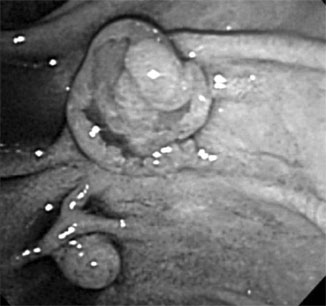
Fig. 2 Duodenoscopy with a side-view endoscope showed the tumor that originated in the minor duodenal papilla. The major papilla existed in the anal side of the minor papilla.
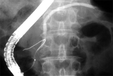
Fig. 3 Endoscopic retrograde pancreatography via the major duodenal papilla revealed no apparent abnormality except a slightly dilated main pancreatic duct and obstruction of the accessory pancreatic duct.
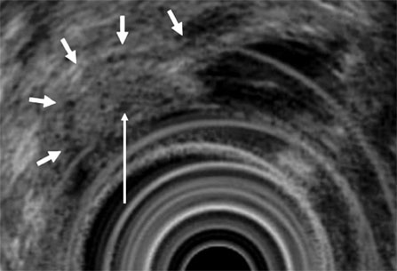
Fig. 4 EUS revealed an elevated hypoechoic mass in the minor duodenal papilla. Also, EUS showed an elevated hypoechoic mass (long arrow) in the minor papilla with retention of muscularis propria (short arrow) of the duodenum.

Fig. 5 Microsopic examination showed a well differentiated adenocarcinoma; tumor cells surround the orifice of the minor papilla with slight invasion into submucosa (arrow) (Elastica Masson staining, original magnification ×20).
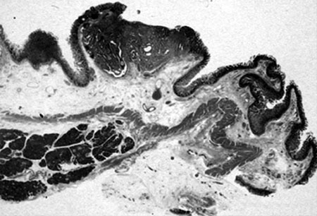
Fig. 6 Microscopic findings showed no infiltration into the pancreas parenchyma. (Hematoxylin Eosin staining, original magnification ×3).
Reference
-
1. Malone MJ, Silverman ML, Braasch JW, Jin GL, Dayal Y. Early somatostatinoma of the papilla of the duct of Santorini. Arch Surg. 1985. 120:1381–1383.
Article2. Stömmer PE, Stolte M, Seifert E. Somatostatinoma of Vater's papilla and of the minor papilla. Cancer. 1987. 60:232–235.
Article3. Singh VV, Bhutani MS, Draganov P. Carcinoid of the minor papilla in incomplete pancreas divisum presenting as acute relapsing pancreatitis. Pancreas. 2003. 27:96–97.
Article4. Sugiyama M, Kimura W, Muto T, Yahagi N, Ichinose M, Miki K. Endoscopic resection of adenoma of the minor papilla. Hepatogastroenterology. 1999. 46:189–192.5. Nakamura T, Ozawa T, Kitagawa M, Takehira Y, Yamada M, Yasumi K, et al. Endoscopic resection of gangliocytic paraganglioma of the minor duodenal papilla: case report and review. Gastrointest Endosc. 2002. 55:270–273.
Article6. Fukuda A, Yazumi S, Sawada M, Seno H, Nabeshima M, Fujii H, et al. Adenomyoma of the minor duodenal papilla. Gastrointest Endosc. 2005. 61:475–479.
Article7. Yamao K, Ohhashi K, Furukawa T, Mizutani S, Matsumoto S, Banno T, et al. Primary carcinoma of the duodenal minor papilla. Gastrointest Endosc. 1998. 48:634–636.
Article8. Ohta T, Nagakawa T, Kobayashi H, Kayahara M, Ueno K, Konishi I, et al. Histomorphological study on the minor duodenal papilla. Gastroenterol Jpn. 1991. 26:356–362.
Article9. Nakao A, Harada A, Nonami T, Kishimoto W, Takeda S, Ito K, et al. Prognosis of cancer of the duodenal papilla of Vater in relation to clinicopathological tumor extension. Hepatogastroenterology. 1994. 41:73–78.10. Goldberg M, Zamir O, Hadary A, Nissan S. Wide local excision as an alternative treatment for periampullary carcinoma. Am J Gastroenterol. 1987. 82:1169–1171.11. Ponchon T, Berger F, Chavaillon A, Bory R, Lambert R. Contribution of endoscopy to diagnosis and treatment of tumors of the ampulla of Vater. Cancer. 1989. 64:161–167.12. Jung S, Kim MH, Seo DW, Lee SK. Endoscopic snare papillectomy of adenocarcinoma of the major duodenal papilla. Gastrointest Endosc. 2001. 54:622.
Article13. Binmoeller KF, Boaventura S, Ramsperger K, Soehendra N. Endoscopic snare excision of benign adenomas of the papilla of Vater. Gastrointest Endosc. 1993. 39:127–131.14. Catalano MF, Linder JD, Chak A, Sivak MV Jr, Raijman I, Geenen JE, et al. Endoscopic management of adenoma of the major duodenal papilla. Gastrointest Endosc. 2004. 59:225–232.
Article
- Full Text Links
-
- Actions
-
Cited
- CITED
-
- Close
- Share
-
- Similar articles
-
- Endoscopic Papillectomy for Synchronous Major and Minor Duodenal Papilla Neuroendocrine Tumors
- Two Cases of Endoscopic Papillectomy for Neuroendocrine Tumor Arising from Minor Papilla
- Endoscopic Approach via the Minor Papilla for the Treatment of Pancreatic Stones
- A Case of Primary Duodenal Adenocarcinoma Treated by Endoscopic Mucosal Resection
- A Case of a Neuroendocrine Carcinoma in the Minor Papilla
