Korean J Ophthalmol.
2004 Dec;18(2):89-99. 10.3341/kjo.2004.18.2.89.
Correlation Between Frequency Doubling Technology Perimetry and Scanning Laser Polarimetry in Glaucoma Suspects and Glaucomatous Eyes
- Affiliations
-
- 1Department of Ophthalmology, University of Soonchunhyang, College of Medicine, Korea.
- 2Unit for Consulting Biostatistics, Asan Medical Center, Korea.
- 3Department of Ophthalmology, University of Ulsan, College of Medicine, Asan Medical Center, Seoul, Korea.
- KMID: 754373
- DOI: http://doi.org/10.3341/kjo.2004.18.2.89
Abstract
- The aim of this study was to determine the relationship between the frequency doubling technology (FDT) screening algorithm and parapapillary retinal nerve fiber layer (RNFL) thickness in the eyes of glaucoma suspects and patients with open angle glaucoma. FDT C20-1 screening program and a scanning laser polarimetry (SLP) system (GDx-NFA) was used to assess 53 glaucomatous eyes, 53 glaucoma suspects and 36 normal control eyes. In glaucomatous eyes, there were correlations between the FDT the screening algorithm and RNFL retardation values in several polarimetric indices, most significantly "inferior thickness" (r = -0.321, P = 0.029). In the eyes of glaucoma suspects, however, we observed no correlation between the FDT results and RNFL retardation values (r = 0.080, P > 0.05, "inferior thickness"). In glaucomatous eyes, the abnormal scores obtained with FDT screening program correlated negatively with RNFL retardation values, as measured by SLP. Despite poor correlation between the FDT abnormal score and RNFL retardation value in glaucoma suspects, detection of abnormality using the FDT screening protocol may aid in the assessment of early glaucomatous structural damage.
Keyword
MeSH Terms
Figure
-
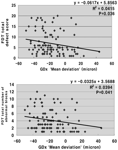
Fig. 1 Correlation between FDT abnormality and GDx "mean deviation".

Fig. 2 Correlation between FDT hemifield defect score and corresponding quadrant GDx deviation.
Reference
-
1. Sommer A, Katz J, Quigley HA, Miller NR, Robin AL, Richter RC, Witt KA. Clinically detectable nerve fiber atrophy precedes the onset of glaucomatous field loss. Arch Ophthalmol. 1991. 109:77–83.2. Quigley HA, Reacher M, Katz J, Strahlman E, Gilbert D, Scott R. Quantitative grading of nerve fiber layer photographs. Ophthalmology. 1993. 100:1800–1807.3. Quigley HA. Examination of the retinal nerve fiber layer in the recognition of early glaucoma damage. Trans Am Ophthalmol Soc. 1986. 84:920–966.4. Weinreb RN, Shakiba S, Zangwill L. Scanning laser polarimetry to measure the nerve fiber layer of normal and glaucomatous eyes. Am J Ophthalmol. 1995. 119:627–636.5. Zangwill L, Berry CA, Garden VS, Weinreb RN. Reproducibility of retardation measurements with the nerve fiber analyzer II. J Glaucoma. 1997. 6:384–389.6. Anton A, Zangwill L, Emdadi A, Weinreb RN. Nerve fiber layer measurements with scanning laser polarimetry in ocular hypertension. Arch Ophthalmol. 1997. 115:331–334.7. Tjon-Fo-Sang MJ, de Vries J, Lemij HG. Measurement by nerve fiber analyzer of retinal nerve fiber layer thickness in normal subjects and patients with ocular hypertension. Am J Ophthalmol. 1996. 122:220–227.8. Weinreb RN, Zangwill L, Berry CC, Bathija R, Sample PA. Detection of glaucoma with scanning laser polarimetry. Arch Ophthalmol. 1998. 116:1583–1589.9. Choplin NT, Landy DC, Dreher AW. Differentiating patients with glaucoma suspects and normal subjects by nerve fiber layer assessment with scanning laser polarimetry. Ophthalmology. 1998. 105:2068–2076.10. Horn FK, Jonas JB, Martus P, Mardin CY, Budde WM. Polarimetric measurement of retinal nerve fiber layer thickness in glaucoma diagnosis. J Glaucoma. 1999. 8:353–362.11. Kondo Y, Yamamoto T, Sato Y, Matsubara M, Kitazawa Y. A frequency-doubling perimetry study in normal-tension glaucoma with hemifield defect. J Glaucoma. 1998. 7:261–265.12. Reyes RD, Tomita G, Kitazawa Y. Retinal nerve fiber layer thickness within the area of apparently normal visual field in normal tension glaucoma with hemifield defect. J Glaucoma. 1998. 7:329–335.13. Wu LL, Suzuki Y, Kunimatsu S, Araie M, Iwase A, Tomita G. Frequency doubling technology and confocal scanning ophthalmoscopic optic disc analysis in open-angle glaucoma with hemifield defects. J Glaucoma. 2001. 10:256–260.14. Quigley HA. Identification of glaucoma-related visual field abnormality with the screening protocol of frequency doubling technology. Am J Ophthalmol. 1998. 125:819–829.15. Lachenmayr B, Airaksinen PJ, Drance SM, Wijsman K. Correlation of retinal nerve-fiber-layer loss, changes at the optic nerve head and various psychophysical criteria in glaucoma. Graefe's Arch Clin Exp Ophthalmol. 1991. 229:133–138.16. Horn FK, Jonas JB, Junemann A, Korth M, Junemann A, Grundler A. The full field flicker test in early diagnosis of chronic open-angle glaucoma. Am J Ophthalmol. 1997. 123:313–319.17. Weinreb RN, Shakiba S, Sample PA, Shahrokni S, van Horn S, Garden VS, Asawaphureekorn S, Zangwill L. Association between quantitative nerve fiber layer measurement and visual field loss in glaucoma. Am J Ophthalmol. 1995. 120:732–738.18. Eid TM, Spaeth GL, Katz LJ, Azura-Blanco A, Agusberger J, Nicholl J. Quantitative estimation of retinal nerve fiber layer height in glaucoma and the relationship with optic nerve head topography and visual field. J Glaucoma. 1997. 6:221–230.19. Tjon-Fo-Sang MJ, Lemij HG. The sensitivity and specificity of nerve fiber layer measurements in glaucoma as determined with scanning laser polarimetry. Am J Ophthalmol. 1997. 123:62–69.20. Teesalu P, Airaksinen PJ, Tuulonen A. Blue-on-yellow visual field and retinal nerve fiber layer in ocular hypertension and glaucoma. Ophthalmology. 1998. 105:2077–2081.21. Abecia E, Honrubia FM. Retinal nerve fiber defects and automated perimetry evaluation in ocular hypertensives. Int Ophthalmol. 1992. 16:239–242.22. Sample PA, Bosworth CF, Blumenthal EZ, Girkin C, Weinreb RN. Visual function-specific perimetry for indirect comparison of different ganglion cell populations in glaucoma. Invest Ophthalmol Vis Sci. 2000. 41:1783–1790.23. Paczka JA, Friedman DS, Quigley HA, Barron Y, Vitale S. Diagnostic capabilities of frequency-doubling technology, scanning laser polarimetry, and nerve fiber layer photographs to distinguish glaucomatous damage. Am J Ophthalmol. 2001. 131:188–197.24. Kelly DH. Frequency doubling in visual responses. J Opt Soc Am. 1996. 110:486–489.25. Johnson CA, Samuels SJ. Screening for glaucomatous visual field loss with frequency-doubling perimetry. Invest Ophthalmol Vis Sci. 1997. 38:413–425.26. Dandona L, Hendrickson A, Quigley HA. Selective effects of experimental glaucoma on axonal transport by retinal ganglion cells to the dorsal lateral geniculate nucleus. Invest Ophthalmol Vis Sci. 1991. 32:1593–1599.27. Chaturvedi N, Hedley-Whyte ET, Dreyer EB. Lateral geniculate nucleus in glaucoma. Am J Ophthalmol. 1993. 116:182–188.28. Glovinsky Y, Quigley HA, Pease ME. Foveal ganglion cell loss is size dependent in experimental glaucoma. Invest Ophthalmol Vis Sci. 1993. 34:395–400.29. Burnstein Y, Ellish NJ, Magbalon M, Higginbotham EJ. Comparison of frequency doubling perimetry with humphrey visual field analysis in a glaucoma practice. Am J Ophthalmol. 2000. 129:328–333.30. Sponsel WE, Arango S, Trigo Y, Mensah J. Clinical classification of glaucomatous visual field loss by frequency doubling perimetry. Am J Ophthalmol. 1998. 125:830–836.31. Kwon YH, Hong SP, Honkanen RA, Alward WL. Correlation of automated visual field parameters and peripapillary nerve fiber layer thickness as measured by scanning laser polarimetry. J Glaucoma. 2000. 9:281–288.32. Quigley HA, Sanchez RM, Dunkelberger GR, L'Hernault NL, Baginski TA. Chronic glaucoma selectively damages large optic nerve fibers. Invest Ophthalmol Vis Sci. 1987. 28:913–920.33. Lauande-Pimentel R, Carvalho RA, Oliveira HC, Goncalves DC, Silva LM, Costa VP. Discrimination between normal and glaucomatous eyes with visual field and scanning laser polarimetry measurements. Br J Ophthalmol. 2001. 85:586–591.34. Greenfield DS, Knighton RW, Huang SR. Effect of corneal polarization axis on assessment of retinal laser polarimetry. Am J Ophthalmol. 2000. 129:715–722.
- Full Text Links
-
- Actions
-
Cited
- CITED
-
- Close
- Share
-
- Similar articles
-
- Study of Contralateral Eye of Normal Tension Glaucoma and a Unilateral Visual Field Defect
- Differentiating Patients with Glaucoma from Glaucoma Suspects by Retinal Nerve Fiber Layer Assessment Using Nerve Fiber Analyzer
- GDx-VCC Performance to Discriminate Normal, Pre-perimetric Glaucomatous Eyes
- The Comparison of The Matrix Perimetry and Humphrey Standard Perimetry in Various Patients Group
- Scanning Laser Polarimetry Using Variable Corneal Compensation in Detection of Localized Visual Field Defects
