Synergistic Activation of LEPR and ADRB2 Induced by Leptin Enhances Reactive Oxygen Specie Generation in Triple-Negative Breast Cancer Cells
- Affiliations
-
- 1School of Medicine, Nankai University, Tianjin, China
- 2Tianjin Key Laboratory of Oral and Maxillofacial Function Reconstruction, Hospital of Stomatology, Nankai University, Tianjin, China
- KMID: 2566864
- DOI: http://doi.org/10.4143/crt.2024.368
Abstract
- Purpose
Leptin interacts not only with leptin receptor (LEPR) but also engages with other receptors. While the pro-oncogenic effects of the adrenergic receptor β2 (ADRB2) are well-established, the role of leptin in activating ADRB2 in triple-negative breast cancer (TNBC) remains unclear.
Materials and Methods
The pro-carcinogenic effects of LEPR were investigated using murine TNBC cell lines, 4T1 and EMT6, and a tumor-bearing mouse model. Expression levels of LEPR, NADPH oxidase 4 (NOX4), and ADRB2 in TNBC cells and tumor tissues were analyzed via western blot and quantitative real-time polymerase chain reaction. Changes in reactive oxygen species (ROS) levels were assessed using flow cytometry and MitoSox staining, while immunofluorescence double-staining confirmed the co-localization of LEPR and ADRB2.
Results
LEPR activation promoted NOX4-derived ROS and mitochondrial ROS production, facilitating TNBC cell proliferation and migration, effects which were mitigated by the LEPR inhibitor Allo-aca. Co-expression of LEPR and ADRB2 was observed on cell membranes, and bioinformatics data revealed a positive correlation between the two receptors. Leptin activated both LEPR and ADRB2, enhancing intracellular ROS generation and promoting tumor progression, which was effectively countered by a specific ADRB2 inhibitor ICI118551. In vivo, leptin injection accelerated tumor growth and lung metastases without affecting appetite, while treatments with Allo-aca or ICI118551 mitigated these effects.
Conclusion
This study demonstrates that leptin stimulates the growth and metastasis of TNBC through the activation of both LEPR and ADRB2, resulting in increased ROS production. These findings highlight LEPR and ADRB2 as potential biomarkers and therapeutic targets in TNBC.
Keyword
Figure
-

Fig. 1. Leptin upregulates LEPR expression in TNBC cells. (A, B) Bioinformatics data from Kaplan-Meier database (https://kmplot.com/analysis/index.php?p=service) showed OS curve (A) and DMFS curve (B) among TNBC patients stratified by high and low LEPR expression levels. Higher LEPR expression was associated with poor prognosis. (C, D) Quantitative PCR and western blot were performed to analyze the expression of LEPR mRNA (C) and LEPR protein (D) in 4T1 and EMT6 cells following stimulation with increasing concentrations of leptin (0, 10, 50, 100, 200 ng/mL) for 24 hours, respectively. n=3-4. (E, F) Quantitative LEPR mRNA (E) and LEPR protein (F) expression in 4T1 and EMT6 cells following 24 hours of leptin (100 ng/mL) and/or Allo-aca (Allo, 100 nM) treatment. n=3. DMFS, distant metastases-free survival; HR, hazard ratio; LEPR, leptin receptor; OS, overall survival; PCR, polymerase chain reaction; SEM, standard error of mean; TNBC, triple-negative breast cancer. All data are presented as the mean±SEM. ns, no significance; *p < 0.05, **p < 0.01, ***p < 0.001.
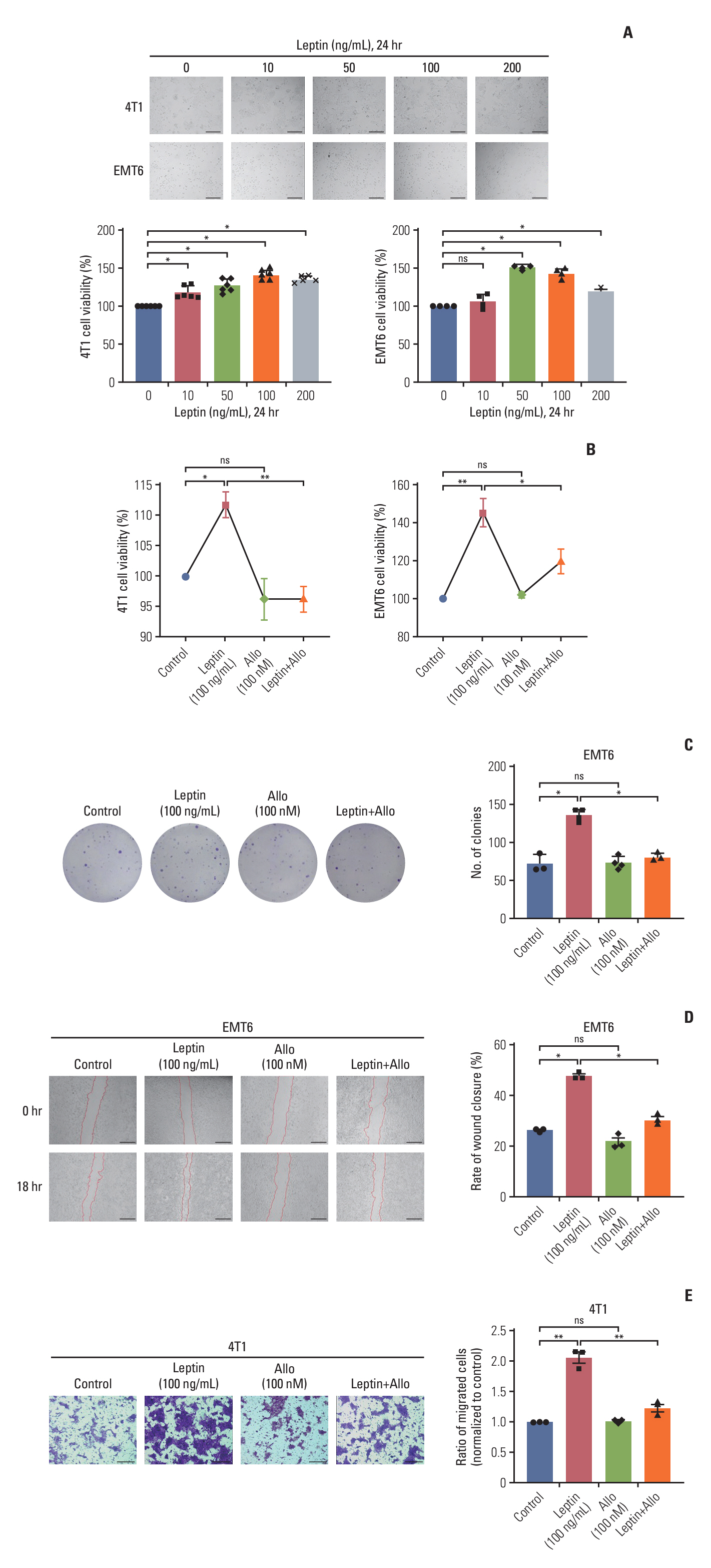
Fig. 2. LEPR activation promotes the proliferation and migration of TNBC cells. (A) CCK-8 assay was employed to assess the proliferative capacity of 4T1 and EMT6 cells upon exposure to leptin (0, 10, 50, 100, 200 ng/mL). n=3. Scale bars=200 μm. (B, C) CCK-8 assay (B) and colony formation assay (C) were conducted to evaluate the proliferative ability of 4T1 and EMT6 cells treated with leptin (100 ng/mL) and/or Allo (100 nM). n=3. (D, E) Wound closure assay and transwell migration assay were employed to investigate the migratory ability of EMT6 (scale bars=500 μm) and 4T1 (scale bars=200 μm) cells following treatment with leptin and/or Allo, respectively. n=3. CCK-8, Cell Counting Kit-8; LEPR, leptin receptor; SEM, standard error of mean; TNBC, triple-negative breast cancer. All data are presented as the mean±SEM. ns, no significance; *p < 0.05, **p < 0.01.

Fig. 3. Leptin induces NOX4-derived ROS production and mtROS generation. (A) Flow cytometry analysis of ROS levels in 4T1 and EMT6 cells treated with varying concentrations of leptin (0, 10, 50, 100, and 200 ng/mL), assessed using the fluorescent probe DCFH-DA. n=3. (B) Flow cytometry analysis of ROS levels in 4T1 and EMT6 cells treated with leptin (100 ng/mL) and/or Allo (100 nM), utilizing the fluorescent probe DCFH-DA. n=3. (C) Schematic diagram of the two main sources of ROS generation. (D, E) Quantitative PCR (D) and western blotting (E) were employed to evaluate the expression of NOX4 in 4T1 and EMT6 cells following stimulation with increasing concentrations of leptin (0, 10, 50, 100, and 200 ng/mL) for 24 hours. n=3. (F, G) Quantitative PCR (F) and western blotting (G) were employed to assess the expression of NOX4 in 4T1 and EMT6 cells treated with leptin (100 ng/mL) and/or Allo (100 nM). n=3. (H, I) MitoSOX staining assay to detect mtROS levels in 4T1 and EMT6 cells treated with leptin (100 ng/mL) and/or Allo (100 nM). n=3. mtROS, mitochondrial ROS; NOX4, NADPH oxidase 4; PCR, polymerase chain reaction; ROS, reactive oxygen species; SEM, standard error of mean. All data are presented as the mean±SEM. ns, no significance; *p < 0.05, **p < 0.01, *** p < 0.001.

Fig. 4. Leptin upregulates ADRB2 expression in TNBC cells. (A) Quantitative PCR was utilized to assess the expression levels of ADRB2 in 4T1 (n=3) and EMT6 (n=6) cells following stimulation with 50 ng/mL or 100 ng/mL leptin. (B) Western blotting was utilized to assess the expression levels of ADRB2 in 4T1 and EMT6 cells following stimulation with 50 ng/mL or 100 ng/mL leptin. n=4. (C, D) Quantitative PCR (C) and western blotting (D) were employed to analyze the expression of ADRB2 in 4T1 and EMT6 cells treated with leptin (100 ng/mL) and/or Allo (100 nM). n=3. (E) Representative images of immunofluorescence double staining showing the expression and co-localization of LEPR (Alexa Fluor 488 Conjugated) and ADRB2 (Alexa Fluor 594-conjugated) in the presence of leptin and/or Allo, or the ADRB2 inhibitor ICI118551 (ICI). (F-J) Statistical curves depicting the fluorescence intensity of LEPR and ADRB2 in the co-localization images for different experimental groups. (K) Quantitative analysis of LEPR-ADRB2 co-localization on cell membranes in each group, evaluated using Pearson’s correlation coefficient. Data are presented as mean±SEM, n=3. ADRB2, adrenergic receptor β2; LEPR, leptin receptor; PCR, polymerase chain reaction; SEM, standard error of mean; TNBC, triple-negative breast cancer. All data are presented as the mean±SEM. ns, no significance; *p < 0.05, **p < 0.01.

Fig. 5. ADRB2 mediates the leptin-induced ROS generation. (A) Flow cytometry analysis of ROS levels in 4T1 and EMT6 cells treated with leptin and/or the ADRB2 inhibitor ICI, assessed using the fluorescent probe DCFH-DA. (B) Western blotting analyzed the expression of ADRB2 and NOX4 in 4T1 cells treated with leptin (100 ng/mL) and/or ICI (10 μM). n=3. (C) MitoSOX staining assay was utilized to detect mtROS levels in EMT6 cells treated with leptin (100 ng/mL) and/or ICI (10 μM). ADRB2, adrenergic receptor β2; mtROS, mitochondrial ROS; NOX4, NADPH oxidase 4; ROS, reactive oxygen species; SEM, standard error of mean. All data are presented as the mean±SEM. ns, no significance; *p < 0.05.

Fig. 6. LEPR and ADRB2 in leptin-induced breast cancer progression in vivo. (A) Female BALB/c mice were subcutaneously inoculated with 1×10⁵ 4T1 cells into the mammary fat pad to establish the breast tumor animal model. Drug administration commenced 7 days postinoculation and continued for 21 days. Tumors, blood samples, and lungs were collected for analysis. (B) Representative image of tumors and tumor growth curves in different experimental groups. n=5. (C) Tumor weights were measured on day 28. n=5. (D, E) Food intake (D) and body weight curves (E) in different groups. n=5. (F) Metastatic lung nodules were visualized using H&E staining in different groups. (G) GSH content was assessed in both serum and tumor samples collected from different groups. n=3-5. (H, I) Real-time qPCR (H) and western blotting (I) were used to analyze the expression of LEPR, ADRB2, and NOX4 in tumors from different groups. n=3. ADRB2, adrenergic receptor β2; GSH, glutathione; LEPR, leptin receptor; NOX4, NADPH oxidase 4; qPCR, quantitative real-time polymerase chain reacion; SEM, standard error of mean. All data are presented as the mean±SEM. ns, no significance; *p < 0.05, **p < 0.01, ***p < 0.001.
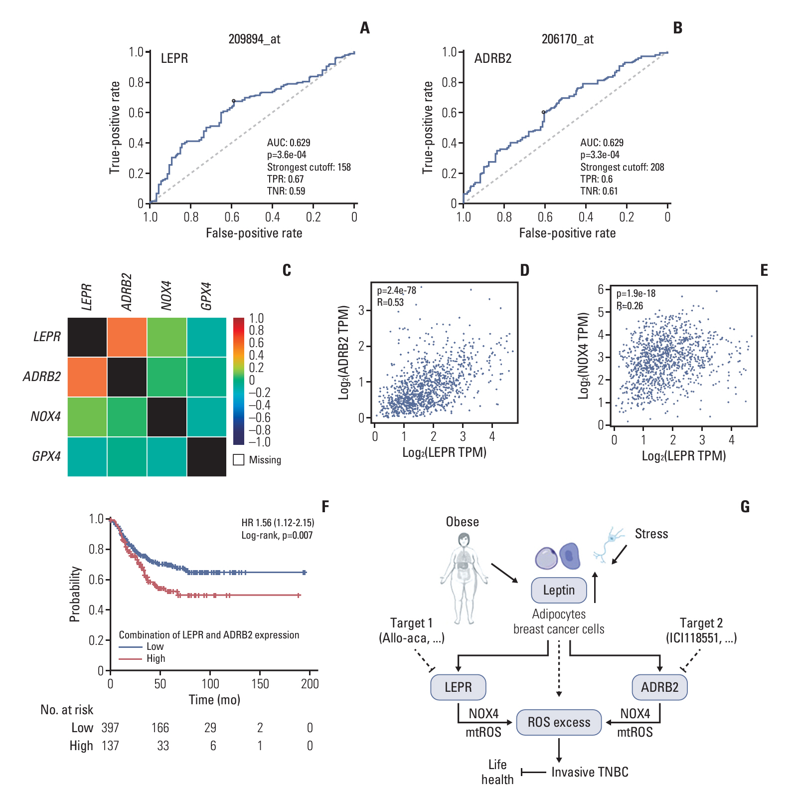
Fig. 7. The clinical significance of LEPR and ADRB2. (A, B) Receiver operating characteristic curve analysis was conducted to evaluate the prognostic significance of LEPR (A) and ADRB2 (B) in breast cancer patients. p < 0.01. (C-E) The correlation between LEPR, ADRB2, NOX4, and GPX4 was analyzed using the bc-GenExMiner database (C) and GEPIA database (D, E). (F) Kaplan-Meier plotter was utilized to illustrate RFS in TNBC patients with high expression of LEPR and ADRB2 combined. p=0.0071. (G) The schematic diagram of the study work. ADRB2, adrenergic receptor β2; AUC, area under the curve; GPX4, glutathione peroxidase 4; HR, hazard ratio; LEPR, leptin receptor; mtROS, mitochondrial reactive oxygen species; NOX4, NADPH oxidase 4; RFS, relapse-free survival; TNBC, triple-negative breast cancer; TNR, true-negative ratio; TPM, transcripts per million; TPR, true-positive ratio.
Reference
-
References
1. Lipsyc-Sharf M, Ballman KV, Campbell JD, Muss HB, Perez EA, Shulman LN, et al. Age, body mass index, tumor subtype, and racial and ethnic disparities in breast cancer survival. JAMA Netw Open. 2023; 6:e2339584.
Article2. Picon-Ruiz M, Morata-Tarifa C, Valle-Goffin JJ, Friedman ER, Slingerland JM. Obesity and adverse breast cancer risk and outcome: mechanistic insights and strategies for intervention. CA Cancer J Clin. 2017; 67:378–97.
Article3. Zhang Y, Proenca R, Maffei M, Barone M, Leopold L, Friedman JM. Positional cloning of the mouse obese gene and its human homologue. Nature. 1994; 372:425–32.
Article4. Ahima RS, Flier JS. Leptin. Annu Rev Physiol. 2000; 62:413–37.
Article5. Tartaglia LA, Dembski M, Weng X, Deng N, Culpepper J, Devos R, et al. Identification and expression cloning of a leptin receptor, OB-R. Cell. 1995; 83:1263–71.
Article6. Ahima RS, Osei SY. Leptin signaling. Physiol Behav. 2004; 81:223–41.
Article7. Ishikawa M, Kitayama J, Nagawa H. Enhanced expression of leptin and leptin receptor (OB-R) in human breast cancer. Clin Cancer Res. 2004; 10:4325–31.
Article8. Garcia-Estevez L, Calvo I, Perez S, Gallegos I, Diaz E, Sampayo-Cordero M, et al. Predictive role of leptin receptor (ObR) overexpression in patients with early breast cancer receiving neoadjuvant systemic treatment. Cancers (Basel). 2021; 13:3269.
Article9. Sweeney G. Leptin signalling. Cell Signal. 2002; 14:655–63.
Article10. Cleary MP, Phillips FC, Getzin SC, Jacobson TL, Jacobson MK, Christensen TA, et al. Genetically obese MMTV-TGF-alpha/Lep(ob)Lep(ob) female mice do not develop mammary tumors. Breast Cancer Res Treat. 2003; 77:205–15.
Article11. Valentine JM, Ahmadian M, Keinan O, Abu-Odeh M, Zhao P, Zhou X, et al. beta3-Adrenergic receptor downregulation leads to adipocyte catecholamine resistance in obesity. J Clin Invest. 2022; 132:e153357.
Article12. Arner P, Andersson DP, Backdahl J, Dahlman I, Ryden M. Weight gain and impaired glucose metabolism in women are predicted by inefficient subcutaneous fat cell lipolysis. Cell Metab. 2018; 28:45–54.
Article13. Zeng W, Pirzgalska RM, Pereira MM, Kubasova N, Barateiro A, Seixas E, et al. Sympathetic neuro-adipose connections mediate leptin-driven lipolysis. Cell. 2015; 163:84–94.
Article14. Chauveau C, Devedjian JC, Delecourt C, Jeanfils J, Hardouin P, Broux O. Leptin receptors and beta2-adrenergic receptor mRNA expression in brain injury-related heterotopic ossification. J Recept Signal Transduct Res. 2008; 28:347–59.15. Kamiya A, Hayama Y, Kato S, Shimomura A, Shimomura T, Irie K, et al. Genetic manipulation of autonomic nerve fiber innervation and activity and its effect on breast cancer progression. Nat Neurosci. 2019; 22:1289–305.
Article16. Qin JF, Jin FJ, Li N, Guan HT, Lan L, Ni H, et al. Adrenergic receptor beta2 activation by stress promotes breast cancer progression through macrophages M2 polarization in tumor microenvironment. BMB Rep. 2015; 48:295–300.
Article17. Chang A, Botteri E, Gillis RD, Lofling L, Le CP, Ziegler AI, et al. Beta-blockade enhances anthracycline control of metastasis in triple-negative breast cancer. Sci Transl Med. 2023; 15:eadf1147.
Article18. Mahbouli S, Der Vartanian A, Ortega S, Rouge S, Vasson MP, Rossary A. Leptin induces ROS via NOX5 in healthy and neoplastic mammary epithelial cells. Oncol Rep. 2017; 38:3254–64.
Article19. Rios Garcia M, Steinbauer B, Srivastava K, Singhal M, Mattijssen F, Maida A, et al. Acetyl-CoA carboxylase 1-dependent protein acetylation controls breast cancer metastasis and recurrence. Cell Metab. 2017; 26:842–55.
Article20. Saxena NK, Taliaferro-Smith L, Knight BB, Merlin D, Anania FA, O’Regan RM, et al. Bidirectional crosstalk between leptin and insulin-like growth factor-I signaling promotes invasion and migration of breast cancer cells via transactivation of epidermal growth factor receptor. Cancer Res. 2008; 68:9712–22.
Article21. Feola A, Ricci S, Kouidhi S, Rizzo A, Penon A, Formisano P, et al. Multifaceted breast cancer: the molecular connection with obesity. J Cell Physiol. 2017; 232:69–77.
Article22. Giordano C, Vizza D, Panza S, Barone I, Bonofiglio D, Lanzino M, et al. Leptin increases HER2 protein levels through a STAT3-mediated up-regulation of Hsp90 in breast cancer cells. Mol Oncol. 2013; 7:379–91.
Article23. Hu J, Lu R, Zhang Y, Li W, Hu Q, Chen C, et al. beta-adrenergic receptor inhibition enhances oncolytic herpes virus propagation through STAT3 activation in gastric cancer. Cell Biosci. 2021; 11:174.24. Kang Y, Nagaraja AS, Armaiz-Pena GN, Dorniak PL, Hu W, Rupaimoole R, et al. Adrenergic stimulation of DUSP1 impairs chemotherapy response in ovarian cancer. Clin Cancer Res. 2016; 22:1713–24.
Article25. Garofalo C, Koda M, Cascio S, Sulkowska M, Kanczuga-Koda L, Golaszewska J, et al. Increased expression of leptin and the leptin receptor as a marker of breast cancer progression: possible role of obesity-related stimuli. Clin Cancer Res. 2006; 12:1447–53.
Article26. Kim HS. Leptin and leptin receptor expression in breast cancer. Cancer Res Treat. 2009; 41:155–63.
Article27. Sanchez-Jimenez F, Perez-Perez A, de la Cruz-Merino L, Sanchez-Margalet V. Obesity and breast cancer: role of leptin. Front Oncol. 2019; 9:596.28. Garcia-Estevez L, Gonzalez-Martinez S, Moreno-Bueno G. The leptin axis and its association with the adaptive immune system in breast cancer. Front Immunol. 2021; 12:784823.29. Ray A. Adipokine leptin in obesity-related pathology of breast cancer. J Biosci. 2012; 37:289–94.
Article30. Munzberg H, Bjornholm M, Bates SH, Myers MG Jr. Leptin receptor action and mechanisms of leptin resistance. Cell Mol Life Sci. 2005; 62:642–52.31. Kimura M, Tateishi N, Shiota T, Yoshie F, Yamauchi H, Suzuki M, et al. Long-term exercise down-regulates leptin receptor mRNA in the arcuate nucleus. Neuroreport. 2004; 15:713–6.
Article32. Yang G, Ge H, Boucher A, Yu X, Li C. Modulation of direct leptin signaling by soluble leptin receptor. Mol Endocrinol. 2004; 18:1354–62.
Article33. Maamra M, Bidlingmaier M, Postel-Vinay MC, Wu Z, Strasburger CJ, Ross RJ. Generation of human soluble leptin receptor by proteolytic cleavage of membrane-anchored receptors. Endocrinology. 2001; 142:4389–93.
Article34. Moloney JN, Cotter TG. ROS signalling in the biology of cancer. Semin Cell Dev Biol. 2018; 80:50–64.
Article35. Zhao X, Liu M, Li C, Liu X, Zhao J, Ma H, et al. High dose Vitamin C inhibits PD-L1 by ROS-pSTAT3 signal pathway and enhances T cell function in TNBC. Int Immunopharmacol. 2024; 126:111321.
Article36. Zhang Z, Zhang H, Li D, Zhou X, Qin Q, Zhang Q. Caspase-3-mediated GSDME induced pyroptosis in breast cancer cells through the ROS/JNK signalling pathway. J Cell Mol Med. 2021; 25:8159–68.
Article37. Shan M, Qin J, Jin F, Han X, Guan H, Li X, et al. Autophagy suppresses isoprenaline-induced M2 macrophage polarization via the ROS/ERK and mTOR signaling pathway. Free Radic Biol Med. 2017; 110:432–43.
Article38. Yue R, Zhou BO, Shimada IS, Zhao Z, Morrison SJ. Leptin receptor promotes adipogenesis and reduces osteogenesis by regulating mesenchymal stromal cells in adult bone marrow. Cell Stem Cell. 2016; 18:782–96.
Article39. Lu Y, Zhao H, Liu Y, Zuo Y, Xu Q, Liu L, et al. Chronic stress activates plexinA1/VEGFR2-JAK2-STAT3 in vascular endothelial cells to promote angiogenesis. Front Oncol. 2021; 11:709057.
Article40. Rahmouni K, Sigmund CD, Haynes WG, Mark AL. Hypothalamic ERK mediates the anorectic and thermogenic sympathetic effects of leptin. Diabetes. 2009; 58:536–42.
Article41. Jin M, Wang Y, Zhou T, Li W, Wen Q. Norepinephrine/beta(2)-adrenergic receptor pathway promotes the cell proliferation and nerve growth factor production in triple-negative breast cancer. J Breast Cancer. 2023; 26:268–85.
Article
- Full Text Links
-
- Actions
-
Cited
- CITED
-
- Close
- Share
-
- Similar articles
-
- Hydroxyzine Induces Cell Death in Triple-Negative Breast Cancer Cells via Mitochondrial Superoxide and Modulation of Jak2/STAT3 Signaling
- Domperidone Exerts Antitumor Activity in Triple-Negative Breast Cancer Cells by Modulating Reactive Oxygen Species and JAK/STAT3 Signaling
- Effects of Polyamines on TNFalpha- or Tamoxifen-induced Apoptosis in Human Breast Cancer Cells
- Reactive Oxygen Species are Involved in Y-27632-induced Neurite Outgrowth in PC12 Cells
- Role of Calmodulin in the Generation of Reactive Oxygen Species and Apoptosis Induced by Tamoxifen in HepG2 Human Hepatoma Cells
