Korean J Orthod.
2024 Sep;54(5):274-283. 10.4041/kjod23.206.
Upper airway dimensions and craniofacial morphology: A correlation study using cone beam computed tomography
- Affiliations
-
- 1Department of Orthodontics, Catholic University of Portugal, Viseu, Portugal
- 2Department of Orthodontics, University of Porto, Porto, Portugal
- KMID: 2559817
- DOI: http://doi.org/10.4041/kjod23.206
Abstract
Objective
To determine the correlation between dentoskeletal parameters related to craniofacial morphology and the upper airway (UA) volume.
Methods
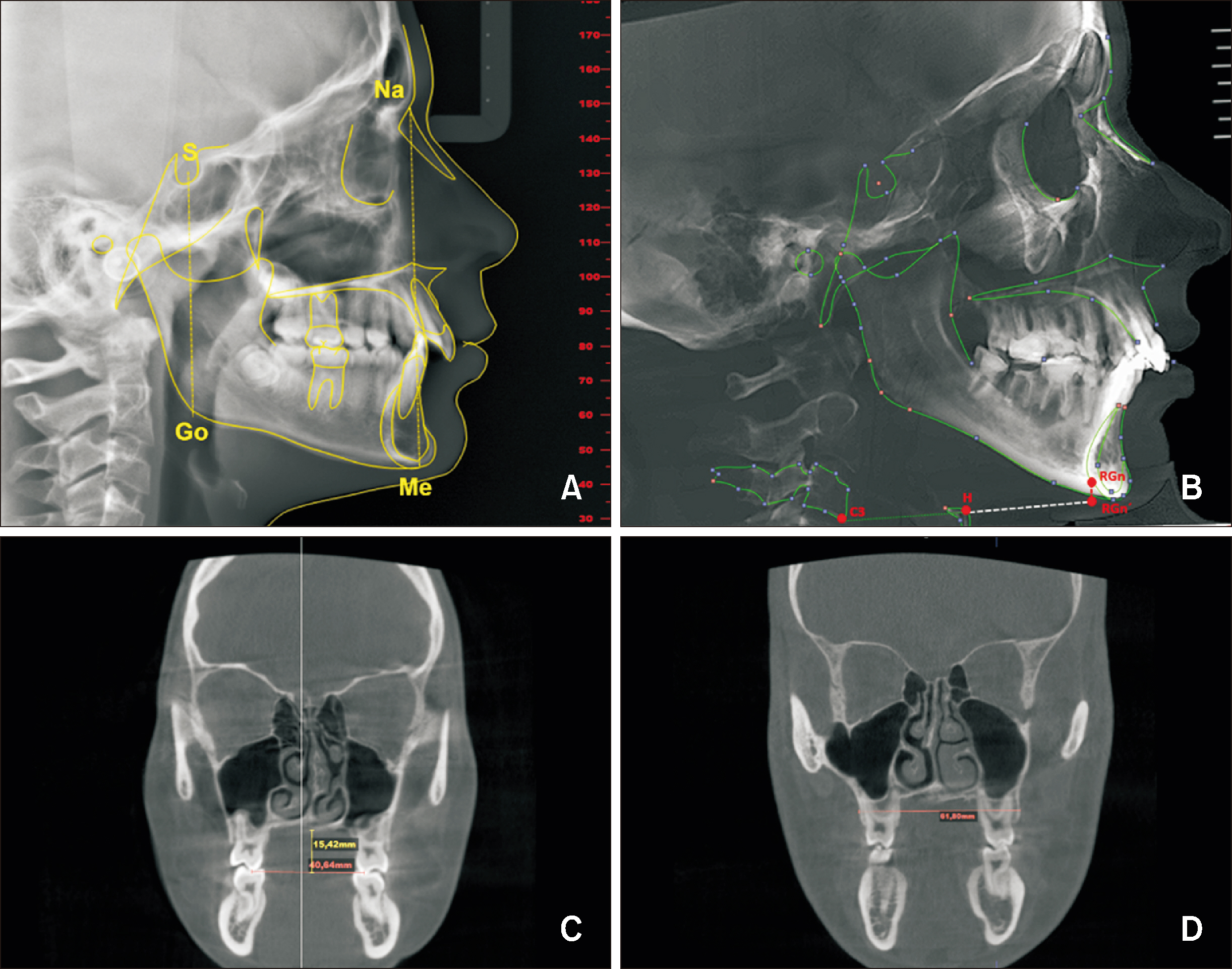
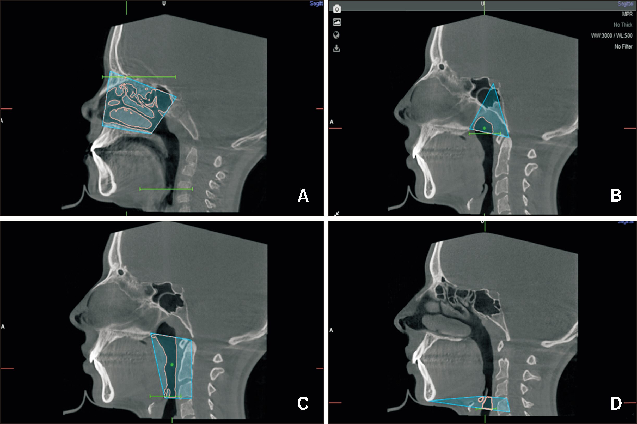
Cone-beam computed tomography images of 106 randomly selected orthodontic patients were analyzed using NemoFab Ortho software. The dentoskeletal variables assessed were anterior facial height (AFH), posterior facial height (PFH), PFH/AFH ratio, hyoid position, maxillary width (MW), and palatal depth. The UA volume (evaluation in anatomical regions and as a whole) was also assessed using the same software. We also evaluated potential differences in UA variables between age and sex groups. The correlation between the dentoskeletal parameters and UA volume was calculated using the Pearson correlation coefficient (R). Analysis of variance and Student’s t test were performed to assess differences between age and sex for UA variables. Statistical analyses were performed using SPSS software (version 26 for Windows).
Results
This study found that PFH, AFH, and MW were the dentoskeletal parameters most strongly correlated with UA volume. However, the ANB angle did not show any significant correlation with UA volume. Additionally, differences in UA volumes were observed between age groups. Sex differences were found in both the “8–12” and “≥ 16” age groups for oropharyngeal and pharyngeal volumes.
Conclusions
In conclusion, our findings indicate a significant correlation between UA volume and dentoskeletal parameters, particularly those related to facial height and MW.
Keyword
Figure
Reference
-
1. Solow B, Siersbaek-Nielsen S, Greve E. 1984; Airway adequacy, head posture, and craniofacial morphology. Am J Orthod. 86:214–23. https://doi.org/10.1016/0002-9416(84)90373-7. DOI: 10.1016/0002-9416(84)90373-7.2. McNamara JA. 1981; Influence of respiratory pattern on craniofacial growth. Angle Orthod. 51:269–300. https://pubmed.ncbi.nlm.nih.gov/6947703/.3. Klein JC. 1986; Nasal respiratory function and craniofacial growth. Arch Otolaryngol Head Neck Surg. 112:843–9. https://doi.org/10.1001/archotol.1986.03780080043009. DOI: 10.1001/archotol.1986.03780080043009.4. Principato JJ. 1991; Upper airway obstruction and craniofacial morphology. Otolaryngol Head Neck Surg. 104:881–90. https://doi.org/10.1177/019459989110400621. DOI: 10.1177/019459989110400621. PMID: 1908986.5. Indriksone I, Jakobsone G. 2015; The influence of craniofacial morphology on the upper airway dimensions. Angle Orthod. 85:874–80. https://doi.org/10.2319/061014-418.1. DOI: 10.2319/061014-418.1. PMID: 25363812. PMCID: PMC8610410.6. Diwakar R, Kochhar AS, Gupta H, Kaur H, Sidhu MS, Skountrianos H, et al. 2021; Effect of craniofacial morphology on pharyngeal airway volume measured using cone-beam computed tomography (CBCT)-a retrospective pilot study. Int J Environ Res Public Health. 18:5040. https://doi.org/10.3390/ijerph18095040. DOI: 10.3390/ijerph18095040. PMID: 34068732. PMCID: PMC8126215.7. Moss ML, Salentijn L. 1969; The primary role of functional matrices in facial growth. Am J Orthod. 55:566–77. https://doi.org/10.1016/0002-9416(69)90034-7. DOI: 10.1016/0002-9416(69)90034-7.8. Page DC, Mahony D. 2010; The airway, breathing and orthodontics. Todays FDA. 22:43–7. https://pubmed.ncbi.nlm.nih.gov/20443530/.9. Almuzian M, Ju X, Almukhtar A, Ayoub A, Al-Muzian L, McDonald JP. 2018; Does rapid maxillary expansion affect nasopharyngeal airway? A prospective Cone Beam Computerised Tomography (CBCT) based study. Surgeon. 16:1–11. https://doi.org/10.1016/j.surge.2015.12.006. DOI: 10.1016/j.surge.2015.12.006.10. Dalmau E, Zamora N, Tarazona B, Gandia JL, Paredes V. 2015; A comparative study of the pharyngeal airway space, measured with cone beam computed tomography, between patients with different craniofacial morphologies. J Craniomaxillofac Surg. 43:1438–46. https://doi.org/10.1016/j.jcms.2015.06.016. DOI: 10.1016/j.jcms.2015.06.016. PMID: 26189145.11. Di Carlo G, Polimeni A, Melsen B, Cattaneo PM. 2015; The relationship between upper airways and craniofacial morphology studied in 3D. A CBCT study. Orthod Craniofac Res. 18:1–11. https://doi.org/10.1111/ocr.12053. DOI: 10.1111/ocr.12053.12. Dastan F, Ghaffari H, Shishvan HH, Zareiyan M, Akhlaghian M, Shahab S. 2021; Correlation between the upper airway volume and the hyoid bone position, palatal depth, nasal septum deviation, and concha bullosa in different types of malocclusion: a retrospective cone-beam computed tomography study. Dent Med Probl. 58:509–14. https://doi.org/10.17219/dmp/130099. DOI: 10.17219/dmp/130099. PMID: 34850611.13. Tamburrino RK, Boucher NS, Vanarsdall RL, Secchi A. 2010; The transverse dimension: diagnosis and relevance to functional occlusion. RWISO J. 2010:13–23. https://www.learncco.com/wp-content/uploads/2022/02/Transverse-Dimension.-Diagnosis-and-Relevance-to-Functional-Occlusion-Tamburrino-et-all.pdf.14. Lotfi V, Ghoneima A, Lagravere M, Kula K, Stewart K. 2018; Three-dimensional evaluation of airway volume changes in two expansion activation protocols. Int Orthod. 16:144–57. https://doi.org/10.1016/j.ortho.2018.01.001. DOI: 10.1016/j.ortho.2018.01.001.15. Walter SD, Eliasziw M, Donner A. 1998; Sample size and optimal designs for reliability studies. Stat Med. 17:101–10. https://doi.org/10.1002/(sici)1097-0258(19980115)17:1<101::aid-sim727>3.0.co;2-e. DOI: 10.1002/(SICI)1097-0258(19980115)17:1<101::AID-SIM727>3.3.CO;2-5.16. Tseng YC, Tsai FC, Chou ST, Hsu CY, Cheng JH, Chen CM. 2021; Evaluation of pharyngeal airway volume for different dentofacial skeletal patterns using cone-beam computed tomography. J Dent Sci. 16:51–7. https://doi.org/10.1016/j.jds.2020.07.015. DOI: 10.1016/j.jds.2020.07.015.17. Gulsen A, Okay C, Aslan BI, Uner O, Yavuzer R. 2006; The relationship between craniofacial structures and the nose in Anatolian Turkish adults: a cephalometric evaluation. Am J Orthod Dentofacial Orthop. 130:131.e15–25. https://doi.org/10.1016/j.ajodo.2006.01.020. DOI: 10.1016/j.ajodo.2006.01.020. PMID: 16905054.18. Nehra K, Sharma V. 2009; Nasal morphology as an indicator of vertical maxillary skeletal pattern. J Orthod. 36:160–6. https://doi.org/10.1179/14653120723148. DOI: 10.1179/14653120723148.19. Grauer D, Cevidanes LS, Styner MA, Ackerman JL, Proffit WR. 2009; Pharyngeal airway volume and shape from cone-beam computed tomography: relationship to facial morphology. Am J Orthod Dentofacial Orthop. 136:805–14. https://doi.org/10.1016/j.ajodo.2008.01.020. DOI: 10.1016/j.ajodo.2008.01.020. PMID: 19962603. PMCID: PMC2891076.20. Cheng JH, Hsiao SY, Chen CM, Hsu KJ. 2020; Relationship between hyoid bone and pharyngeal airway in different skeletal patterns. J Dent Sci. 15:286–93. https://doi.org/10.1016/j.jds.2020.05.012. DOI: 10.1016/j.jds.2020.05.012. PMID: 32952886. PMCID: PMC7486506.21. Acharya A, Mishra P, Shrestha RM. 2022; Pharyngeal airway space dimensions and hyoid bone position in various craniofacial morphologies. J Indian Orthod Soc. 56:150–7. https://doi.org/10.1177/03015742211007621. DOI: 10.1177/03015742211007621.22. Yamashita AL, Iwaki Filho L, Leite PCC, Navarro RL, Ramos AL, Previdelli ITS, et al. 2017; Three-dimensional analysis of the pharyngeal airway space and hyoid bone position after orthognathic surgery. J Craniomaxillofac Surg. 45:1408–14. https://doi.org/10.1016/j.jcms.2017.06.016. DOI: 10.1016/j.jcms.2017.06.016. PMID: 28743605.23. Shokri A, Miresmaeili A, Ahmadi A, Amini P, Falah-Kooshki S. 2018; Comparison of pharyngeal airway volume in different skeletal facial patterns using cone beam computed tomography. J Clin Exp Dent. 10:e1017–28. https://doi.org/10.4317/jced.55033. DOI: 10.4317/jced.55033. PMID: 30386509. PMCID: PMC6203907.24. Shokri A, Mollabashi V, Zahedi F, Tapak L. 2020; Position of the hyoid bone and its correlation with airway dimensions in different classes of skeletal malocclusion using cone-beam computed tomography. Imaging Sci Dent. 50:105–15. https://doi.org/10.5624/isd.2020.50.2.105. DOI: 10.5624/isd.2020.50.2.105. PMID: 32601585. PMCID: PMC7314608.25. Nejaim Y, Aps JKM, Groppo FC, Haiter Neto F. 2018; Evaluation of pharyngeal space and its correlation with mandible and hyoid bone in patients with different skeletal classes and facial types. Am J Orthod Dentofacial Orthop. 153:825–33. https://doi.org/10.1016/j.ajodo.2017.09.018. Erratum in: Am J Orthod Dentofacial Orthop 2018;154:162. https://doi.org/10.1016/j.ajodo.2018.06.005. DOI: 10.1016/j.ajodo.2018.06.005. PMID: 29853240.26. Kitahara T, Hoshino Y, Maruyama K, In E, Takahashi I. 2010; Changes in the pharyngeal airway space and hyoid bone position after mandibular setback surgery for skeletal Class III jaw deformity in Japanese women. Am J Orthod Dentofacial Orthop. 138:708.e1–10. discussion 708–9. https://doi.org/10.1016/j.ajodo.2010.06.014. DOI: 10.1016/j.ajodo.2010.06.014. PMID: 21130322.27. Eggensperger N, Smolka W, Iizuka T. 2005; Long-term changes of hyoid bone position and pharyngeal airway size following mandibular setback by sagittal split ramus osteotomy. J Craniomaxillofac Surg. 33:111–7. https://doi.org/10.1016/j.jcms.2004.10.004. DOI: 10.1016/j.jcms.2004.10.004.28. Kusumaningrum A, Budiardjo SB, Setyanto DB. 2019; Correlation between palatal depth and duration of upper airway obstruction since diagnosis in children. Pesqui Bras Odontopediatria e Clín Integr. 19:1–5. https://www.scielo.br/j/pboci/a/pW5bSn5cNdZGZmGphxKm7dJ/?lang=en. DOI: 10.4034/PBOCI.2019.191.116.29. Miranda-Viana M, Freitas DQ, Machado AH, Gomes AF, Nejaim Y. 2021; Do the dimensions of the hard palate have a relationship with the volumes of the upper airways and maxillary sinuses? A CBCT study. BMC Oral Health. 21:356. https://doi.org/10.1186/s12903-021-01724-8. DOI: 10.1186/s12903-021-01724-8. PMID: 0dd4711a07be4de7b09abe000542904a.30. Li Q, Tang H, Liu X, Luo Q, Jiang Z, Martin D, et al. 2020; Comparison of dimensions and volume of upper airway before and after mini-implant assisted rapid maxillary expansion. Angle Orthod. 90:432–41. https://doi.org/10.2319/080919-522.1. DOI: 10.2319/080919-522.1. PMID: 33378437. PMCID: PMC8032299.31. Itikawa CE, Valera FCP, Matsumoto MAN, Lima WTA. 2012; Effect of rapid maxillary expansion on the dimension of the nasal cavity and on facial morphology assessed by acoustic rhinometry and rhinomanometry. Dental Press J Orthod. 17:129–33. https://doi.org/10.1590/S2176-94512012000400024. DOI: 10.1590/S2176-94512012000400024. PMID: af8355dae40e40f987fce2ce635aced9.32. Yacout YM, El-Harouni NM, Madian AM. 2022; Dimensional changes of upper airway after slow vs rapid miniscrew-supported maxillary expansion in adolescents: a cone-beam computed tomography study. BMC Oral Health. 22:529. https://doi.org/10.1186/s12903-022-02581-9. DOI: 10.1186/s12903-022-02581-9. PMID: 36424571. PMCID: PMC9686034. PMID: bc830205fb4847e6a05f2cf46f3c31a1.33. Loftus PA, Wise SK, Nieto D, Panella N, Aiken A, DelGaudio JM. 2016; Intranasal volume increases with age: computed tomography volumetric analysis in adults. Laryngoscope. 126:2212–5. https://doi.org/10.1002/lary.26064. DOI: 10.1002/lary.26064. PMID: 27261245.34. Ganjaei KG, Soler ZM, Mappus ED, Worley ML, Rowan NR, Garcia GJM, et al. 2019; Radiologic changes in the aging nasal cavity. Rhinology. 57:117–24. https://doi.org/10.4193/Rhin18.096. DOI: 10.4193/Rhin18.096. PMID: 30352446. PMCID: PMC6543530.35. Saati S, Ramezani K, Ramezani N, Alafchi B. 2021; Evaluation of pharyngeal airway volume and nasal septum deviation relation in different sagittal and vertical craniofacial patterns through cone beam computed tomography. J Oral Maxillofac Surge Med Pathol. 33:107–14. https://doi.org/10.1016/j.ajoms.2020.09.001. DOI: 10.1016/j.ajoms.2020.09.001.
- Full Text Links
- Actions
-
Cited
- CITED
-
- Close
- Share
- Similar articles
-
- Pharyngeal airway analysis of different craniofacial morphology using cone-beam computed tomography (CBCT)
- Position of the hyoid bone and its correlation with airway dimensions in different classes of skeletal malocclusion using cone-beam computed tomography
- Three dimensional analysis of the upper airway and facial morphology in children with Class II malocclusion using cone-beam computed tomography
- Cone-beam computed tomography assessment of upper airway dimensions in patients at risk of obstructive sleep apnea identified using STOP-Bang scores
- Factors Influencing Upper Airway Dimensions in Skeletal Class Ⅱ Children and Adolescents: A CBCT Study


