Assessment of Esophageal Reconstruction via Bioreactor Cultivation of a Synthetic Scaffold in a Canine Model
- Affiliations
-
- 1Department of Otorhinolaryngology-Head and Neck Surgery, Biomedical Research Institute, Seoul National University Hospital, Seoul, Korea
- 2Department of Biomedical Engineering, Inje University, Gimhae, Korea
- 3Department of Nature-Inspired Nanoconvergence Systems, Korea Institute of Machinery and Materials, Daejeon, Korea
- 4Department of Otorhinolaryngology-Head and Neck Surgery, Seoul National University College of Medicine, Seoul, Korea
- 5Department of Radiology, Seoul National University, College of Medicine, Seoul, Korea
- 6Department of Pathology, Seoul National University, College of Medicine, Seoul, Korea
- KMID: 2542358
- DOI: http://doi.org/10.21053/ceo.2022.01522
Abstract
Objectives
. Using tissue-engineered materials for esophageal reconstruction is a technically challenging task in animals that requires bioreactor training to enhance cellular reactivity. There have been many attempts at esophageal tissue engineering, but the success rate has been limited due to difficulty in initial epithelialization in the special environment of peristalsis. The purpose of this study was to evaluate the potential of an artificial esophagus that can enhance the regeneration of esophageal mucosa and muscle through the optimal combination of a double-layered polymeric scaffold and a custom-designed mesenchymal stem cell-based bioreactor system in a canine model.
Methods
. We fabricated a novel double-layered scaffold as a tissue-engineered esophagus using an electrospinning technique. Prior to transplantation, human-derived mesenchymal stem cells were seeded into the lumen of the scaffold, and bioreactor cultivation was performed to enhance cellular reactivity. After 3 days of cultivation using the bioreactor system, tissue-engineered artificial esophagus was transplanted into a partial esophageal defect (5×3 cm-long resection) in a canine model.
Results
. Scanning electron microscopy (SEM) showed that the electrospun fibers in a tubular scaffold were randomly and circumferentially located toward the inner and outer surfaces. Complete recovery of the esophageal mucosa was confirmed by endoscopic analysis and SEM. Esophagogastroduodenoscopy and computed tomography also showed that there were no signs of leakage or stricture and that there was a normal lumen with complete epithelialization. Significant regeneration of the mucosal layer was observed by keratin-5 immunostaining. Alpha-smooth muscle actin immunostaining showed significantly greater esophageal muscle regeneration at 12 months than at 6 months.
Conclusion
. Custom-designed bioreactor cultured electrospun polyurethane scaffolds can be a promising approach for esophageal tissue engineering.
Figure
-

Fig. 1 Schematic illustration of the process used to fabricate two-layered tubular scaffolds and esophageal transplantation involving the tissue-engineered technique. (A) Two-layered tubular scaffolds were prepared by electrospinning at different rotation rates of the mandrel. (B) Human adipose-derived mesenchymal stem cells (hMSCs) were inoculated on the inner wall of tubular scaffolds with different fibrous structures for regeneration of esophageal mucosa. (C) These scaffolds were incubated in a bioreactor system. (D) The tissue-engineered esophageal scaffolds were then implanted into partial esophageal defects in a beagle model. PEO, polyethylene oxide; PU, polyurethane.
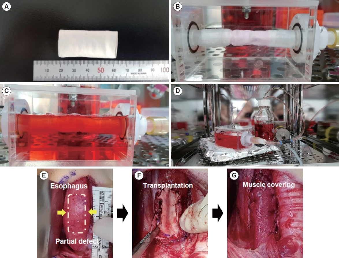
Fig. 2 Bioreactor cultivation of mesenchymal stem cell-inoculated tubular scaffolds and transplantation into esophageal defects in a beagle model. (A) The length and diameter of the prepared two-layered tubular scaffold are 4.5 cm and 2 cm, respectively. After human adipose-derived mesenchymal stem cells were seeded on the two-layered tubular scaffold, it was incubated in a horizontal rotation system for 1 day and then transferred to the bioreactor chamber (B). After filling the chamber with the culture medium (C), the mechanical stimuli were applied at a predetermined time (D). Scaffolds cultured with a bioreactor system were cut to fit the partial defect site of the beagle esophagus and implanted into the esophageal defect (E–G). The graft was covered with a sternocleidomastoid muscle flap for stable regeneration (G).
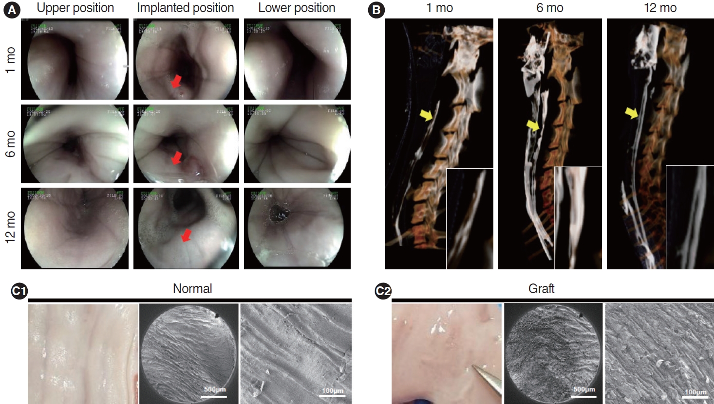
Fig. 3 After transplantation, esophagogastroduodenoscopy was performed at 1, 6, and 12 months to observe the surface conditions inside the esophagus (A). The implanted sites (red arrows) were completely covered with newly formed mucosal layers, showing no stenosis or inflammation. Three-dimensional computed tomography scans were performed at 1, 6, and 12 months after implantation to observe microleakage at the transplanted sites (yellow arrows) (B). No leakage of contrast medium was observed at the graft sites. The morphology of the regenerated mucosal surface at 12 months after transplantation was examined by gross images and scanning electron microscopy analysis of the collected graft sites (C).

Fig. 4 Whole histology of the regenerated esophagus 6 and 12 months after transplantation (A) (H&E; scale bar, 2 mm). At 6 months after transplantation, regeneration of the mucosal layer was clearly observed (black arrows), but regeneration of the muscle layer was incomplete. It was confirmed that the muscle layer was also regenerated at 12 months. In addition, a newly regenerated esophageal gland (indicated by yellow arrows) was observed in the thickly formed submucosa (B) (H&E; scale bar, 200 μm). At 12 months, the esophageal gland was similar to normal (blue arrows). The histological morphology of the esophageal epithelium and submucosa was clearly characterized by Masson’s trichrome staining (C; scale bar, 500 μm).
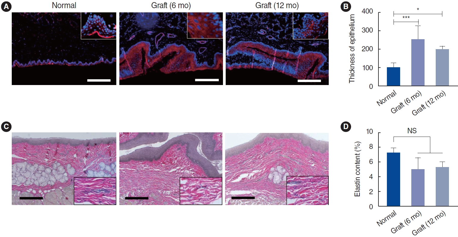
Fig. 5 Re-epithelialization and elastin distribution at 6 and 12 months after scaffold implantation into a partial full-thickness esophageal defect. (A) Keratin-5 immunostaining demonstrated the regenerated esophageal epithelium at 6 and 12 months after implantation (scale bar, 200 μm). (B) The regenerated epithelium was significantly thicker than the normal epithelium (*P<0.05 and ***P<0.001). (C) The regeneration of elastin fibers, indicating the mechanical elasticity of esophageal tissue, was confirmed by elastin immunostaining (scale bar, 200 μm). At 12 months after implantation, elastin fibers with morphologies similar to normal were abundantly observed. (D) No significant differences were found in elastin fibers between the transplanted group (6 and 12 months) and the normal group. NS, not significant.
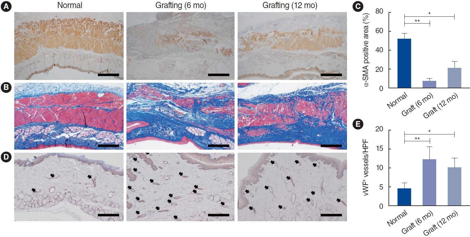
Fig. 6 Immunohistochemical staining of regenerative esophageal muscle and neovascularization at 6 and 12 months after implantation. (A) Representative images of alpha-smooth muscle actin (α-SMA) immunostaining in the reconstructed esophagus after surgery (scale bar, 500 μm). α-SMA-positive signals exhibited newly regenerated muscle adjacent to the implanted sites. (B) Masson’s trichrome shows the morphology of the regenerated esophageal muscle and the distribution of collagen remodeling (scale bar, 500 μm). (C) Quantitative analysis of the α-SMA-positive area. (D) Representative image showing the regenerated blood vessels by von Willebrand factor (vWF) expression. The arrows represent vWF-positive vessels (scale bar, 200 μm). (E) Statistical analysis of the number of vWF-positive blood vessels per high power field. *P<0.05, **P<0.01.
Reference
-
1. Luc G, Charles G, Gronnier C, Cabau M, Kalisky C, Meulle M, et al. Decellularized and matured esophageal scaffold for circumferential esophagus replacement: proof of concept in a pig model. Biomaterials. 2018; Aug. 175:1–18.
Article2. Chung EJ. Bioartificial esophagus: where are we now? Adv Exp Med Biol. 2018; Nov. 1064:313–32.
Article3. Irino T, Tsekrekos A, Coppola A, Scandavini CM, Shetye A, Lundell L, et al. Long-term functional outcomes after replacement of the esophagus with gastric, colonic, or jejunal conduits: a systematic literature review. Dis Esophagus. 2017; Dec. 30(12):1–11.
Article4. Totonelli G, Maghsoudlou P, Fishman JM, Orlando G, Ansari T, Sibbons P, et al. Esophageal tissue engineering: a new approach for esophageal replacement. World J Gastroenterol. 2012; Dec. 18(47):6900–7.
Article5. Kim IG, Wu Y, Park SA, Cho H, Choi JJ, Kwon SK, et al. Tissue-engineered esophagus via bioreactor cultivation for circumferential esophageal reconstruction. Tissue Eng Part A. 2019; Nov. 25(21–22):1478–92.
Article6. Park H, Kim IG, Wu Y, Cho H, Shin JW, Park SA, et al. Experimental investigation of esophageal reconstruction with electrospun polyurethane nanofiber and 3D printing polycaprolactone scaffolds using a rat model. Head Neck. 2021; Mar. 43(3):833–48.7. Kim SD, Kim IG, Tran HN, Cho H, Janarthanan G, Noh I, et al. Three-dimensional printed design of antibiotic-releasing esophageal patches for antimicrobial activity prevention. Tissue Eng Part A. 2021; Dec. 27(23–24):1490–502.
Article8. Chung EJ, Ju HW, Park HJ, Park CH. Three-layered scaffolds for artificial esophagus using poly(ɛ-caprolactone) nanofibers and silk fibroin: an experimental study in a rat model. J Biomed Mater Res A. 2015; Jun. 103(6):2057–65.
Article9. Wu Y, Kang YG, Cho H, Kim IG, Chung EJ, Shin JW. Combinational effects of mechanical forces and substrate surface characteristics on esophageal epithelial differentiation. J Biomed Mater Res A. 2019; Mar. 107(3):552–60.
Article10. Wu Y, Kang YG, Kim IG, Kim JE, Lee EJ, Chung EJ, et al. Mechanical stimuli enhance simultaneous differentiation into oesophageal cell lineages in a double-layered tubular scaffold. J Tissue Eng Regen Med. 2019; Aug. 13(8):1394–405.
Article11. Hosseini V, Ahadian S, Ostrovidov S, Camci-Unal G, Chen S, Kaji H, et al. Engineered contractile skeletal muscle tissue on a microgrooved methacrylated gelatin substrate. Tissue Eng Part A. 2012; Dec. 18(23–24):2453–65.
Article12. Shah R, Knowles JC, Hunt NP, Lewis MP. Development of a novel smart scaffold for human skeletal muscle regeneration. J Tissue Eng Regen Med. 2016; Feb. 10(2):162–71.
Article13. Heher P, Maleiner B, Pruller J, Teuschl AH, Kollmitzer J, Monforte X, et al. A novel bioreactor for the generation of highly aligned 3D skeletal muscle-like constructs through orientation of fibrin via application of static strain. Acta Biomater. 2015; Sep. 24:251–65.
Article14. Tan JY, Chua CK, Leong KF, Chian KS, Leong WS, Tan LP. Esophageal tissue engineering: an in-depth review on scaffold design. Biotechnol Bioeng. 2012; Jan. 109(1):1–15.
Article15. Chian KS, Leong MF, Kono K. Regenerative medicine for oesophageal reconstruction after cancer treatment. Lancet Oncol. 2015; Feb. 16(2):e84–92.
Article16. Del Gaudio C, Baiguera S, Ajalloueian F, Bianco A, Macchiarini P. Are synthetic scaffolds suitable for the development of clinical tissue-engineered tubular organs? J Biomed Mater Res A. 2014; Jul. 102(7):2427–47.
Article17. Yamamoto Y, Nakamura T, Shimizu Y, Takimoto Y, Matsumoto K, Kiyotani T, et al. Experimental replacement of the thoracic esophagus with a bioabsorbable collagen sponge scaffold supported by a silicone stent in dogs. ASAIO J. 1999; Jul–Aug. 45(4):311–6.
Article18. Mallis P, Chachlaki P, Katsimpoulas M, Stavropoulos-Giokas C, Michalopoulos E. Optimization of decellularization procedure in rat esophagus for possible development of a tissue engineered construct. Bioengineering (Basel). 2018; Dec. 6(1):3.
Article19. Dua KS, Hogan WJ, Aadam AA, Gasparri M. In-vivo oesophageal regeneration in a human being by use of a non-biological scaffold and extracellular matrix. Lancet. 2016; Jul. 388(10039):55–61.
Article20. Hu J, Sun X, Ma H, Xie C, Chen YE, Ma PX. Porous nanofibrous PLLA scaffolds for vascular tissue engineering. Biomaterials. 2010; Nov. 31(31):7971–7.
Article21. Kim IG, Hwang MP, Du P, Ko J, Ha CW, Do SH, et al. Bioactive cell-derived matrices combined with polymer mesh scaffold for osteogenesis and bone healing. Biomaterials. 2015; May. 50:75–86.
Article22. Kim IG, Ko J, Lee HR, Do SH, Park K. Mesenchymal cells condensation-inducible mesh scaffolds for cartilage tissue engineering. Biomaterials. 2016; Apr. 85:18–29.
Article23. Oshima T, Gedda K, Koseki J, Chen X, Husmark J, Watari J, et al. Establishment of esophageal-like non-keratinized stratified epithelium using normal human bronchial epithelial cells. Am J Physiol Cell Physiol. 2011; Jun. 300(6):C1422–9.
Article24. Paunescu V, Deak E, Herman D, Siska IR, Tanasie G, Bunu C, et al. In vitro differentiation of human mesenchymal stem cells to epithelial lineage. J Cell Mol Med. 2007; May–Jun. 11(3):502–8.
Article25. Mammoto T, Ingber DE. Mechanical control of tissue and organ development. Development. 2010; May. 137(9):1407–20.
Article26. Giroux V, Lento AA, Islam M, Pitarresi JR, Kharbanda A, Hamilton KE, et al. Long-lived keratin 15+ esophageal progenitor cells contribute to homeostasis and regeneration. J Clin Invest. 2017; Jun. 127(6):2378–91.
Article27. Maxson S, Lopez EA, Yoo D, Danilkovitch-Miagkova A, Leroux MA. Concise review: role of mesenchymal stem cells in wound repair. Stem Cells Transl Med. 2012; Feb. 1(2):142–9.
Article28. Navas A, Magana-Guerrero FS, Dominguez-Lopez A, Chavez-Garcia C, Partido G, Graue-Hernandez EO, et al. Anti-inflammatory and anti-fibrotic effects of human amniotic membrane mesenchymal stem cells and their potential in corneal repair. Stem Cells Transl Med. 2018; Dec. 7(12):906–17.
Article
- Full Text Links
-
- Actions
-
Cited
- CITED
-
- Close
- Share
-
- Similar articles
-
- Effect of serum-derived albumin scaffold and canine adipose tissue-derived mesenchymal stem cells on osteogenesis in canine segmental bone defect model
- The Effect of Polyurethane Scaffold Surface Treatments on the Adhesion of Chondrocytes Subjected to Interstitial Perfusion Culture
- From In Vitro to Perioperative Vascular Tissue Engineering: Shortening Production Time by Traceable Textile-Reinforcement
- Strategies for Constructing Tissue-Engineered Fat for Soft Tissue Regeneration
- Secondary Esophageal Reconstruction for Esophageal Atresia
