Prog Med Phys.
2022 Dec;33(4):158-163. 10.14316/pmp.2022.33.4.158.
Determination of Absorbed Dose for Gafchromic EBT3 Film Using Texture Analysis of Scanning Electron Microscopy Images: A Feasibility Study
- Affiliations
-
- 1Department of Radiation Oncology, Veterans Health Service Medical Center, Seoul, Korea
- KMID: 2537885
- DOI: http://doi.org/10.14316/pmp.2022.33.4.158
Abstract
- Purpose
We subjected scanning electron microscopic (SEM) images of the active layer of EBT3 film to texture analysis to determine the dose-response curve.
Methods
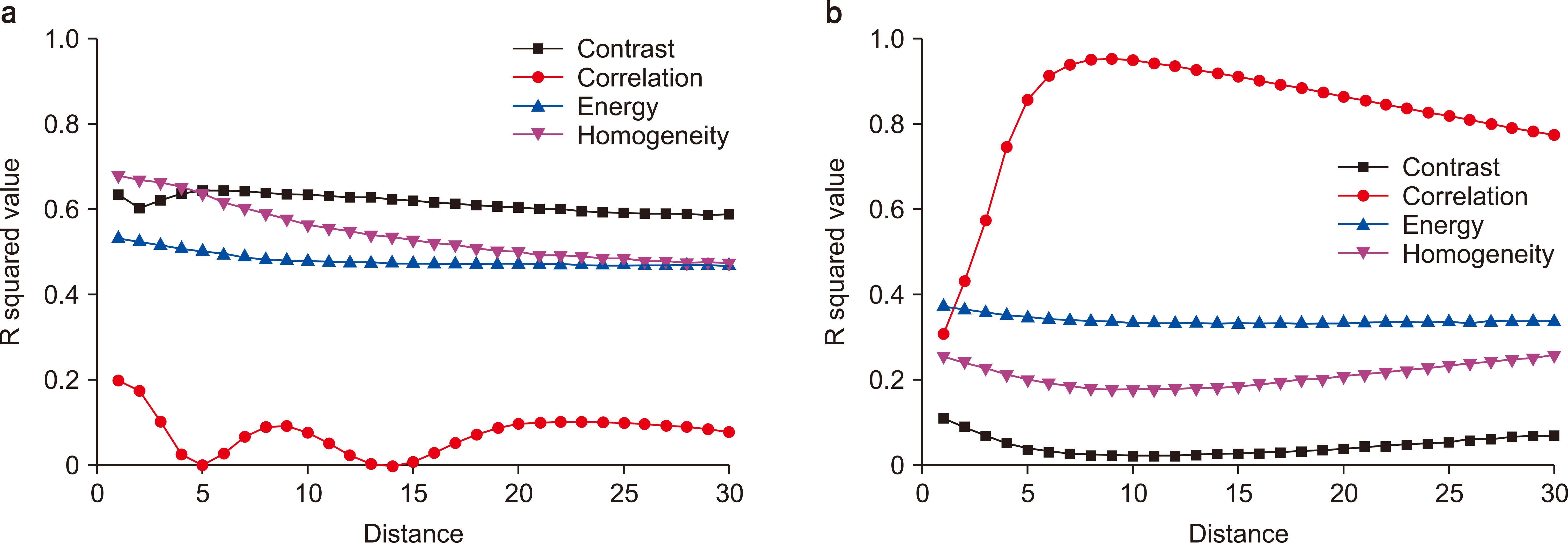
Uncoated Gafchromic EBT3 films were prepared for direct surface SEM scanning. Absorbed doses of 0–20 Gy were delivered to the film’s surface using a 6 MV TrueBeam STx photon beam. The film’s surface was scanned using a SEM under 100× and 3,000× magnification. Four textural features (Homogeneity, Correlation, Contrast, and Energy) were calculated based on the gray level co-occurrence matrix (GLCM) using the SEM images corresponding to each dose. We used R-square to evaluate the linear relationship between delivered doses and textural features of the film’s surface.
Results
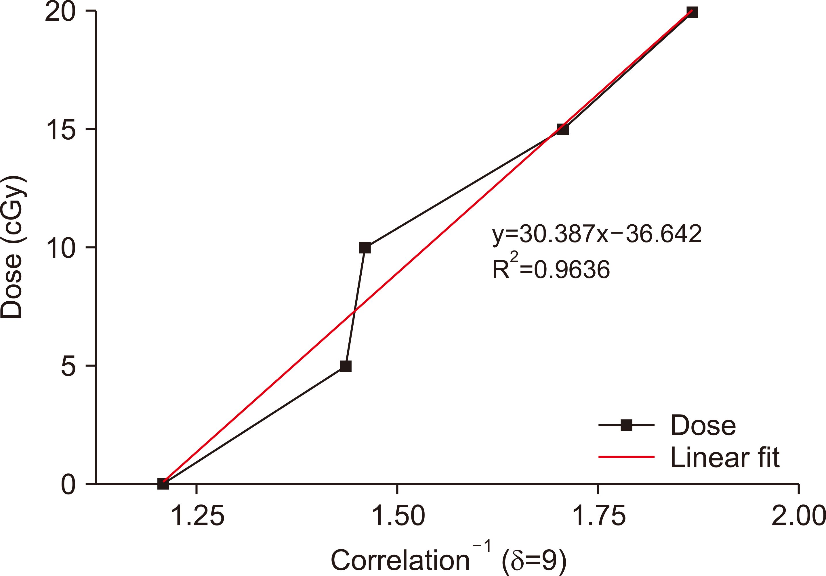
Correlation resulted in higher linearity and dose-response curve sensitivity than Homogeneity, Contrast, or Energy. The R-square value was 0.964 for correlation using 3,000× magnified SEM images with 9-pixel offsets. Dose verification was used to determine the difference between the prescribed and measured doses for 0, 5, 10, 15, and 20 Gy as 0.09, 1.96, −2.29, 0.17, and 0.08 Gy, respectively.
Conclusions
Texture analysis can be used to accurately convert microscopic structural changes to the EBT3 film’s surface into absorbed doses. Our proposed method is feasible and may improve the accuracy of film dosimetry used to protect patients from excess radiation exposure.
Figure
Reference
-
References
1. Casanova Borca V, Pasquino M, Russo G, Grosso P, Cante D, Sciacero P, et al. 2013; Dosimetric characterization and use of GAFCHROMIC EBT3 film for IMRT dose verification. J Appl Clin Med Phys. 14:4111. DOI: 10.1120/jacmp.v14i2.4111. PMID: 23470940. PMCID: PMC5714357.2. Zeidan OA, Stephenson SA, Meeks SL, Wagner TH, Willoughby TR, Kupelian PA, et al. 2006; Characterization and use of EBT radiochromic film for IMRT dose verification. Med Phys. 33:4064–4072. DOI: 10.1118/1.2360012. PMID: 17153386.
Article3. Marrazzo L, Zani M, Pallotta S, Arilli C, Casati M, Compagnucci A, et al. 2015; GafChromic® EBT3 films for patient specific IMRT QA using a multichannel approach. Phys Med. 31:1035–1042. DOI: 10.1016/j.ejmp.2015.08.010. PMID: 26429383.
Article4. Ocadiz A, Livingstone J, Donzelli M, Bartzsch S, Nemoz C, Kefs S, et al. 2019; Film dosimetry studies for patient specific quality assurance in microbeam radiation therapy. Phys Med. 65:227–237. DOI: 10.1016/j.ejmp.2019.09.071. PMID: 31574356.
Article5. Stella G, Cavalli N, Bonanno E, Zirone L, Borzì GR, Pace M, et al. 2022; SBRT/SRS patient-specific QA using GAFchromicTM EBT3 and FilmQATM Pro software. J Radiosurg SBRT. 8:37–45. PMID: 35387411. PMCID: PMC8930055.6. Liu HW, Gräfe J, Khan R, Olivotto I, Barajas JE. 2015; Role of in vivo dosimetry with radiochromic films for dose verification during cutaneous radiation therapy. Radiat Oncol. 10:12. DOI: 10.1186/s13014-014-0325-0. PMID: 25582565. PMCID: PMC4300174.
Article7. Moylan R, Aland T, Kairn T. 2013; Dosimetric accuracy of Gafchromic EBT2 and EBT3 film for in vivo dosimetry. Australas Phys Eng Sci Med. 36:331–337. DOI: 10.1007/s13246-013-0206-0. PMID: 23801092.
Article8. Hartmann B, Martisiková M, Jäkel O. 2010; Homogeneity of Gafchromic EBT2 film. Med Phys. 37:1753–1756. DOI: 10.1118/1.3368601. PMID: 20443496. PMCID: PMC5716526.9. Mizuno H, Takahashi Y, Tanaka A, Hirayama T, Yamaguchi T, Katou H, et al. 2012; Homogeneity of GAFCHROMIC EBT2 film among different lot numbers. J Appl Clin Med Phys. 13:3763. DOI: 10.1120/jacmp.v13i4.3763. PMID: 22766947. PMCID: PMC5716526.
Article10. Martisíková M, Ackermann B, Jäkel O. 2008; Analysis of uncertainties in Gafchromic EBT film dosimetry of photon beams. Phys Med Biol. 53:7013–7027. DOI: 10.1088/0031-9155/53/24/001. PMID: 19015581.
Article11. Saur S, Frengen J. 2008; GafChromic EBT film dosimetry with flatbed CCD scanner: a novel background correction method and full dose uncertainty analysis. Med Phys. 35:3094–3101. DOI: 10.1118/1.2938522. PMID: 18697534.
Article12. Marroquin EY, Herrera González JA, Camacho López MA, Barajas JE, García-Garduño OA. 2016; Evaluation of the uncertainty in an EBT3 film dosimetry system utilizing net optical density. J Appl Clin Med Phys. 17:466–481. DOI: 10.1120/jacmp.v17i5.6262. PMID: 27685125. PMCID: PMC5874103.
Article13. Palmer AL, Bradley D, Nisbet A. 2014; Evaluation and implementation of triple-channel radiochromic film dosimetry in brachytherapy. J Appl Clin Med Phys. 15:4854. DOI: 10.1120/jacmp.v15i4.4854. PMID: 25207417. PMCID: PMC5875501.
Article14. Holm KM, Yukihara EG, Ahmed MF, Greilich S, Jäkel O. 2021; Triple channel analysis of Gafchromic EBT3 irradiated with clinical carbon-ion beams. Phys Med. 87:123–130. DOI: 10.1016/j.ejmp.2021.06.009. PMID: 34146794.
Article15. Ghalati MK, Nunes A, Ferreira H, Serranho P, Bernardes R. 2022; Texture analysis and its applications in biomedical imaging: a survey. IEEE Rev Biomed Eng. 15:222–246. DOI: 10.1109/RBME.2021.3115703. PMID: 34570709.
Article16. Colen RR, Rolfo C, Ak M, Ayoub M, Ahmed S, Elshafeey N, et al. 2021; Radiomics analysis for predicting pembrolizumab response in patients with advanced rare cancers. J Immunother Cancer. 9:e001752. DOI: 10.1136/jitc-2020-001752. PMID: 33849924. PMCID: PMC8051405.
Article17. Brynolfsson P, Nilsson D, Torheim T, Asklund T, Karlsson CT, Trygg J, et al. 2017; Haralick texture features from apparent diffusion coefficient (ADC) MRI images depend on imaging and pre-processing parameters. Sci Rep. 7:4041. DOI: 10.1038/s41598-017-04151-4. PMID: 28642480. PMCID: PMC5481454.
Article18. Rui W, Qiao N, Wu Y, Zhang Y, Aili A, Zhang Z, et al. 2022; Radiomics analysis allows for precise prediction of silent corticotroph adenoma among non-functioning pituitary adenomas. Eur Radiol. 32:1570–1578. DOI: 10.1007/s00330-021-08361-3. PMID: 34837512.
Article19. Shih CT, Hsu JT, Han RP, Hsieh BT, Chang SJ, Wu J. 2013; A novel method of estimating dose responses for polymer gels using texture analysis of scanning electron microscopy images. PLoS One. 8:e67281. DOI: 10.1371/journal.pone.0067281. PMID: 23843998. PMCID: PMC3699568.
Article20. Almond PR, Biggs PJ, Coursey BM, Hanson WF, Huq MS, Nath R, et al. 1999; AAPM's TG-51 protocol for clinical reference dosimetry of high-energy photon and electron beams. Med Phys. 26:1847–1870. DOI: 10.1118/1.598691. PMID: 10505874.
Article21. Arjomandy B, Tailor R, Zhao L, Devic S. 2012; EBT2 film as a depth-dose measurement tool for radiotherapy beams over a wide range of energies and modalities. Med Phys. 39:912–921. DOI: 10.1118/1.3678989. PMID: 22320801.
Article22. Volotskova O, Fang X, Keidar M, Chandarana H, Das IJ. 2019; Microstructure changes in radiochromic films due to magnetic field and radiation. Med Phys. 46:293–301. DOI: 10.1002/mp.13248. PMID: 30341911.
Article
- Full Text Links
- Actions
-
Cited
- CITED
-
- Close
- Share
- Similar articles
-
- Development of Water-Filled Phantom and Comparision Film Dosimetry for Five Phantoms in Gamma Knife
- Comparative Studies on Absorbed Dose by Geant4-based Simulation Using DICOM File and Gafchromic EBT2 Film
- Linear Energy Transfer Dependence Correction of Spread-Out Bragg Peak Measured by EBT3 Film for Dynamically Scanned Proton Beams
- Measurement of Proton Beam Dose-Averaged Linear Energy Transfer Using a Radiochromic Film
- Feasibility Study of a Custom-made Film for End-to-End Quality Assurance Test of Robotic Intensity Modulated Radiation Therapy System


