Linear Energy Transfer Dependence Correction of Spread-Out Bragg Peak Measured by EBT3 Film for Dynamically Scanned Proton Beams
- Affiliations
-
- 1Samsung Advanced Institute for Health Sciences & Technology (SAIHST), Sungkyunkwan University, Seoul, Korea
- 2Department of Radiation Oncology, Samsung Medical Center (SMC), Seoul, Korea
- KMID: 2510474
- DOI: http://doi.org/10.14316/pmp.2020.31.4.135
Abstract
- Purpose
Gafchromic films for proton dosimetry are dependent on linear energy transfers (LETs), resulting in dose underestimation for high LETs. Despite efforts to resolve this problem for singleenergy beams, there remains a need to do so for multi-energy beams. Here, a bimolecular reaction model was applied to correct the under-response of spread-out Bragg peaks (SOBPs).
Methods
For depth-dose measurements, a Gafchromic EBT3 film was positioned in water perpendicular to the ground. The gantry was rotated at 15° to avoid disturbances in the beam path.A set of films was exposed to a uniformly scanned 112-MeV pristine proton beam with six different dose intensities, ranging from 0.373 to 4.865 Gy, at a 2-cm depth. Another set of films was irradiated with SOBPs with maximum energies of 110, 150, and 190 MeV having modulation widths of 5.39, 4.27, and 5.34 cm, respectively. The correction function was obtained using 150.8-MeV SOBP data. The LET of the SOBP was then analytically calculated. Finally, the model was validated for a uniform cubic dose distribution and compared with multilayered ionization chamber data.
Results
The dose error in the plateau region was within 4% when normalized with the maximum dose. The discrepancy of the range was <1 mm for all measured energies. The highest errors occurred at 70 MeV owing to the steep gradient with the narrowest Bragg peak.
Conclusions
With bimolecular model-based correction, an EBT3 film can be used to accurately verify the depth dose of scanned proton beams and could potentially be used to evaluate the depth-dose distribution for patient plans.
Figure
-

Fig. 1 (a) Experimental setup of the film fixed by an in-house-designed film holder in a water tank, (b) Gantry tilted as 3 degree, (c) 1 cm of the gap distance between water surface and the edge of the film.
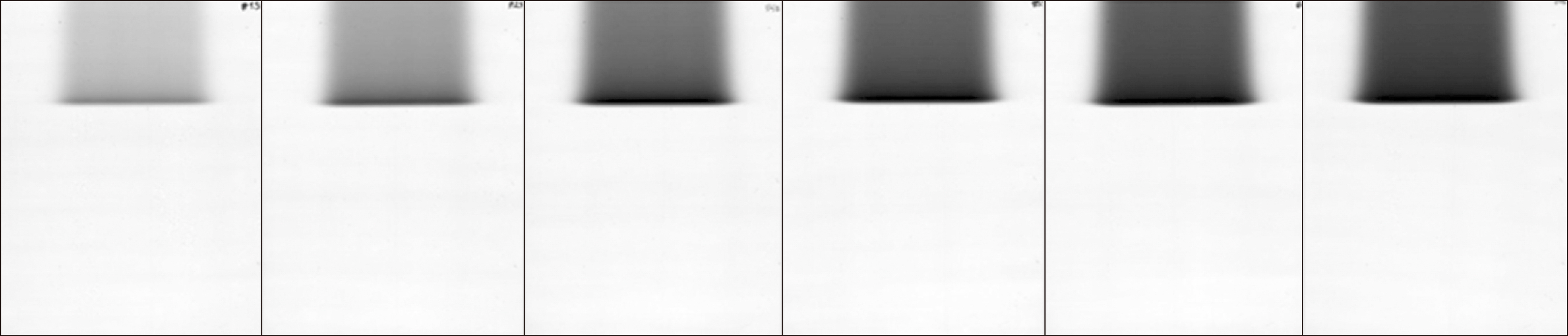
Fig. 2 Scanned film irradiated by 112-MeV beams, showing pristine Bragg peaks with six different dose intensities.

Fig. 3 Analytically calculated linear energy transfer (LET) of spread-out Bragg peaks (SOBPs) with maximum energies of 110, 150, and 190 MeV.

Fig. 4 Dose distribution of cubic beam irradiated obliquely with a gantry angle of 15° in treatment planning system.
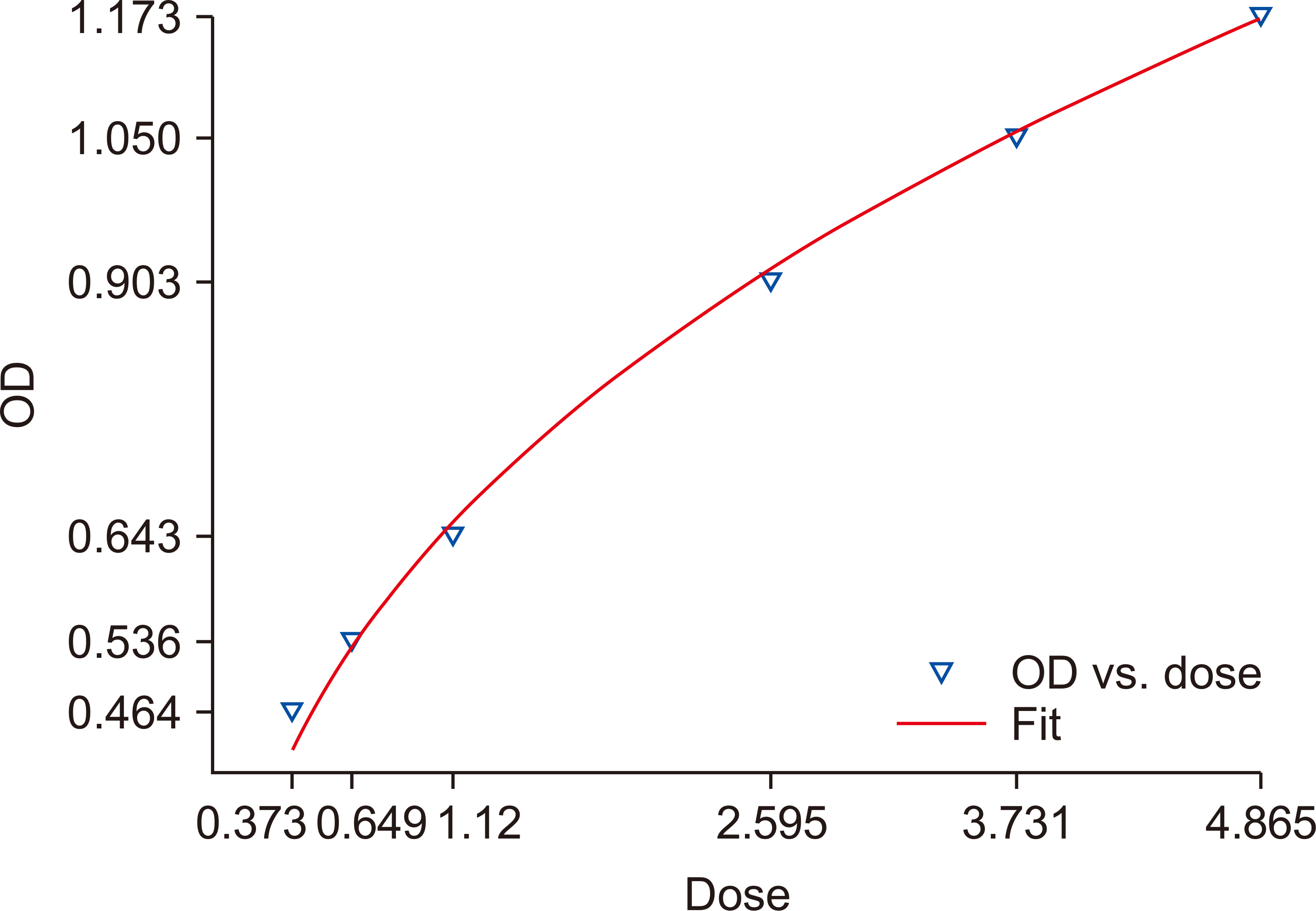
Fig. 5 Characteristic curve of 112-MeV pristine Bragg peak for six different dose intensities at a 2-cm depth.

Fig. 6 (a) D1/2 calculated using spread-out Bragg peak (SOBP) data with a maximum energy of 150 MeV represented along the water depth and the fitted curve based on a fifth-degree Gaussian curve. (b) D1/2 converted to a function along the linear energy transfer (LET).

Fig. 7 Pristine Bragg peaks (70, 92, 112, 150, 170, and 190 MeV from left to right) measured using the EBT3 film (corrected; solid line) and those measured using the MLIC (Zebra; inverted triangles).
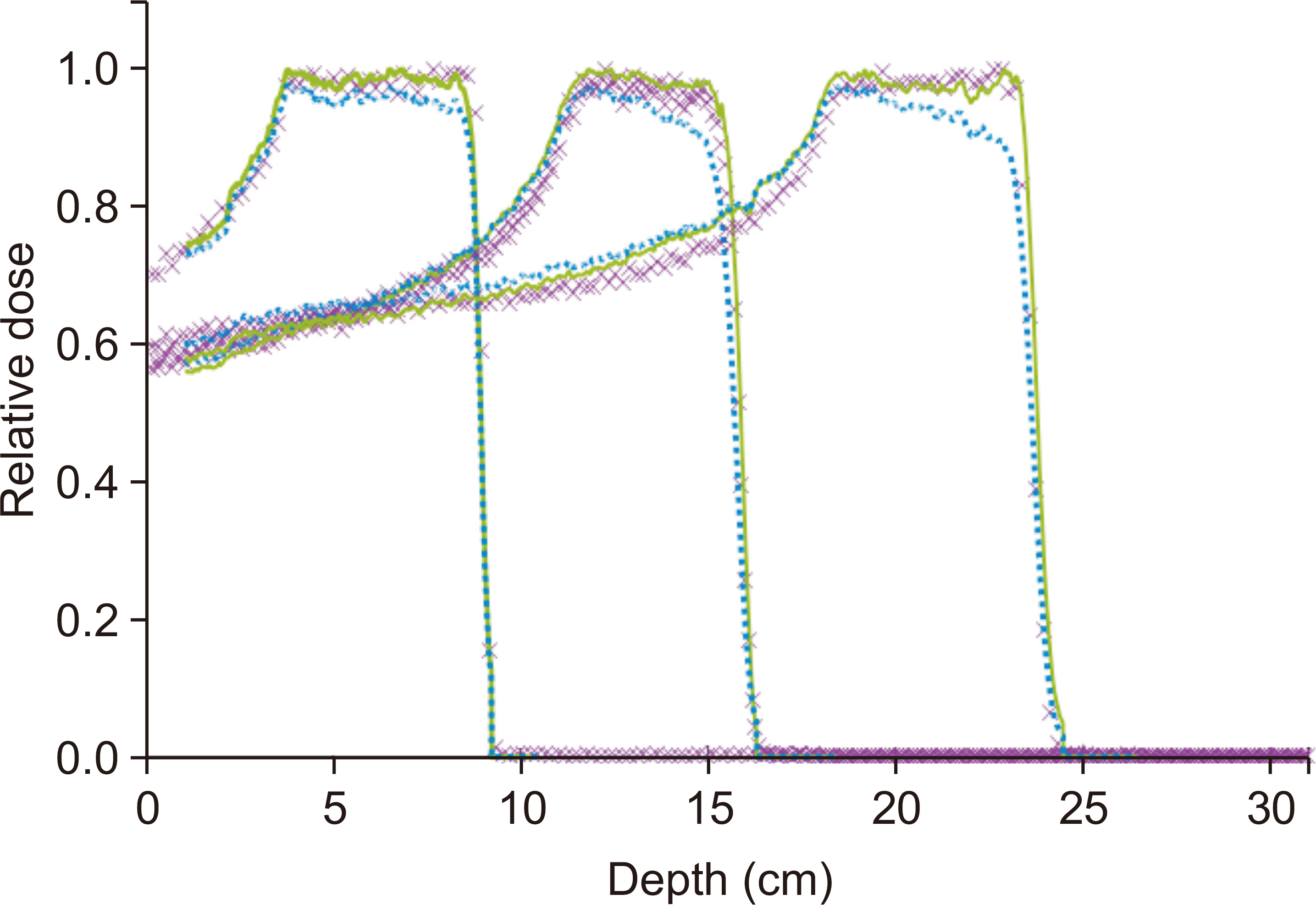
Fig. 8 Depth profile of spread-out Bragg peak (SOBP) measured using the EBT3 film (uncorrected shown as dotted blue line and corrected as solid green line) compared with the multilayer ionization chamber (MLIC) data (purple ‘x' symbol) for maximum energies of 110, 150, and 190 MeV.
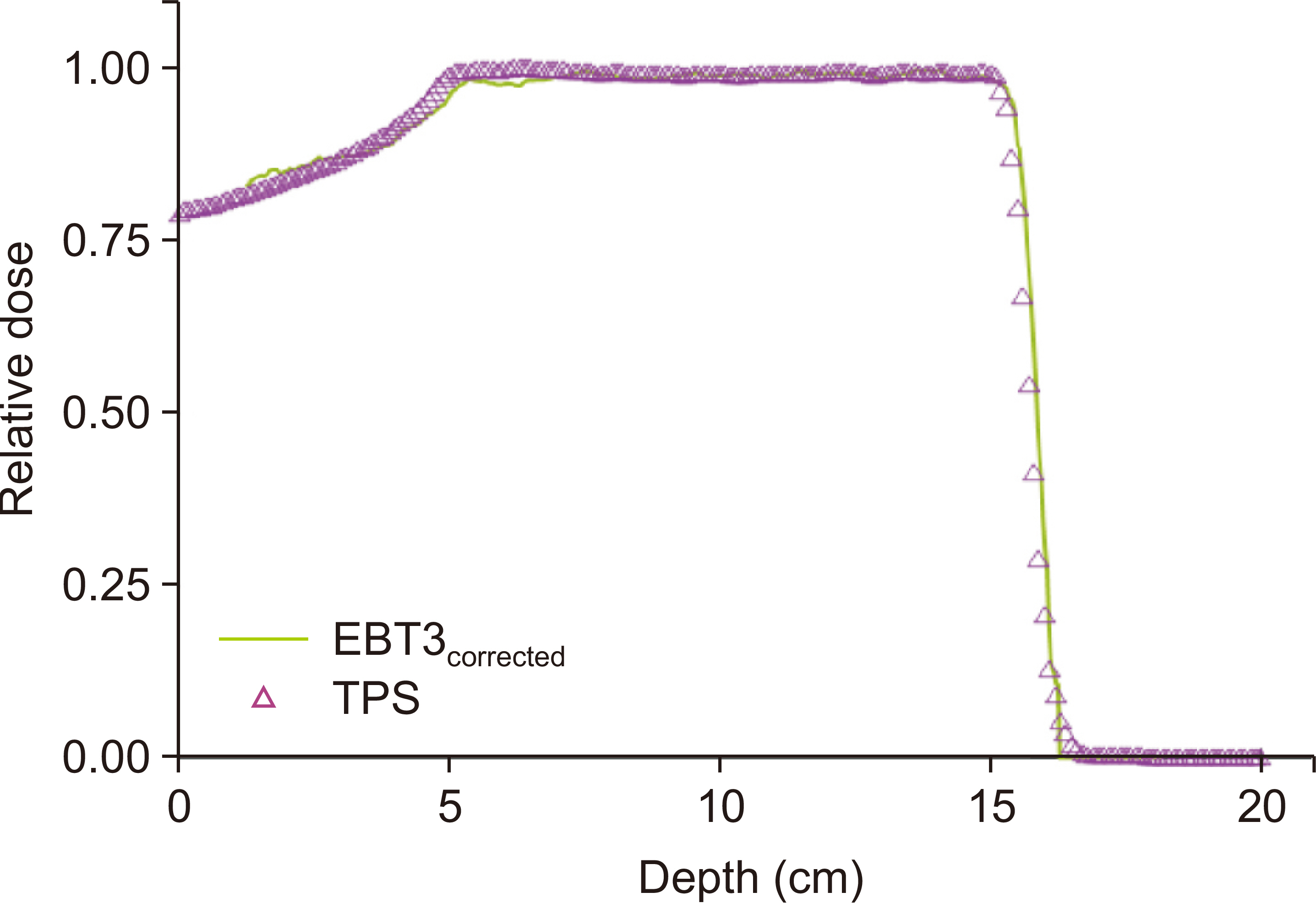
Fig. 9 Depth profile of the cubic treatment planning system (TPS) plan measured with film compared with the TPS data.
Cited by 1 articles
-
Measurement of Proton Beam Dose-Averaged Linear Energy Transfer Using a Radiochromic Film
Seohyeon An, Sang-il Pak, Seonghoon Jeong, Soonki Min, Tae Jeong Kim, Dongho Shin, Youngkyung Lim, Jong Hwi Jeong, Haksoo Kim, Se Byeong Lee
Prog Med Phys. 2022;33(4):80-87. doi: 10.14316/pmp.2022.33.4.80.
Reference
-
References
1. International Commission on Radiation Units & Measurements. 2009. ICRU Report 78: Prescribing, recording, and reporting proton-beam therapy. International Commission on Radiation Units & Measurements;Bethesda: p. 60–62.2. Schaffner B, Pedroni E, Lomax A. 1999; Dose calculation models for proton treatment planning using a dynamic beam delivery system: an attempt to include density heterogeneity effects in the analytical dose calculation. Phys Med Biol. 44:27–41. DOI: 10.1088/0031-9155/44/1/004. PMID: 10071873.
Article3. Sorriaux J, Kacperek A, Rossomme S, Lee JA, Bertrand D, Vynckier S, et al. 2013; Evaluation of Gafchromic® EBT3 films characteristics in therapy photon, electron and proton beams. Phys Med. 29:599–606. DOI: 10.1016/j.ejmp.2012.10.001. PMID: 23107430.
Article4. Reinhardt S, Hillbrand M, Wilkens JJ, Assmann W. 2012; Comparison of Gafchromic EBT2 and EBT3 films for clinical photon and proton beams. Med Phys. 39:5257–5262. DOI: 10.1118/1.4737890. PMID: 22894450.
Article5. Arjomandy B, Tailor R, Anand A, Sahoo N, Gillin M, Prado K, et al. 2010; Energy dependence and dose response of Gafchromic EBT2 film over a wide range of photon, electron, and proton beam energies. Med Phys. 37:1942–1947. DOI: 10.1118/1.3373523. PMID: 20527528.
Article6. Kirby D, Green S, Palmans H, Hugtenburg R, Wojnecki C, Parker D. 2010; LET dependence of GafChromic films and an ion chamber in low-energy proton dosimetry. Phys Med Biol. 55:417–433. DOI: 10.1088/0031-9155/55/2/006. PMID: 20019399.
Article7. Zhao L, Das IJ. 2010; Gafchromic EBT film dosimetry in proton beams. Phys Med Biol. 55:N291–N301. DOI: 10.1088/0031-9155/55/10/N04. PMID: 20427858.
Article8. Perles LA, Mirkovic D, Anand A, Titt U, Mohan R. 2013; LET dependence of the response of EBT2 films in proton dosimetry modeled as a bimolecular chemical reaction. Phys Med Biol. 58:8477–8491. DOI: 10.1088/0031-9155/58/23/8477. PMID: 24240474.
Article9. Park SA, Kwak JW, Yoon MG, Shin DH, Lee SB, Cho KH, et al. 2011; Dose verification of proton beam therapy using the Gafchromic EBT film. Radiat Meas. 46:717–721.
Article10. Gambarini G, Regazzoni V, Grisotto S, Artuso E, Giove D, Borroni M, et al. 2014. Measurements of spatial distributionof absorbed dose in protontherapy with Gafchromic EBT3. Paper presented at: International Symposium on Solid State Dosimetry. p. 742–755. 2014 Apr 13-16; Cusco, Peru.11. Fiorini F, Kirby D, Borghesi M, Doria D, Jeynes JC, Kakolee KF, et al. 2011; Dosimetry and spectral analysis of a radiobiological experiment using laser-driven proton beams. Phys Med Biol. 56:6969–6982. DOI: 10.1088/0031-9155/56/21/013. PMID: 22008825.
Article12. Fiorini F, Kirby D, Thompson J, Green S, Parker DJ, Jones B, et al. 2014; Under-response correction for EBT3 films in the presence of proton spread out Bragg peaks. Phys Med. 30:454–461. DOI: 10.1016/j.ejmp.2013.12.006. PMID: 24461335.
Article13. Lyman T. 1914; Victor schumann. Astrophys J. 38:1–4.
Article14. Candler C. 1960; The photographic process as a diatomic reaction. Aust J Phys. 13:419–436.
Article15. Candler C. 1961; The track density of a charged particle in a photographic emulsion. Il Nuovo Cimento (1955-1965). 21:1–6.
Article16. van Battum LJ, Heijmen BJ. 1995; Film dosimetry in water in a 23 MV therapeutic photon beam. Radiother Oncol. 34:152–159. DOI: 10.1016/0167-8140(94)01500-3. PMID: 7597214.
Article17. Kanai T, Kawachi K, Matsuzawa H, Inada T. 1983; Three-dimensional beam scanning for proton therapy. Nucl Instrum Methods Phys Res. 214:491–496.
Article18. Wilkens JJ, Oelfke U. 2003; Analytical linear energy transfer calculations for proton therapy. Med Phys. 30:806–815. DOI: 10.1118/1.1567852. PMID: 12772988.
Article19. Rosenfeld AB, Wroe AJ, Cornelius IM, Reinhard M, Alexiev D. 2004; Analysis of inelastic interactions for therapeutic proton beams using Monte Carlo simulation. IEEE Trans Nucl Sci. 51:3019–3025.
Article20. Paganetti H. 2002; Nuclear interactions in proton therapy: dose and relative biological effect distributions originating from primary and secondary particles. Phys Med Biol. 47:747–764. DOI: 10.1088/0031-9155/47/5/305. PMID: 11931469.
Article
- Full Text Links
-
- Actions
-
Cited
- CITED
-
- Close
- Share
-
- Similar articles
-
- Measurement of Proton Beam Dose-Averaged Linear Energy Transfer Using a Radiochromic Film
- Proton Beam Therapy
- Evaluation of the Secondary Particle Effect in Inhomogeneous Media for Proton Therapy Using Geant4 Based MC Simulation
- Determination of Absorbed Dose for Gafchromic EBT3 Film Using Texture Analysis of Scanning Electron Microscopy Images: A Feasibility Study
- Therapeutic Proton Beam Range Measurement with EBT3 Film and Comparison with Tool for Particle Simulation
