Prog Med Phys.
2022 Dec;33(4):80-87. 10.14316/pmp.2022.33.4.80.
Measurement of Proton Beam Dose-Averaged Linear Energy Transfer Using a Radiochromic Film
- Affiliations
-
- 1Proton Therapy Center, National Cancer Center, Goyang, Seoul, Korea
- 2Department of Physics, Hanyang University, Seoul, Korea
- 3Department of Radiation Oncology, Kyung Hee University Hospital, Seoul, Korea
- KMID: 2537875
- DOI: http://doi.org/10.14316/pmp.2022.33.4.80
Abstract
- Purpose
Proton therapy has different relative biological effectiveness (RBE) compared with X-ray treatment, which is the standard in radiation therapy, and the fixed RBE value of 1.1 is widely used. However, RBE depends on a charged particle’s linear energy transfer (LET); therefore, measuring LET is important. We have developed a LET measurement method using the inefficiency characteristic of an EBT3 film on a proton beam’s Bragg peak (BP) region.
Methods
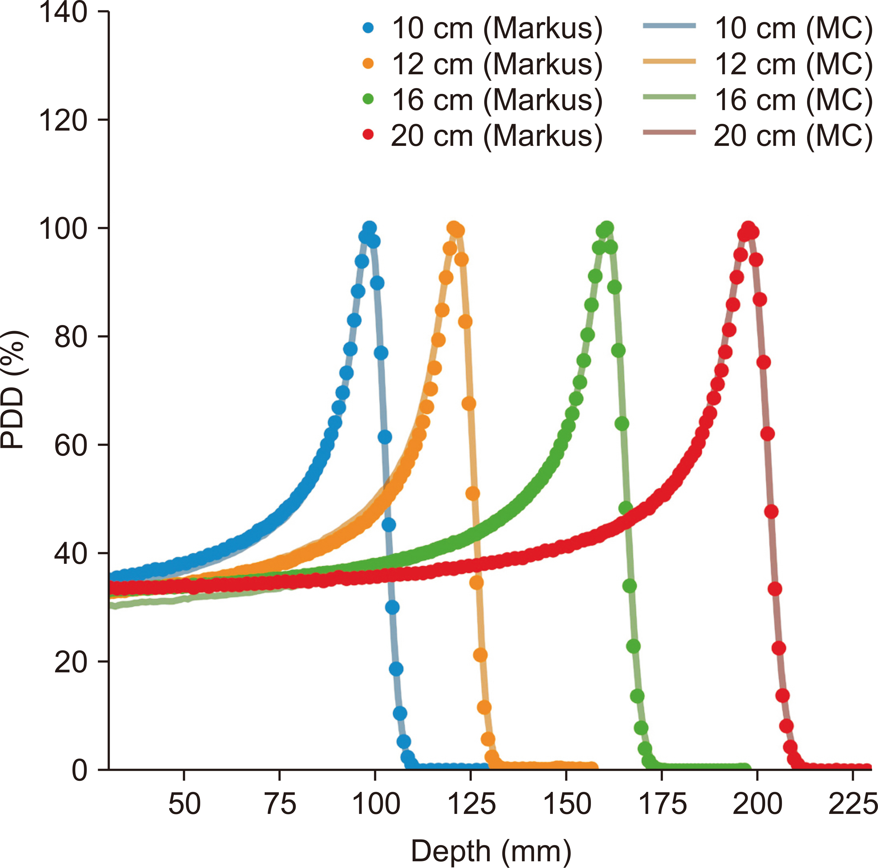
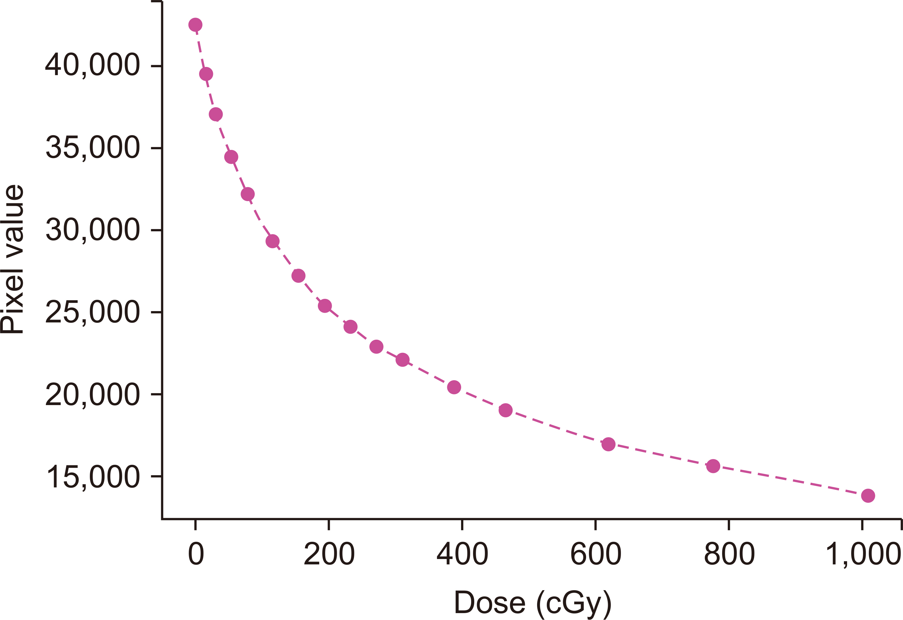
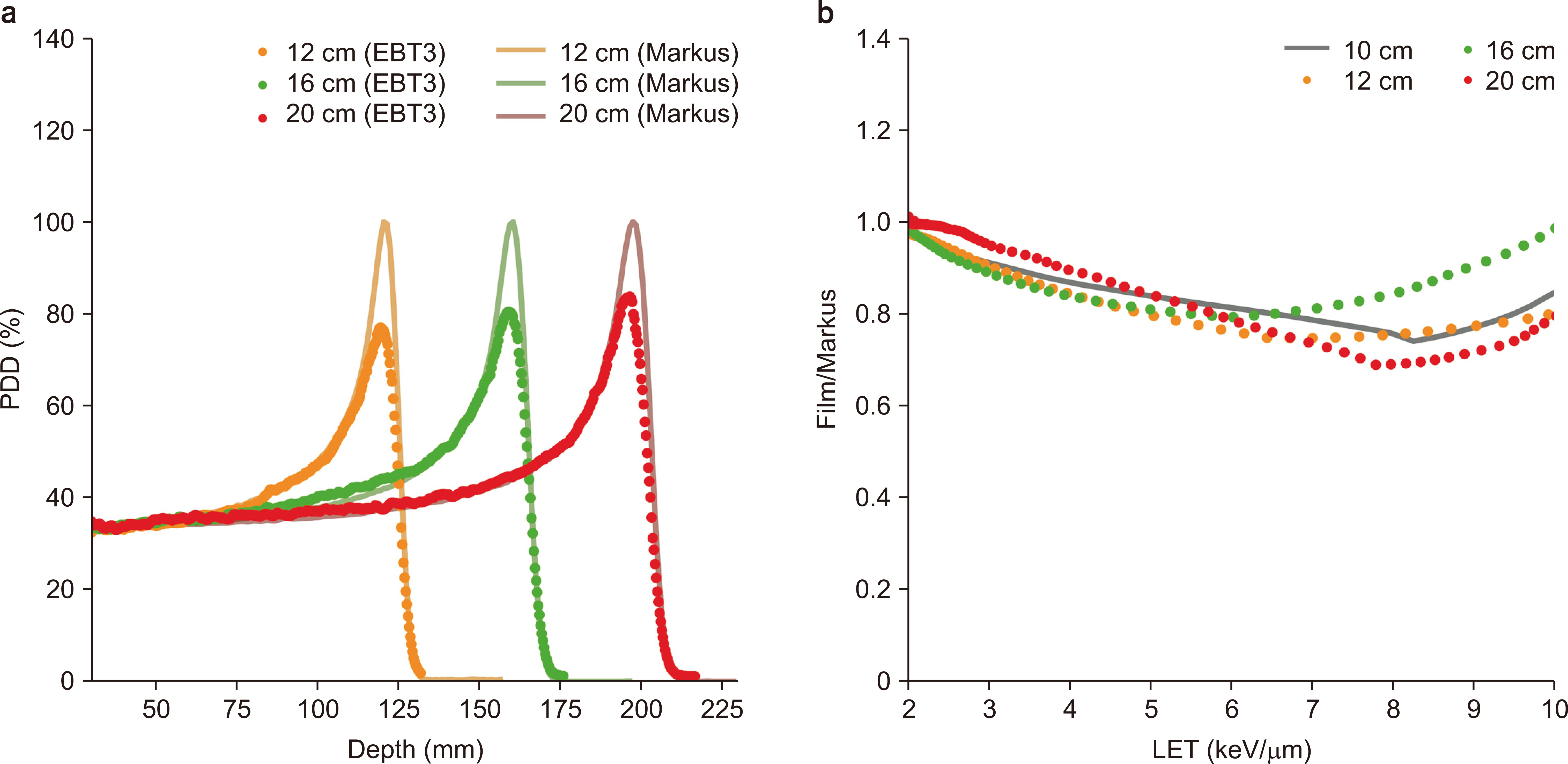
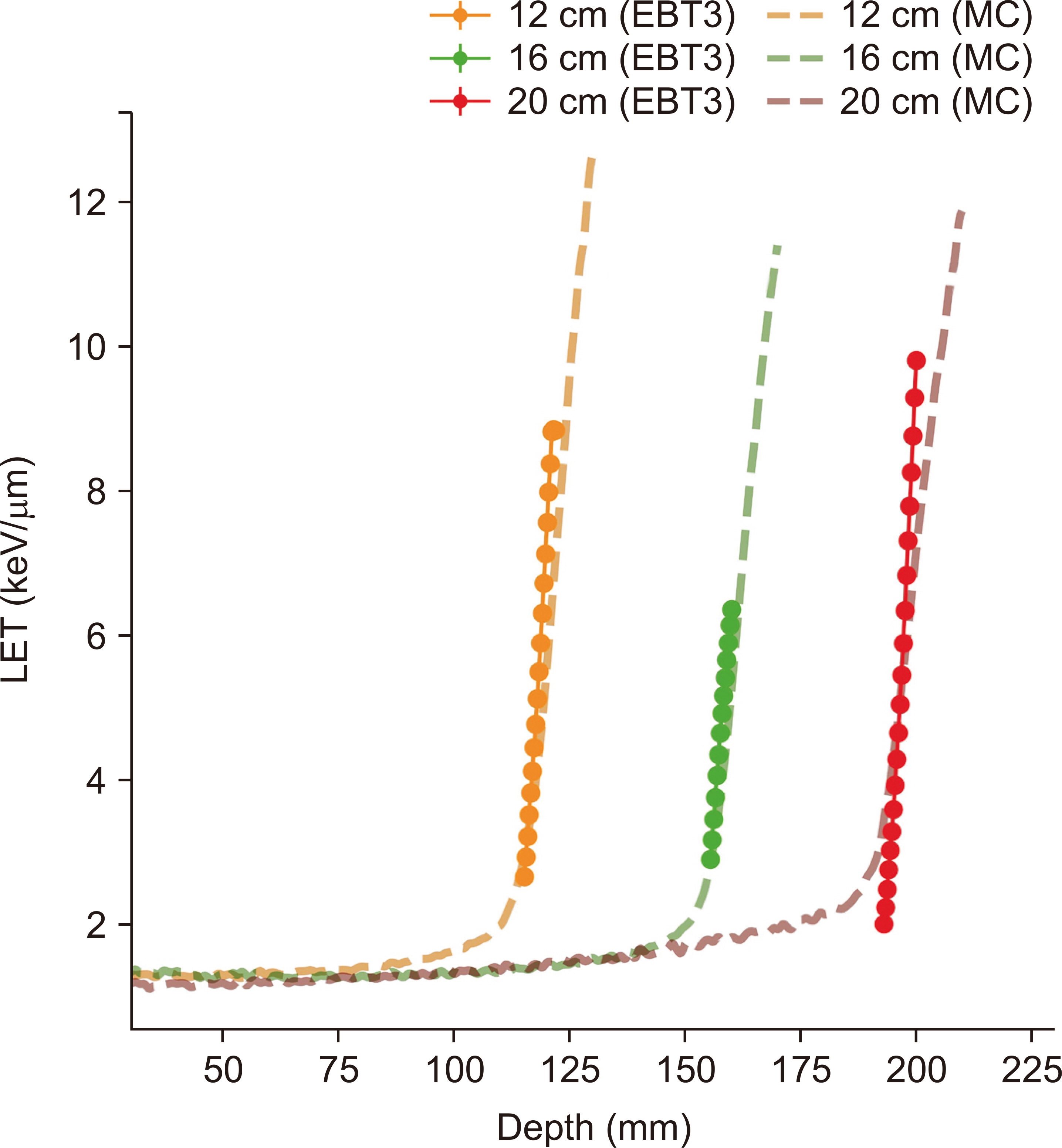
A Gafchromic EBT3 film was used to measure the proton beam LET. It measured the dose at a 10-cm pristine BP proton beam in water to determine the quenching factor of the EBT3 film as a reference beam condition. Monte Carlo (MC) calculations of dose-averaged LET (LET d ) were used to determine the quenching factor and validation. The dose-averaged LETs at the 12-, 16-, and 20-cm pristine BP proton beam in water were calculated with the quenching factor.
Results
Using the passive scattering proton beam nozzle of the National Cancer Center in Korea, the LET d was measured for each beam range. The quenching factor was determined to be 26.15 with 0.3% uncertainty under the reference beam condition. The dose-averaged LETs were measured for each test beam condition.
Conclusions
We developed a method for measuring the proton beam LET using an EBT3 film. This study showed that the magnitude of the quenching effect can be estimated using only one beam range, and the quenching factor determined under the reference condition can be applied to any therapeutic proton beam range.
Keyword
Figure
Reference
-
References
1. Buchsbaum JC, McDonald MW, Johnstone PA, Hoene T, Mendonca M, Cheng CW, et al. 2014; Range modulation in proton therapy planning: a simple method for mitigating effects of increased relative biological effectiveness at the end-of-range of clinical proton beams. Radiat Oncol. 9:2. DOI: 10.1186/1748-717X-9-2. PMID: 24383792. PMCID: PMC3904459.
Article2. Carabe A, Moteabbed M, Depauw N, Schuemann J, Paganetti H. 2012; Range uncertainty in proton therapy due to variable biological effectiveness. Phys Med Biol. 57:1159–1172. DOI: 10.1088/0031-9155/57/5/1159. PMID: 22330133.
Article3. Zhao L, Das IJ. 2010; Gafchromic EBT film dosimetry in proton beams. Phys Med Biol. 55:N291–N301. Erratum in: Phys Med Biol. 2010;55:5617. DOI: 10.1088/0031-9155/55/10/N04. PMID: 20427858.
Article4. Spielberger B, Scholz M, Krämer M, Kraft G. 2001; Experimental investigations of the response of films to heavy-ion irradiation. Phys Med Biol. 46:2889–2897. DOI: 10.1088/0031-9155/46/11/309. PMID: 11720353.
Article5. Martisíková M, Jäkel O. 2010; Dosimetric properties of Gafchromic® EBT films in medical carbon ion beams. Phys Med Biol. 55:5557–5567. DOI: 10.1088/0031-9155/55/18/019. PMID: 20808027.
Article6. Anderson SE, Grams MP, Wan Chan Tseung H, Furutani KM, Beltran CJ. 2019; A linear relationship for the LET-dependence of Gafchromic EBT3 film in spot-scanning proton therapy. Phys Med Biol. 64:055015. DOI: 10.1088/1361-6560/ab0114. PMID: 30673655.
Article7. Kawashima M, Matsumura A, Souda H, Tashiro M. 2020; Simultaneous determination of the dose and linear energy transfer (LET) of carbon-ion beams using radiochromic films. Phys Med Biol. 65:125002. DOI: 10.1088/1361-6560/ab8bf3. PMID: 32320970.
Article8. Lee M, Ahn S, Cheon W, Han Y. 2020; Linear energy transfer dependence correction of spread-out Bragg peak measured by EBT3 film for dynamically scanned proton beams. Prog Med Phys. 31:135–144. DOI: 10.14316/pmp.2020.31.4.135.
Article9. Valdetaro LB, Høye EM, Skyt PS, Petersen JBB, Balling P, Muren LP. 2021; Empirical quenching correction in radiochromic silicone-based three-dimensional dosimetry of spot-scanning proton therapy. Phys Imaging Radiat Oncol. 18:11–18. DOI: 10.1016/j.phro.2021.03.006. PMID: 34258402. PMCID: PMC8254200.
Article10. Kalholm F, Grzanka L, Traneus E, Bassler N. 2021; A systematic review on the usage of averaged LET in radiation biology for particle therapy. Radiother Oncol. 161:211–221. DOI: 10.1016/j.radonc.2021.04.007. PMID: 33894298.
Article11. Birks JB. 1951; Scintillations from organic crystals: specific fluorescence and relative response to different radiations. Proc Phys Soc A. 64:874. DOI: 10.1088/0370-1298/64/10/303.
Article12. Wang LL, Perles LA, Archambault L, Sahoo N, Mirkovic D, Beddar S. 2012; Determination of the quenching correction factors for plastic scintillation detectors in therapeutic high-energy proton beams. Phys Med Biol. 57:7767–7781. DOI: 10.1088/0031-9155/57/23/7767. PMID: 23128412. PMCID: PMC3849705.
Article13. Jeong S, Kim C, An S, Kwon YC, Pak SI, Cheon W, et al. 2022; Determination of the proton LET using thin film solar cells coated with scintillating powder. Med Phys. doi: 10.1002/mp.15977. DOI: 10.1002/mp.15977. PMID: 36135795.
Article14. Perl J, Shin J, Schumann J, Faddegon B, Paganetti H. 2012; TOPAS: an innovative proton Monte Carlo platform for research and clinical applications. Med Phys. 39:6818–6837. DOI: 10.1118/1.4758060. PMID: 23127075. PMCID: PMC3493036.
Article15. Shin WG, Testa M, Kim HS, Jeong JH, Lee SB, Kim YJ, et al. 2017; Independent dose verification system with Monte Carlo simulations using TOPAS for passive scattering proton therapy at the National Cancer Center in Korea. Phys Med Biol. 62:7598–7616. DOI: 10.1088/1361-6560/aa8663. PMID: 28809759.
Article
- Full Text Links
- Actions
-
Cited
- CITED
-
- Close
- Share
- Similar articles
-
- Linear Energy Transfer Dependence Correction of Spread-Out Bragg Peak Measured by EBT3 Film for Dynamically Scanned Proton Beams
- Feasibility Test of Flat-Type Faraday Cup for UltrahighDose-Rate Transmission Proton Beam Therapy
- Proton Beam Therapy
- Evaluation of the Radiochromic Film Dosimetry for a Small Curved Interface
- Measurement of the energy of high-energy electron beam with rapid-processed film


