A Fibular Lengthening Osteotomy Combined with Calcaneal Osteotomy for Post-Traumatic Valgus Ankle Arthritis: A Case Report
- Affiliations
-
- 1Departments of Orthopaedic Surgery, Ilsan Paik Hospital, Inje University College of Medicine, Goyang, Korea
- KMID: 2533197
- DOI: http://doi.org/10.14193/jkfas.2022.26.3.143
Abstract
- Past research has reported that the common causes of ankle arthritis include trauma, congenital deformity, and degeneration. Among them, fracture-induced post-traumatic arthritis is most common. For patients with ankle fractures, an anatomical reduction is performed through surgical treatment. However, insufficient reduction or malunion of the fracture site may change the alignment of the ankle joint, resulting in valgus or varus deformities. Currently, most operative options for valgus arthritis aim to either restore joint alignment and/or reduce the uneven load on the cartilage. In this report, we would like to share our clinical experience of a patient with posttraumatic valgus ankle arthritis caused by severely comminuted fracture and dislocation. A satisfactory outcome could be obtained with combined fibular lengthening osteotomy and medial displacement calcaneal osteotomy.
Keyword
Figure
-
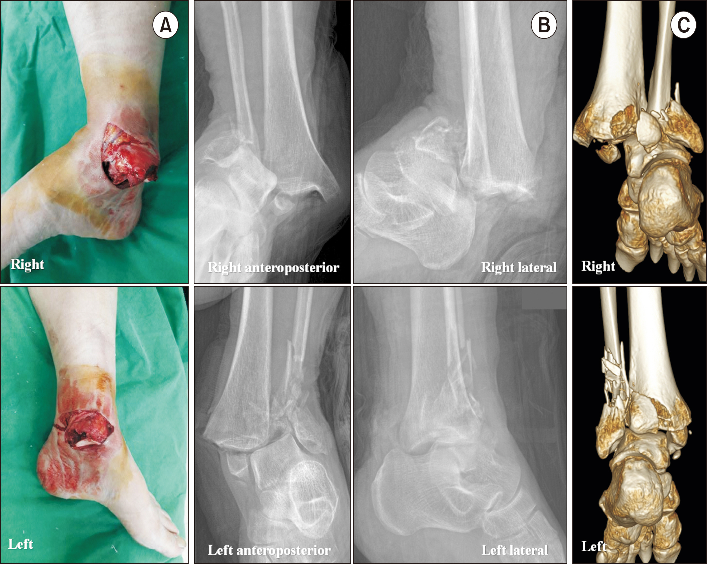
Figure 1 (A) The patient had bilateral medial ankle wound with bony exposure. (B) Initial radiographs showed fracture-dislocation of both ankles. (C) We also found bilateral posterior malleolar fractures with 3-dimensional computed radiographs.

Figure 2 Bilateral intramedullary Steinmann fixation was performed for severe comminuted lateral malleolar fractures. Posterior malleolar fractures were also fixed with cannulated screws through posterolateral approach. Immediate postoperative radiographs of right (A) and left (B) sides.

Figure 3 During the 3-month (A), 6-month (B), and 12-month (C) follow-up, valgus talar tilt gradually increased with relative fibular shortening.
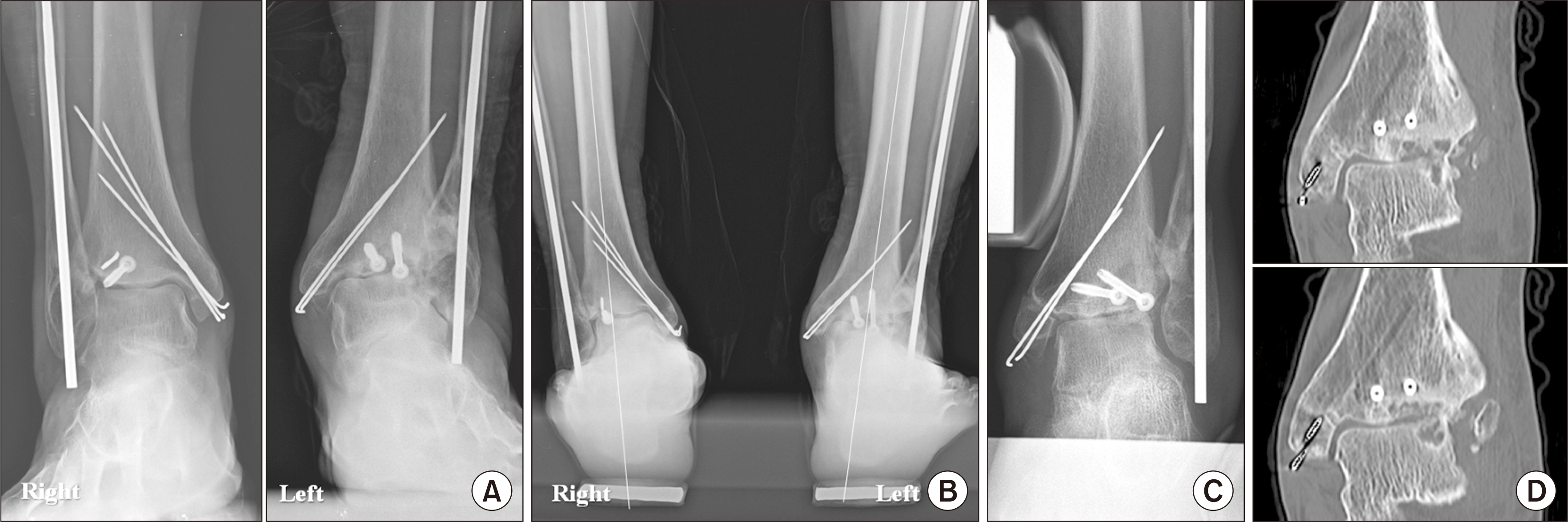
Figure 4 At 18 postoperative months (A), valgus ankle arthritis was developed on the left side while the talar tile on the right side was intact. On the hindfoot alignment view (B), valgus malaligned hindfoot was shown. A valgus deformity was corrected on with varus stress (C). A standard load of 150 N was applied to the ankle joint using the Telos device (METAX, Hungen, Germany). Using 3-dimensional computed tomography (D), we found a bony contact with subchondral cyst formation on the lateral side of talar dome and tibial plafond.

Figure 5 (A) A Z-shaped osteotomy line for fibular lengthening was drawn on the fibula. (B) A cartilage denuded surface on the lateral talar dome was exposed and multiple drilling was performed. On immediate postoperative anteroposterior (C) and lateral (D) radiographs, we found that a successful talar tilt correction was obtained through the fibular lengthening osteotomy combined with medial displacement calcaneal osteotomy. (E) A calcaneus axial radiograph showed a successful medial displacement (10 cm) of calcaneal tuberosity.
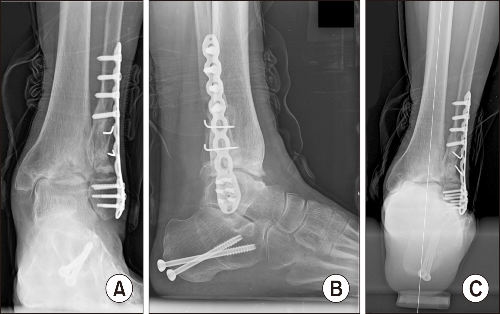
Figure 6 (A, B) At postoperative 18 months, talar tilt was changed to 6.9° valgus with an obvious gap between lateral talar dome and tibial plafond. (C) A hindfoot alignment was also corrected to the neutral position.
Reference
-
References
1. Choi JY, Kim JH, Ko HT, Suh JS. 2015; Single oblique posterolateral approach for open reduction and internal fixation of posterior malleolar fractures with an associated lateral malleolar fracture. J Foot Ankle Surg. 54:559–64. doi: 10.1053/j.jfas.2014.09.043. DOI: 10.1053/j.jfas.2014.09.043. PMID: 25459093.2. Barg A, Pagenstert GI, Leumann AG, Müller AM, Henninger HB, Valderrabano V. 2012; Treatment of the arthritic valgus ankle. Foot Ankle Clin. 17:647–63. doi: 10.1016/j.fcl.2012.08.007. DOI: 10.1016/j.fcl.2012.08.007. PMID: 23158375.
Article3. Gómez-Palomo JM, Martínez-Crespo A, Rodríguez-Delourme I, García-Pérez JR, Martínez-Espinosa M, Montañez-Heredia E. 2020; Fibular-lengthening osteotomy in patient with progressive valgus deformity after an ankle fracture. Am J Case Rep. 21:e920460. doi: 10.12659/AJCR.920460. DOI: 10.12659/AJCR.920460. PMID: 32047142. PMCID: PMC7038640.
Article4. Weber D, Friederich NF, Müller W. 1998; Lengthening osteotomy of the fibula for post-traumatic malunion. Indications, technique and results. Int Orthop. 22:149–52. doi: 10.1007/s002640050229. DOI: 10.1007/s002640050229. PMID: 9728305. PMCID: PMC3619596.5. Bluman EM, Chiodo CP. 2008; Valgus ankle deformity and arthritis. Foot Ankle Clin. 13:443–70. ixdoi: 10.1016/j.fcl.2008.04.008. DOI: 10.1016/j.fcl.2008.04.008. PMID: 18692009.
Article6. Knupp M, Stufkens SA, Bolliger L, Barg A, Hintermann B. 2011; Classification and treatment of supramalleolar deformities. Foot Ankle Int. 32:1023–31. doi: 10.3113/FAI.2011.1023. DOI: 10.3113/FAI.2011.1023. PMID: 22338950.
Article7. Kim J, Kim JB, Lee WC. 2021; Outcomes of joint preservation surgery in valgus ankle arthritis without deltoid ligament insufficiency. Foot Ankle Int. 42:1419–30. doi: 10.1177/10711007211016001. DOI: 10.1177/10711007211016001. PMID: 34109853.
Article8. Brilhault J. 2022; Calcaneal osteotomy for hindfoot deformity. Orthop Traumatol Surg Res. 108(1S):103121. doi: 10.1016/j.otsr.2021.103121. DOI: 10.1016/j.otsr.2021.103121. PMID: 34687951.
Article9. Schon LC, de Cesar Netto C, Day J, Deland JT, Hintermann B, Johnson JE, et al. 2020; Consensus for the indication of a medializing displacement calcaneal osteotomy in the treatment of progressive collapsing foot deformity. Foot Ankle Int. 41:1282–5. doi: 10.1177/1071100720950747. DOI: 10.1177/1071100720950747. PMID: 32844661.
Article10. Ju DG, Debbi EM, Neustein AZ, Moon CN. 2019; Fibular lengthening osteotomy with revision syndesmotic repair for ankle fracture malunion. J Orthop Trauma. 33 Suppl 1:S38–9. doi: 10.1097/BOT.0000000000001534. DOI: 10.1097/BOT.0000000000001534. PMID: 31290833.
Article
- Full Text Links
-
- Actions
-
Cited
- CITED
-
- Close
- Share
-
- Similar articles
-
- Pseudoaneurysm after Proximal Metatarsal Osteotomy for Hallux Valgus Correction: A Case Report
- Operative Treatment for Fibular Shortening after Trauma: A Case Report
- High Tibial Osteotomy With Fibular Shaft Osteotomy
- Surgical treatments of the ankle arthritis
- Hindfoot Alignment Change after High Tibial Valgization Osteotomy in a Patient with an Ipsilateral Fused Ankle: A Case Report
