J Korean Foot Ankle Soc.
2021 Dec;25(4):177-180. 10.14193/jkfas.2021.25.4.177.
Operative Treatment for Fibular Shortening after Trauma: A Case Report
- Affiliations
-
- 1Department of Orthopedic Surgery, Kangdong Sacred Heart Hospital, Hallym University College of Medicine, Seoul,
- 2Department of Orthopedic Surgery, Gospel Hospital, Kosin University College of Medicine, Busan, Korea
- KMID: 2523299
- DOI: http://doi.org/10.14193/jkfas.2021.25.4.177
Abstract
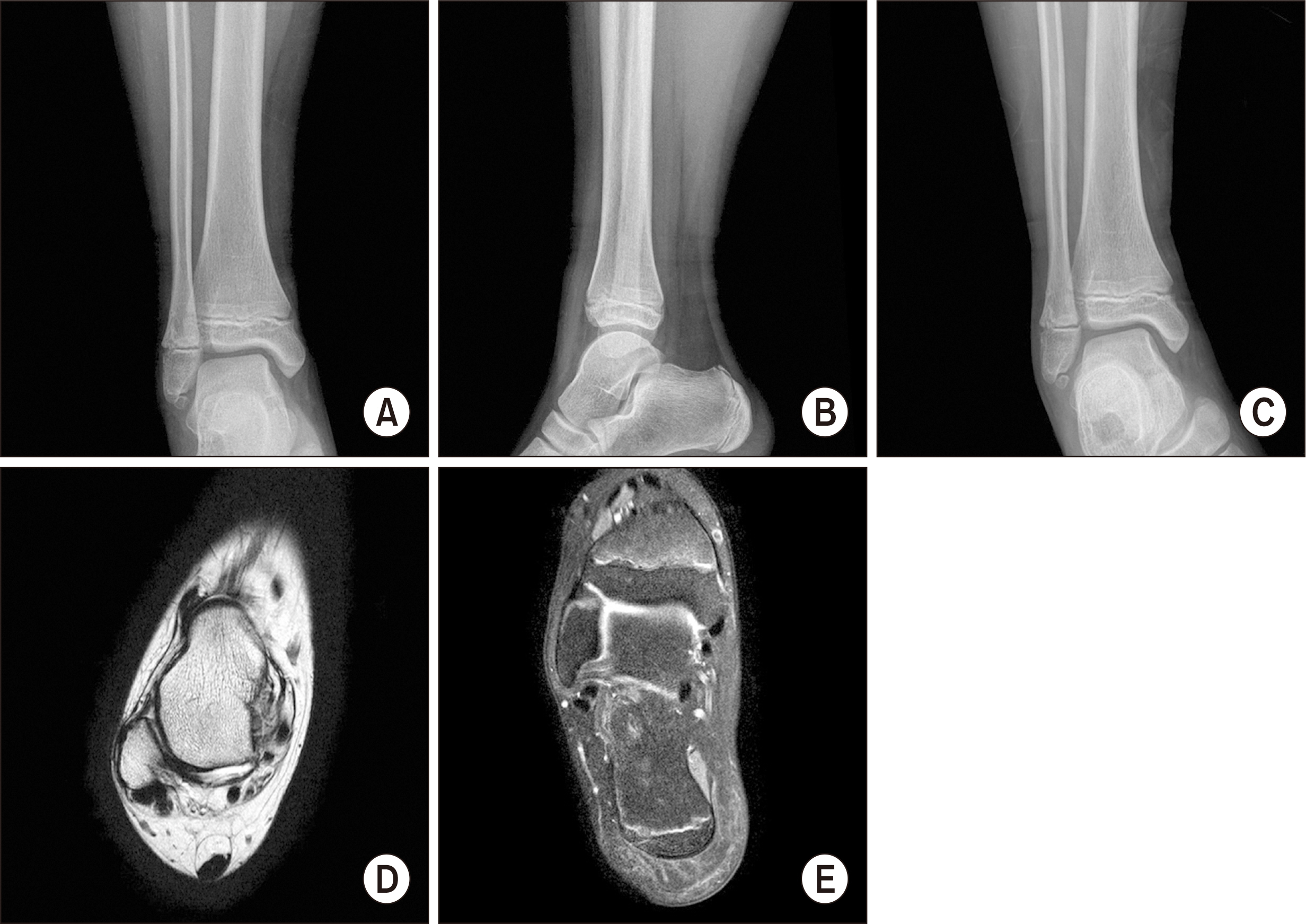
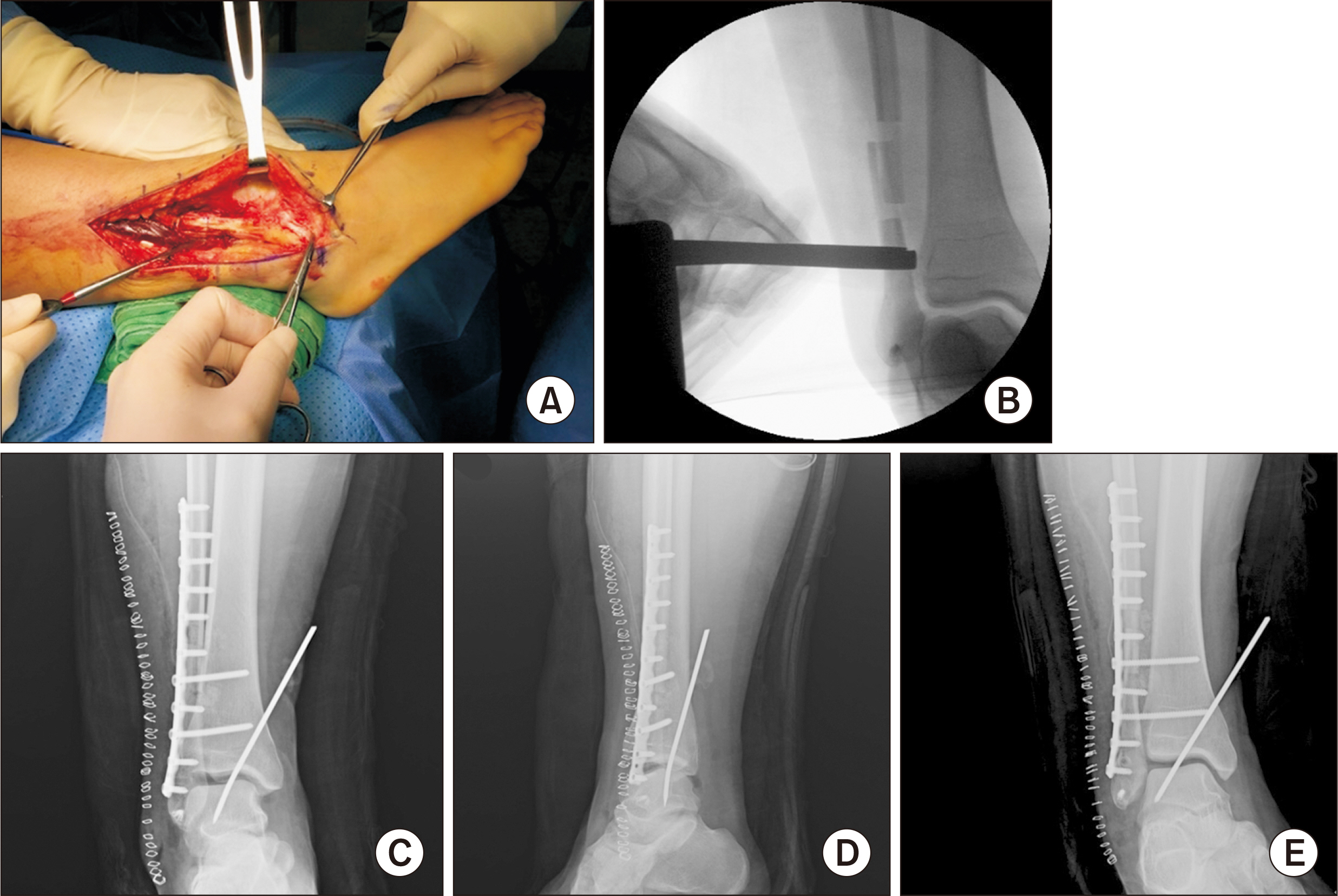
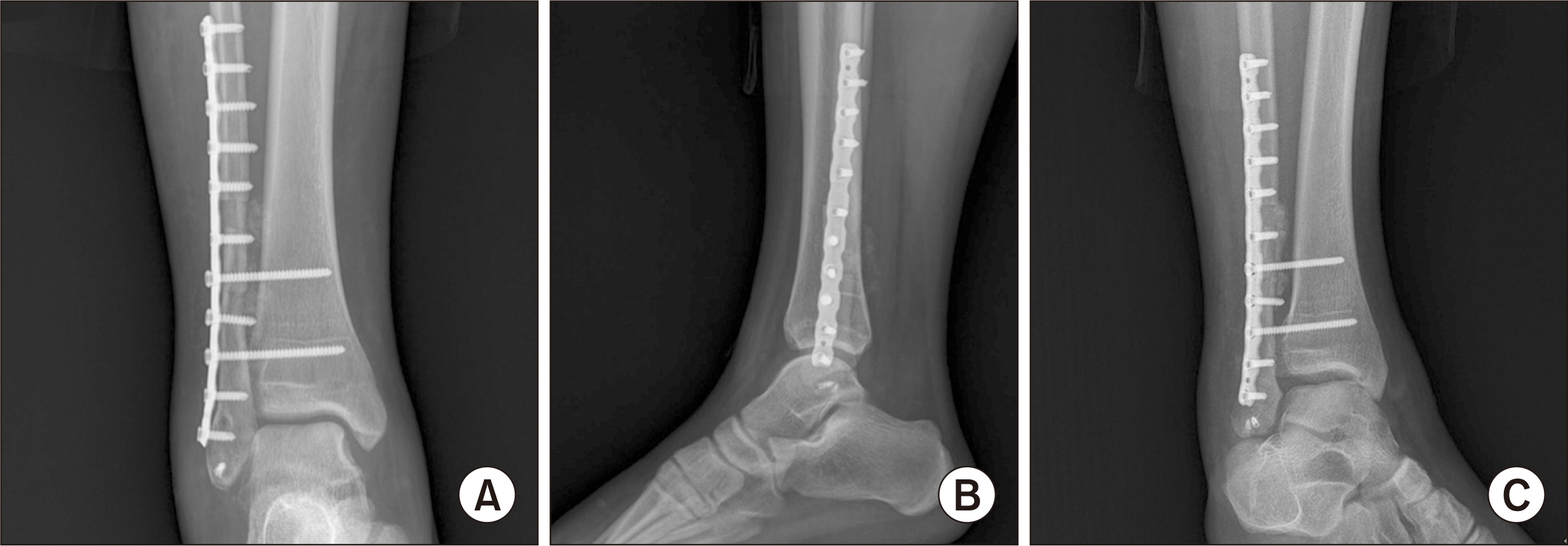
- Pediatric ankle fractures can cause physeal injuries which can lead to the shortening of the fibula. This induces a lateral shift of the talus, valgus tilt, and instability of the ankle joint, which can result in an arthritic change in this joint. Patients with a shortening of the fibula may complain of constant pain and restricted movements in their daily lives and during sports activities. Ankle reconstruction with fibula lengthening Z-osteotomy can provide excellent results if arthritis is absent or minimal, especially in young and active patients. To the best of the authors’ knowledge, this is the first report in South Korea regarding the treatment of fibula shortening following a growth arrest due to injury.
Keyword
Figure
Reference
-
1. Kay RM, Matthys GA. 2001; Pediatric ankle fractures: evaluation and treatment. J Am Acad Orthop Surg. 9:268–78. doi: 10.5435/00124635-200107000-00007. DOI: 10.5435/00124635-200107000-00007. PMID: 11476537.
Article2. Wuerz TH, Gurd DP. 2013; Pediatric physeal ankle fracture. J Am Acad Orthop Surg. 21:234–44. doi: 10.5435/JAAOS-21-04-234. DOI: 10.5435/JAAOS-21-04-234. PMID: 23545729.
Article3. Kim JR, Song KH. 2008; Factors influencing premature physeal closure following physeal injury of the ankle in children. J Korean Orthop Assoc. 43:672–6. doi: 10.4055/jkoa.2008.43.6.672. DOI: 10.4055/jkoa.2008.43.6.672.
Article4. Gómez-Palomo JM, Martínez-Crespo A, Rodríguez-Delourme I, García-Pérez JR, Martínez-Espinosa M, Montañez-Heredia E. 2020; Fibular-lengthening osteotomy in patient with progressive valgus deformity after an ankle fracture. Am J Case Rep. 21:e920460. doi: 10.12659/AJCR.920460. DOI: 10.12659/AJCR.920460. PMID: 32047142. PMCID: PMC7038640.
Article5. Manoudis GN, Kontogeorgakos VA, Badras LS. 2012; Distal fibular lengthening after premature growth arrest: a case report. J Orthop Surg (Hong Kong). 20:409–13. doi: 10.1177/230949901202000332. DOI: 10.1177/230949901202000332. PMID: 23255659.
Article6. Ramsey PL, Hamilton W. 1976; Changes in tibiotalar area of contact caused by lateral talar shift. J Bone Joint Surg Am. 58:356–7. doi: 10.2106/00004623-197658030-00010. DOI: 10.2106/00004623-197658030-00010. PMID: 1262367.
Article7. Weber D, Friederich NF, Müller W. 1998; Lengthening osteotomy of the fibula for post-traumatic malunion. Indications, technique and results. Int Orthop. 22:149–52. doi: 10.1007/s002640050229. DOI: 10.1007/s002640050229. PMID: 9728305. PMCID: PMC3619596.8. Weber D, Weber M. 2016; Corrective osteotomies for malunited malleolar fractures. Foot Ankle Clin. 21:37–48. doi: 10.1016/j.fcl.2015.09.003. DOI: 10.1016/j.fcl.2015.09.003. PMID: 26915777.
Article9. Thordarson DB, Motamed S, Hedman T, Ebramzadeh E, Bakshian S. 1997; The effect of fibular malreduction on contact pressures in an ankle fracture malunion model. J Bone Joint Surg Am. 79:1809–15. doi: 10.2106/00004623-199712000-00006. DOI: 10.2106/00004623-199712000-00006. PMID: 9409794.
Article10. Egger AC, Berkowitz MJ. 2018; Operative treatment of the malunited fibula fracture. Foot Ankle Int. 39:1242–52. doi: 10.1177/1071100718795630. DOI: 10.1177/1071100718795630. PMID: 30165033.
Article
- Full Text Links
- Actions
-
Cited
- CITED
-
- Close
- Share
- Similar articles
-
- A Fibular Lengthening Osteotomy Combined with Calcaneal Osteotomy for Post-Traumatic Valgus Ankle Arthritis: A Case Report
- Vascularized Fibular Bone Graft for Tuberculous Spondylitis: Case Report
- Virtual Surgical Planning and Three-Dimensional Printing for Reconstruction of Mandibular Defect by Fibula Free Flap: A Case Report
- The effects of the flbular stabilization in the treatment of tibio-fibula shaft fracture
- Relationship of Tibial Nonunion with Fibular Nonunion in the Tibio-fibular Shaft Fracture


