J Korean Foot Ankle Soc.
2014 Jun;18(2):80-82. 10.14193/jkfas.2014.18.2.80.
Pseudoaneurysm after Proximal Metatarsal Osteotomy for Hallux Valgus Correction: A Case Report
- Affiliations
-
- 1Foot and Ankle Service, KT Lee's Orthopedic Hospital, Seoul, Korea.
- 2Department of Orthopedic Surgery, Ajou University Hospital, Ajou University School of Medicine, Suwon, Korea. parkyounguk@gmail.com
- KMID: 2181666
- DOI: http://doi.org/10.14193/jkfas.2014.18.2.80
Abstract
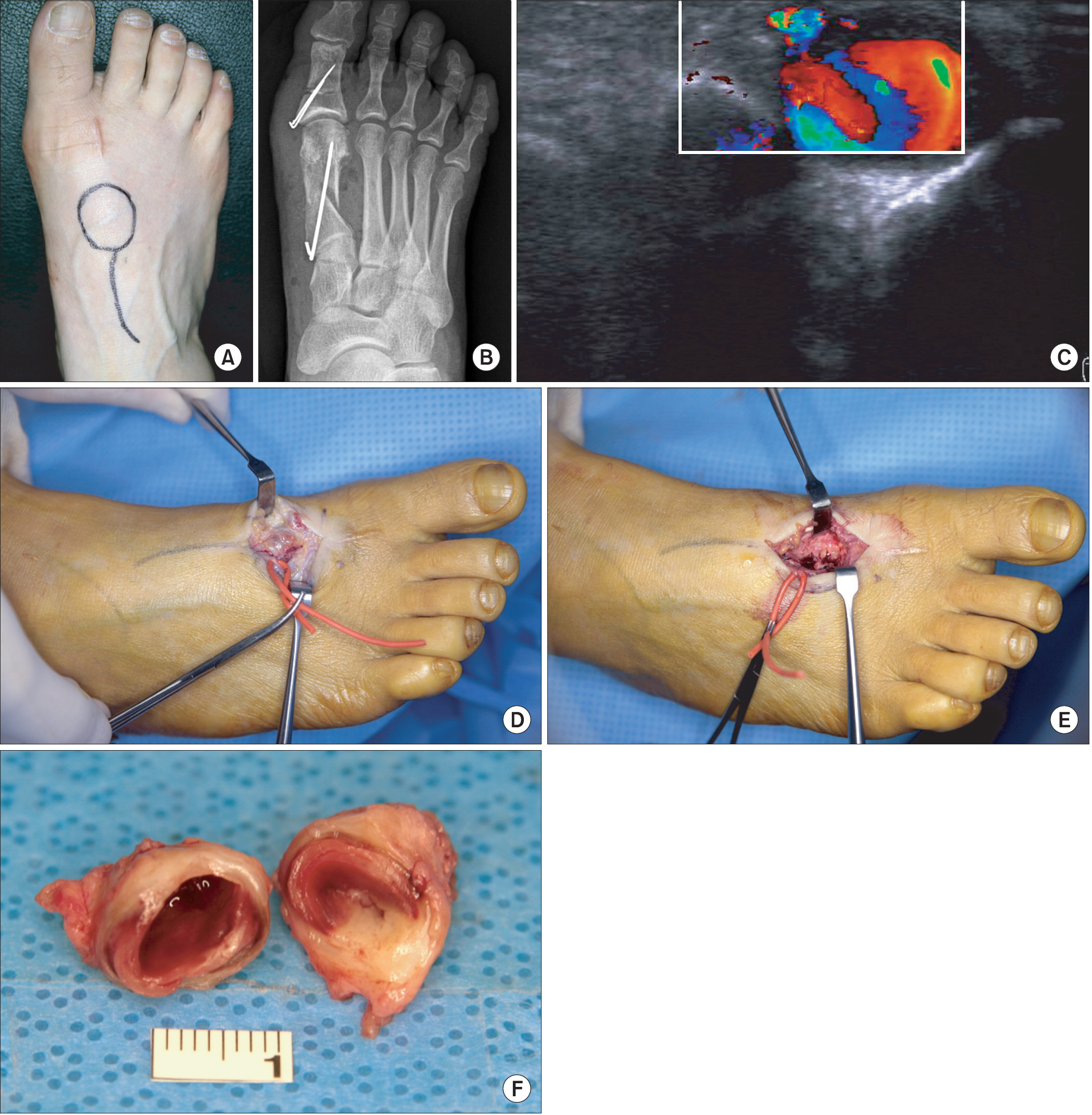
- Occurrence of pseudoaneurysm in the foot and ankle is rare, and is usually caused by traumatic injury or by iatrogenic intervention. Iatrogenic pseudoaneurysms in the foot and ankle have been observed after rearfoot and ankle fusions, ankle arthroscopy, endoscopic and open plantar fasciotomy, tibial osteotomy with limb lengthening, midfoot amputation, and Lapidus procedure. We report on a patient who developed a pseudoaneurysm of the dorsal metatarsal artery following correction of hallux valgus. The patient underwent proximal chevron osteotomy and Akin phalangeal osteotomy. The feeding artery was ligated and the pseudoaneurysm was excised.
Keyword
MeSH Terms
Figure
Reference
-
References
1. Bogokowsky H, Slutzki S, Negri M, Halpern Z. Pseudoaneurysm of the dorsalis pedis artery. Injury. 1985; 16:424–5.
Article2. Economou P, Paton R, Galasko CS. Traumatic pseudoaneurysm of the lateral plantar artery in a child. J Pediatr Surg. 1993; 28:626.
Article3. Higgs SL. Arteriovenous aneurysm of the posterior tibial vessels following operation for stabilizing the foot. Proc R Soc Med. 1931; 24:1378–9.
Article4. Nierenberg G, Hoffman A, Engel A, Stein H. Pseudoaneurysm with an arteriovenous fistula of the tibial vessels after plantar fasciotomy: a case report. Foot Ankle Int. 1997; 18:524–5.
Article5. Bandy WD, Strong L, Roberts T, Dyer R. False aneurysm–a complication following an inversion ankle sprain: a case report. J Orthop Sports Phys Ther. 1996; 23:272–9.
Article6. Thornton BP, Minion DJ, Quick R, Vasconez HC, Endean ED. Pseudoaneurysm of the lateral plantar artery after foot laceration. J Vasc Surg. 2003; 37:672–5.
Article7. Yamaguchi S, Mii S, Yonemitsu Y, Orita H, Sakata H. A traumatic pseudoaneurysm of the dorsalis pedis artery: report of a case. Surg Today. 2002; 32:756–7.
Article8. Wollstein R, Wolf Y, Sklair-Levy M, Matan Y, London E, Nyska M. Obliteration of a late traumatic posterior tibial artery pseudoaneurysm by duplex compression. J Trauma. 2000; 48:1156–8.
Article9. Aiyer S, Thakkar CJ, Samant PD, Verlekar S, Nirawane R. Pseudoaneurysm of the posterior tibial artery following a closed fracture of the calcaneus. A case report. J Bone Joint Surg Am. 2005; 87:2308–12.
Article10. Maydew MS. Dorsalis pedis aneurysm: ultrasound diagnosis. Emerg Radiol. 2007; 13:277–80.
Article
- Full Text Links
- Actions
-
Cited
- CITED
-
- Close
- Share
- Similar articles
-
- Minimally Invasive Proximal Transverse Metatarsal Osteotomy Followed by Intramedullary Plate Fixation for Hallux Valgus Deformity: A Case Report
- Chevron Osteotomy as the Treatment of Moderate to Severe Hallux Valgus Deformity
- Corrective Osteotomies in Hallux Valgus
- Comparison of Proximal Metatarsal Osteotomy andDistal Chevron Osteotomy for Correction of Hallux Valgus
- A Comparison of Operative Treatment of Hallux Valgus with a Proximal Metatarsal Osteotomy and with a Modified Chevron Osteotomy


