Hindfoot Alignment Change after High Tibial Valgization Osteotomy in a Patient with an Ipsilateral Fused Ankle: A Case Report
- Affiliations
-
- 1Department of Orthopedic Surgery, Inje University Ilsan Paik Hospital, Inje University College of Medicine, Goyang, Korea
- KMID: 2556558
- DOI: http://doi.org/10.14193/jkfas.2024.28.2.75
Abstract
- Ankle arthrodesis was performed on a 55-year-old male patient with an active lifestyle who developed severe arthritis in the left ankle. Over the follow-up period, high tibial valgization osteotomy was conducted for painful medial knee joint arthritis with genu varum deformity to correct overall lower limb alignment from varus to valgus with respect to the fused ankle. This study was conducted to investigate how hindfoot alignment would change when the overall alignment of the lower limb shifted from varus to valgus with the ipsilateral ankle in a fused state. Conclusively, while no intrinsic changes in the hindfoot alignment were observed following the alteration of lower limb alignment, the hindfoot naturally adjusted to valgus deviation in response to limb valgus realignment. Moreover, symptoms changed in line with this adjustment. Given the absence of similar case studies or reports, a review of relevant literature is included to contribute to knowledge of this subject.
Keyword
Figure
-
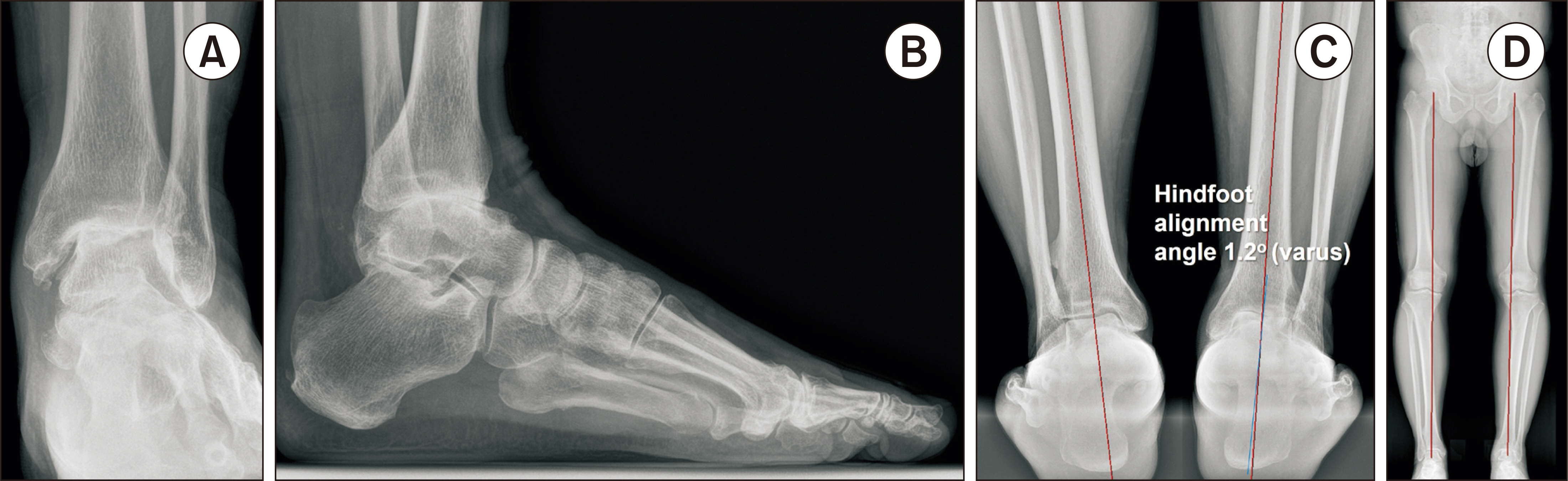
Figure 1 Initial ankle radiographs of the ankle in anteroposterior (A) and lateral (B) views revealed complete loss of joint space between the talus and the tibial plafond, with the presence of loose bodies near the medial aspect. Additionally, hindfoot alignment view (C) showed that the alignment of the hindfoot was in a neutral position (hindfoot alignment angle 1.2 degrees [varus]). Furthermore, in the hip-to-talus view, arthritis with varus deformity of the bilateral knee joints was observed (D).
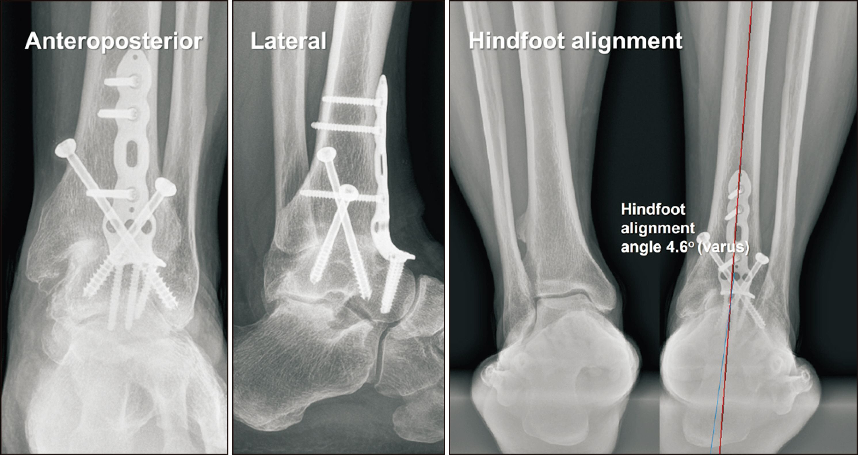
Figure 2 Radiographs taken at postoperative 6 months showed that fusion of the ankle joint was mostly achieved. A hindfoot alignment revealed that there was a slight increase in the varus malalignment of the hindfoot (hindfoot varus angle 4.6 degrees [varus]), indicating that the ankle fused with a slight increase in hindfoot varus.
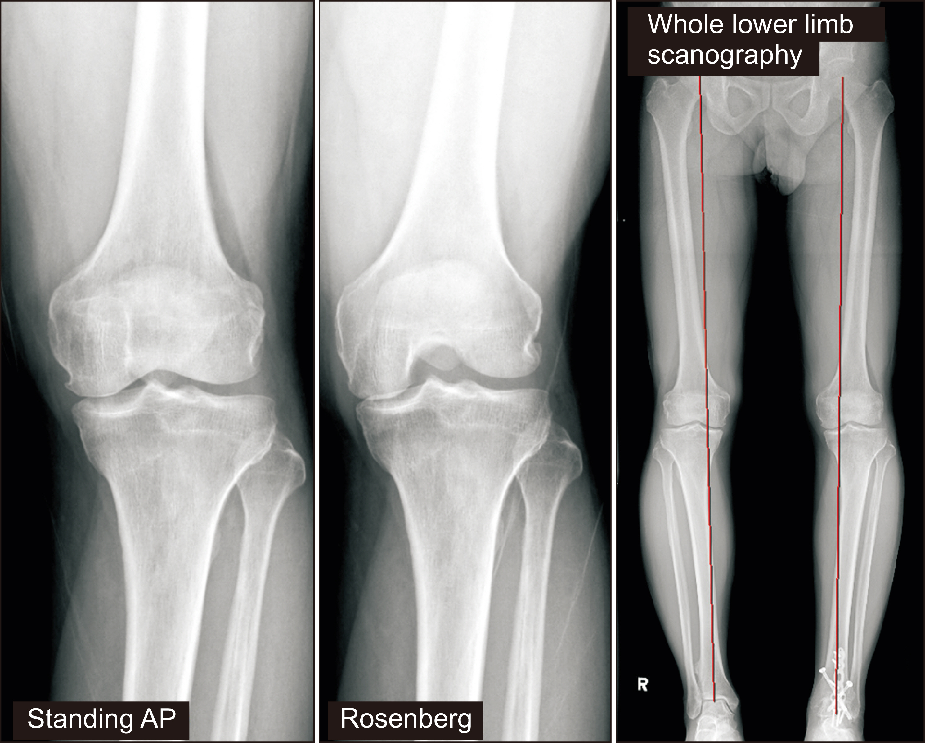
Figure 3 Six months following the ankle arthrodesis, the patient experienced a recurrence of bilateral knee pain that had existed previously without any specific incident. Radiographs revealed narrowing of the joint space in the medial compartments of both knee joints, osteophyte formation, and varus deformity. AP: anteroposterior.
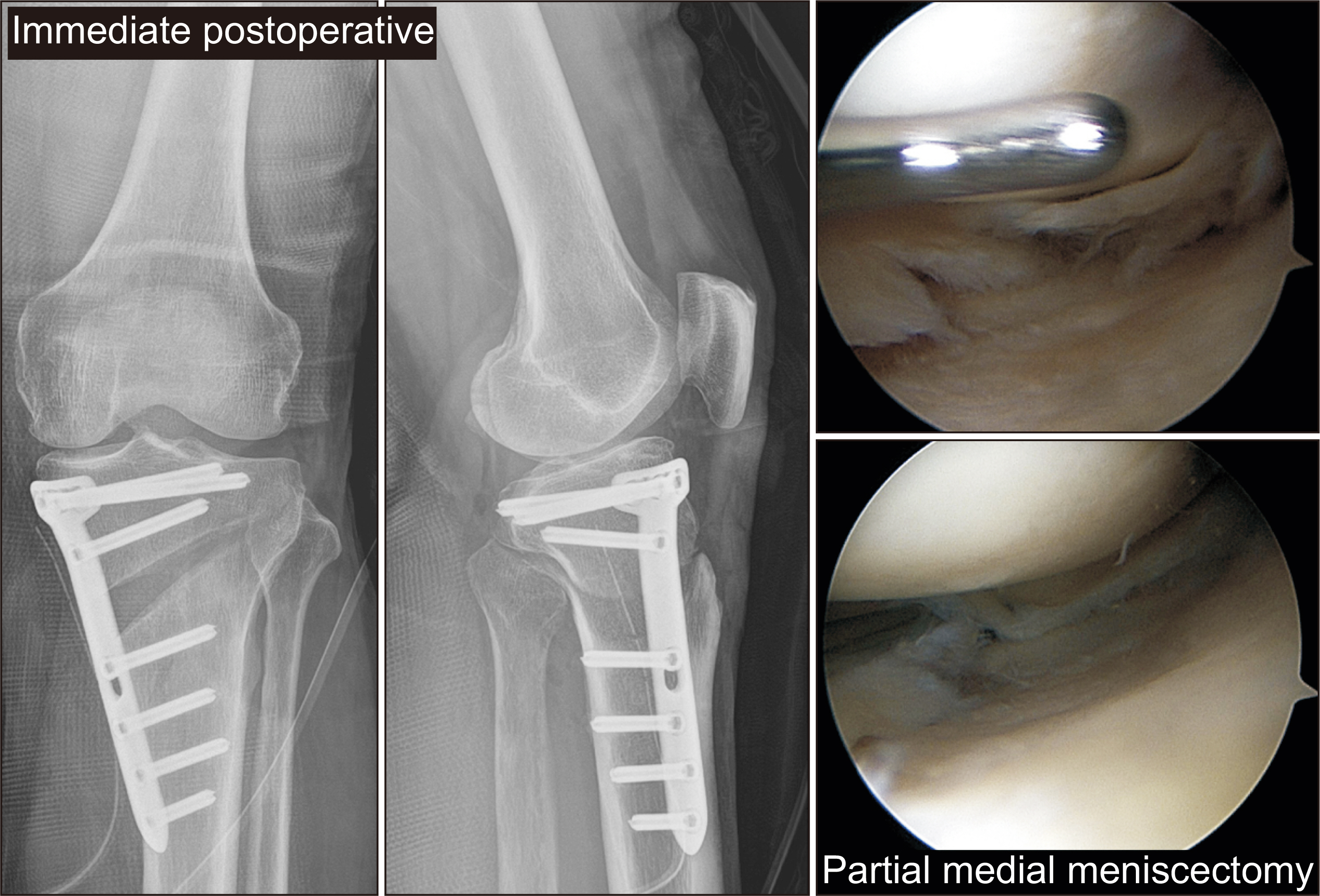
Figure 4 At postoperative 8 months after the ankle arthrodesis, arthroscopic medial meniscectomy and medial opening wedge high tibial osteotomy were performed on the ipsilateral knee joint.
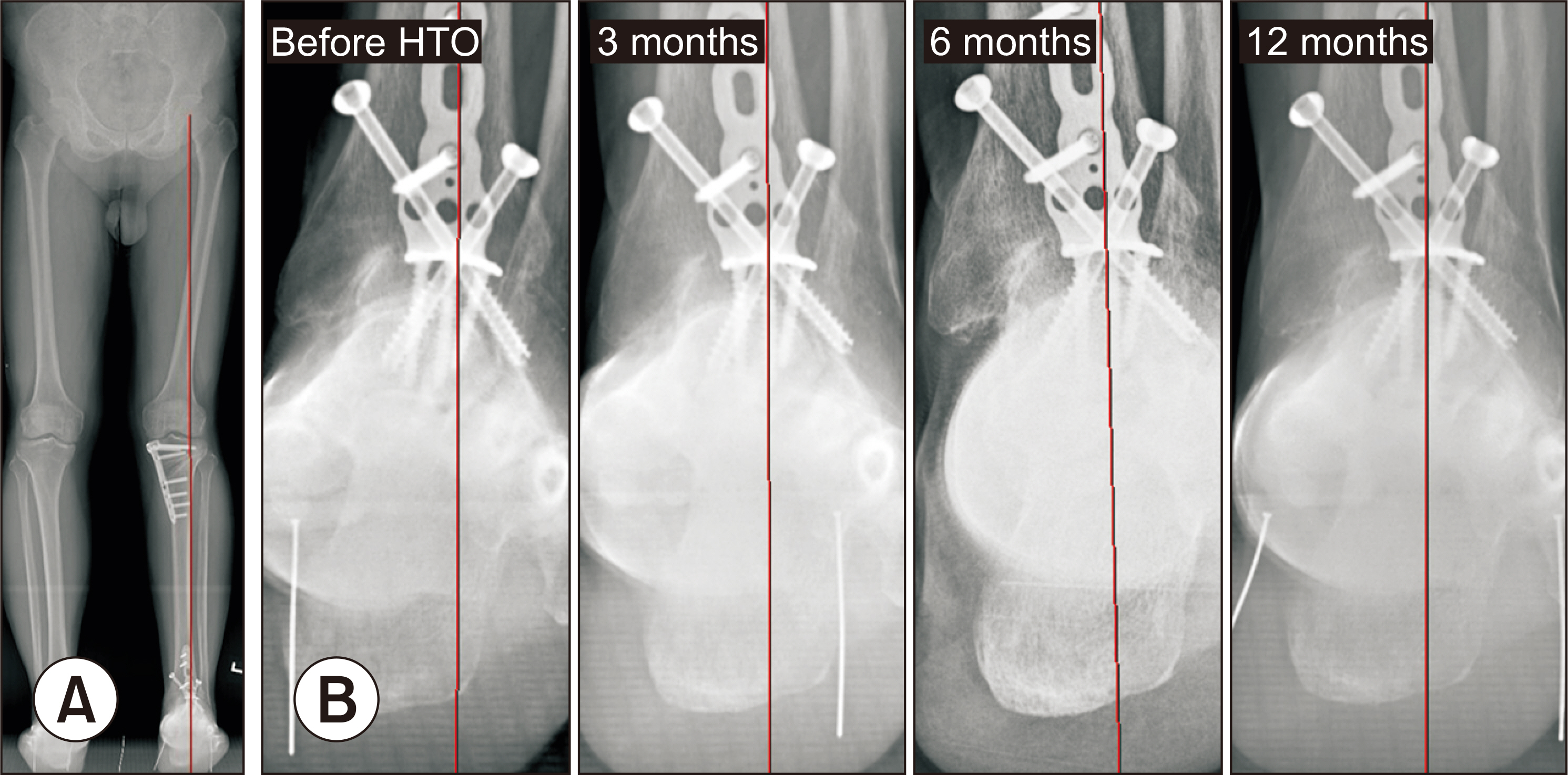
Figure 5 In the hip-to-calcaneus radiographic technique, to assess changes in hindfoot alignment, it is essential to secure a line connecting the center of the femoral head and the center of the tibial plafond. However, in this patient’s case, as the ankle was already fused, it was not possible to establish a line through the center of the tibial plafond. Therefore, alignment was inferred by extending a line from the center of the femoral head to the intersection of the two cannulated screws used during the ankle arthrodesis and assessing the relationship between this line and the most inferior part of the hindfoot (A). For assessing hindfoot alignment, the relationship between this line and the most inferior part of the hindfoot was examined. Post-operative hip-to-calcaneus radiographs revealed an increase in hindfoot valgus following the high tibial osteotomy compared to pre-operative status (B). HTO: high tibial osteotomy.
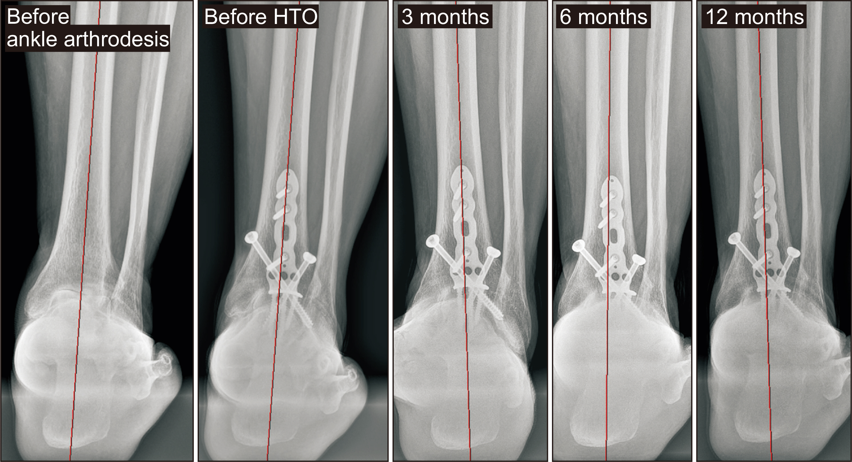
Figure 6 Serial hindfoot alignment views revealed no significant changes in the alignment of the hindfoot up to 12 months after the knee valgization operation, compared to the pre-operative status. HTO: high tibial osteotomy.
Reference
-
1. Cho WS, Cho HS, Byun SE. 2017; Changes in hindfoot alignment after total knee arthroplasty in knee osteoarthritic patients with varus deformity. Knee Surg Sports Traumatol Arthrosc. 25:3596–604. doi: 10.1007/s00167-016-4278-8. DOI: 10.1007/s00167-016-4278-8. PMID: 27527338.
Article2. Hayashi K, Tanaka Y, Kumai T, Sugimoto K, Takakura Y. 2008; Correlation of compensatory alignment of the subtalar joint to the progression of primary osteoarthritis of the ankle. Foot Ankle Int. 29:400–6. doi: 10.3113/FAI.2008.0400. DOI: 10.3113/FAI.2008.0400. PMID: 18442455.
Article3. Yoshimoto K, Noguchi M, Yamada A, Nasu Y. 2019; Compensatory function of the subtalar joint for lower extremity malalignment. Adv Orthop. 2019:7656878. doi: 10.1155/2019/7656878. DOI: 10.1155/2019/7656878. PMID: 30918725. PMCID: PMC6408994.
Article4. Choi JY, Song SJ, Kim SJ, Kim SH, Park JS, Suh JS. 2018; Changes in hindfoot alignment after high or low tibial osteotomy. Foot Ankle Int. 39:1097–105. doi: 10.1177/1071100718773767. DOI: 10.1177/1071100718773767. PMID: 29761747.
Article5. Son HS, Choi JG, Ahn J, Jeong BO. 2021; Hindfoot alignment change after total ankle arthroplasty for varus osteoarthritis. Foot Ankle Int. 42:431–9. doi: 10.1177/1071100720970937. DOI: 10.1177/1071100720970937. PMID: 33218258.
Article6. Lee WC, Moon JS, Lee HS, Lee K. 2011; Alignment of ankle and hindfoot in early stage ankle osteoarthritis. Foot Ankle Int. 32:693–9. doi: 10.3113/FAI.2011.0693. DOI: 10.3113/FAI.2011.0693. PMID: 21972764.
Article7. Krähenbühl N, Siegler L, Deforth M, Zwicky L, Hintermann B, Knupp M. 2019; Subtalar joint alignment in ankle osteoarthritis. Foot Ankle Surg. 25:143–9. doi: 10.1016/j.fas.2017.10.004. DOI: 10.1016/j.fas.2017.10.004. PMID: 29409290.
Article8. Saltzman CL, el-Khoury GY. 1995; The hindfoot alignment view. Foot Ankle Int. 16:572–6. doi: 10.1177/107110079501600911. DOI: 10.1177/107110079501600911. PMID: 8563927.
Article9. Choi JY, Lee HI, Kim JH, Suh JS. 2021; Radiographic measurements on hindfoot alignment view in 1128 asymptomatic subjects. Foot Ankle Surg. 27:366–70. doi: 10.1016/j.fas.2020.04.010. DOI: 10.1016/j.fas.2020.04.010. PMID: 32451174.
Article10. Nha KW, Han JH, Chae SW, Choi JY. 2023; Effect of medial closing wedge distal femoral varization osteotomy on coronal ankle and hindfoot alignment. Foot Ankle Int. 44:330–9. doi: 10.1177/10711007231154208. DOI: 10.1177/10711007231154208. PMID: 36825582.
Article11. Haraguchi N, Ota K, Tsunoda N, Seike K, Kanetake Y, Tsutaya A. 2015; Weight-bearing-line analysis in supramalleolar osteotomy for varus-type osteoarthritis of the ankle. J Bone Joint Surg Am. 97:333–9. doi: 10.2106/JBJS.M.01327. DOI: 10.2106/JBJS.M.01327. PMID: 25695986.
Article
- Full Text Links
-
- Actions
-
Cited
- CITED
-
- Close
- Share
-
- Similar articles
-
- Supramalleolar Osteotomy in Patients with Varus Ankle Osteoarthritis
- A Fibular Lengthening Osteotomy Combined with Calcaneal Osteotomy for Post-Traumatic Valgus Ankle Arthritis: A Case Report
- Supramalleolar Tibial Osteotomy for Medial Compartment Ankle Osteoarthritis
- Proximal Tibiofibular Arthrolysis in High Tibial Osteotomy
- Low Tibial Osteotomy
