Korean J Transplant.
2022 Mar;36(1):73-78. 10.4285/kjt.21.0007.
Living donor liver transplantation in an infant patient with progressive familial intrahepatic cholestasis along with hepatocellular carcinoma: a case report
- Affiliations
-
- 1Department of Surgery, Asan Medical Center, University of Ulsan College of Medicine, Seoul, Korea
- 2Department of Pediatrics, Asan Medical Center, University of Ulsan College of Medicine, Seoul, Korea
- KMID: 2527901
- DOI: http://doi.org/10.4285/kjt.21.0007
Abstract
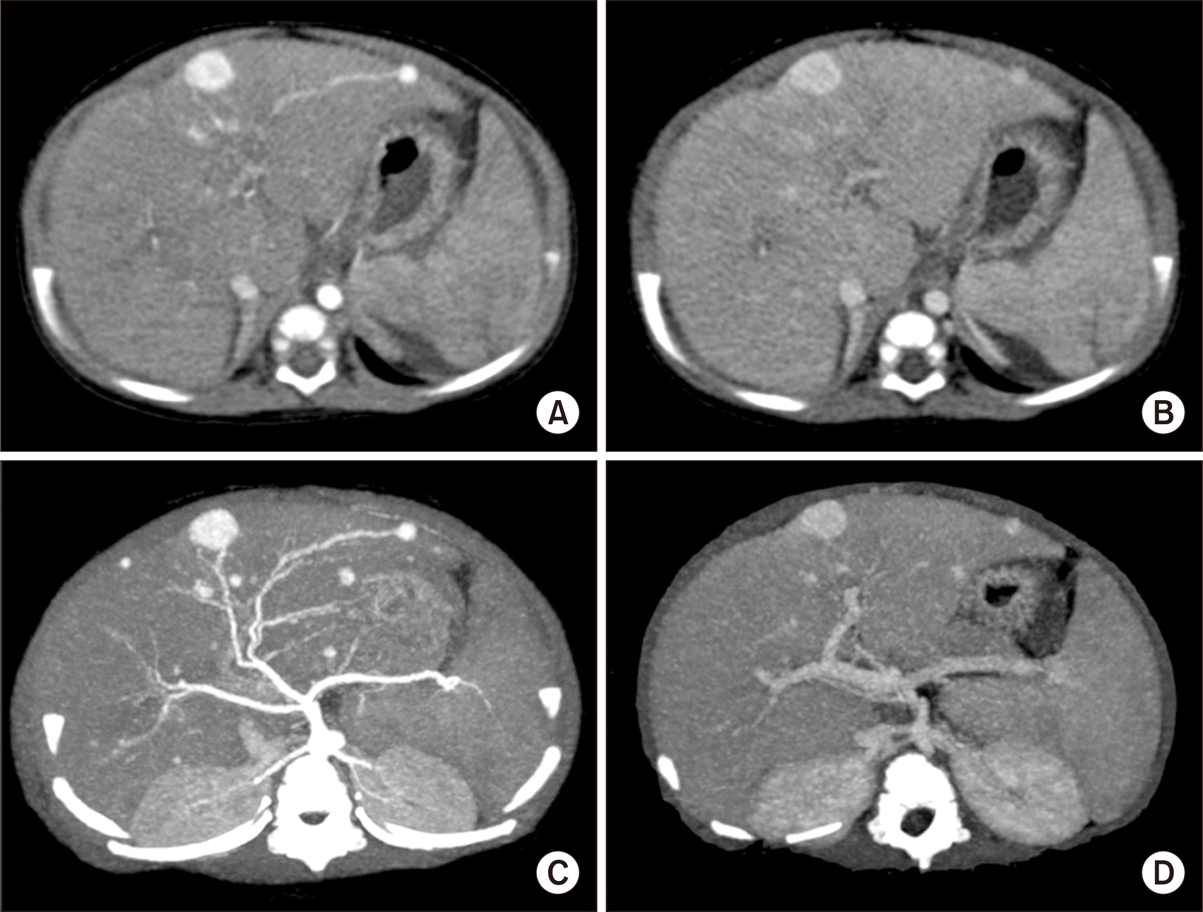
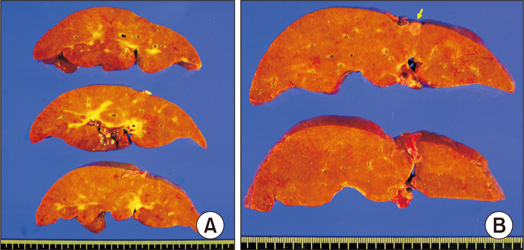
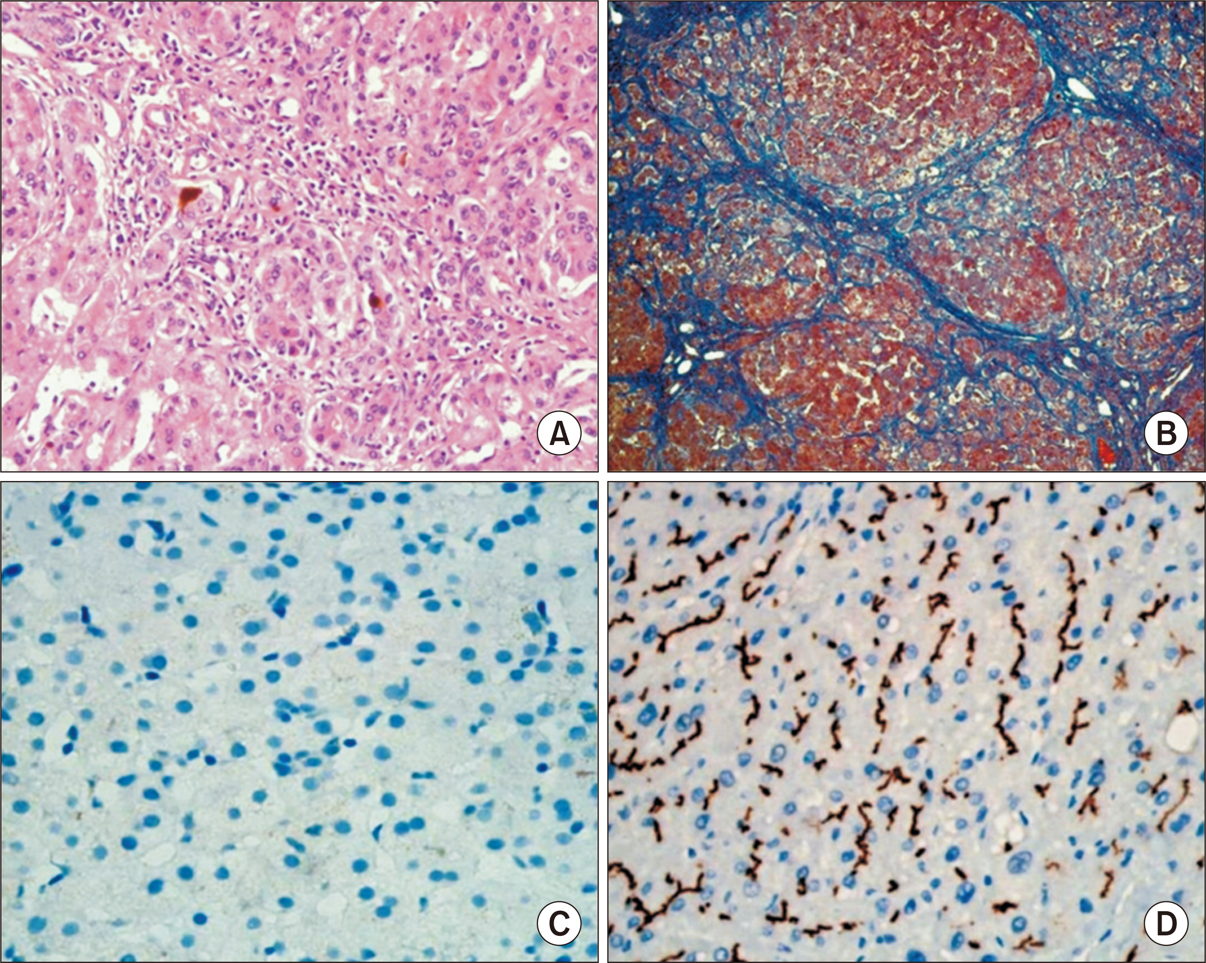
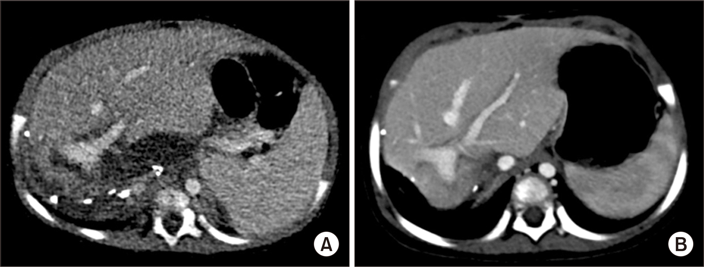
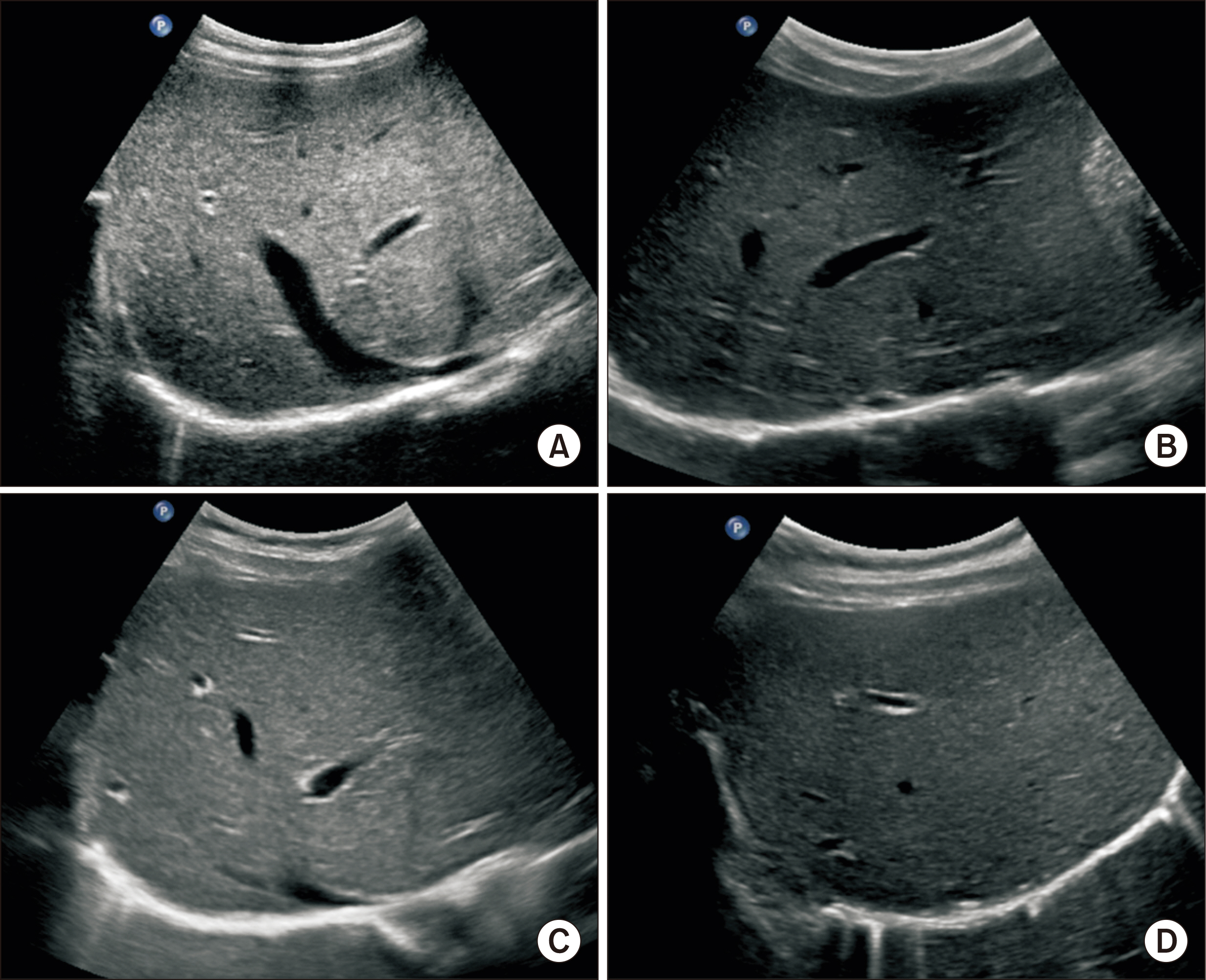
- Progressive familial intrahepatic cholestasis (PFIC) is an autosomal recessive inherited disease requiring liver transplantation (LT). Hepatocellular carcinoma (HCC) is very rare in infants. We present a case of living donor LT using a left lateral section graft performed in a 7-month-old female infant diagnosed with PFIC type II and HCC. No mutation on ABCB11 gene was identified. Because of progressive deterioration of liver function, living donor LT with her mother’s left lateral section graft was performed. Pretransplant serum alpha-fetoprotein (AFP) level was increased to 2,740 ng/mL, but HCC was not taken into account because of its rarity. The explant liver showed micronodular liver cirrhosis, multiple infantile hemangiomas and two HCCs of 0.7 cm and 0.3 cm in size. The patient recovered uneventfully from the LT operation. This patient has been regularly followed up with abdomen ultrasonography and AFP measurement every 6 months. The patient has been continually doing well for 8 years after the LT. In conclusion, LT is currently the only effective treatment for PFIC-associated end-stage liver diseases. HCC can develop at the cirrhotic liver of any cause, thus elevation of HCC tumor markers in pediatric patients is an important clue to perform further investigation before LT.
Figure
Cited by 1 articles
-
Liver transplantation in pediatric patients with progressive familial intrahepatic cholestasis: Single center experience of seven cases
Jung-Man Namgoong, Shin Hwang, Hyunhee Kwon, Suhyeon Ha, Kyung Mo Kim, Seak Hee Oh, Seung-Mo Hong
Ann Hepatobiliary Pancreat Surg. 2022;26(1):69-75. doi: 10.14701/ahbps.21-114.
Reference
-
1. van Mil SW, Klomp LW, Bull LN, Houwen RH. 2001; FIC1 disease: a spectrum of intrahepatic cholestatic disorders. Semin Liver Dis. 21:535–44. DOI: 10.1055/s-2001-19034. PMID: 11745041.
Article2. Sindhi R, Rohan V, Bukowinski A, Tadros S, de Ville de Goyet J, Rapkin L, et al. 2020; Liver transplantation for pediatric liver cancer. Cancers (Basel). 12:720. DOI: 10.3390/cancers12030720. PMID: 32204368. PMCID: PMC7140094.
Article3. Aydogdu S, Cakir M, Arikan C, Tumgor G, Yuksekkaya HA, Yilmaz F, et al. 2007; Liver transplantation for progressive familial intrahepatic cholestasis: clinical and histopathological findings, outcome and impact on growth. Pediatr Transplant. 11:634–40. DOI: 10.1111/j.1399-3046.2007.00722.x. PMID: 17663686.
Article4. Liu Y, Sun LY, Zhu ZJ, Wei L, Qu W, Zeng ZG. 2018; Liver transplantation for progressive familial intrahepatic cholestasis. Ann Transplant. 23:666–73. DOI: 10.12659/AOT.909941. PMID: 30250015. PMCID: PMC6248029.
Article5. Esquivel CO, Gutiérrez C, Cox KL, Garcia-Kennedy R, Berquist W, Concepcion W. 1994; Hepatocellular carcinoma and liver cell dysplasia in children with chronic liver disease. J Pediatr Surg. 29:1465–9. DOI: 10.1016/0022-3468(94)90145-7. PMID: 7844722.
Article6. Brunati A, Feruzi Z, Sokal E, Smets F, Fervaille C, Gosseye S, et al. 2007; Early occurrence of hepatocellular carcinoma in biliary atresia treated by liver transplantation. Pediatr Transplant. 11:117–9. DOI: 10.1111/j.1399-3046.2006.00623.x. PMID: 17239135.
Article7. Iida T, Zendejas IR, Kayler LK, Magliocca JF, Kim RD, Hemming AW, et al. 2009; Hepatocellular carcinoma in a 10-month-old biliary atresia child. Pediatr Transplant. 13:1048–9. DOI: 10.1111/j.1399-3046.2008.01094.x. PMID: 19032418.
Article8. Kim JM, Lee SK, Kwon CH, Joh JW, Choe YH, Park CK. 2012; Hepatocellular carcinoma in an infant with biliary atresia younger than 1 year. J Pediatr Surg. 47:819–21. DOI: 10.1016/j.jpedsurg.2012.01.020. PMID: 22498405.
Article9. Hori T, Egawa H, Miyagawa-Hayashino A, Yorifuji T, Yonekawa Y, Nguyen JH, et al. 2011; Living-donor liver transplantation for progressive familial intrahepatic cholestasis. World J Surg. 35:393–402. DOI: 10.1007/s00268-010-0869-6. PMID: 21125272.
Article10. Hori T, Egawa H, Takada Y, Ueda M, Oike F, Ogura Y, et al. 2011; Progressive familial intrahepatic cholestasis: a single-center experience of living-donor liver transplantation during two decades in Japan. Clin Transplant. 25:776–85. DOI: 10.1111/j.1399-0012.2010.01368.x. PMID: 21158920.
Article11. Maggiore G, Gonzales E, Sciveres M, Redon MJ, Grosse B, Stieger B, et al. 2010; Relapsing features of bile salt export pump deficiency after liver transplantation in two patients with progressive familial intrahepatic cholestasis type 2. J Hepatol. 53:981–6. DOI: 10.1016/j.jhep.2010.05.025. PMID: 20800306.
Article12. Emad A, Fadel S, El Wakeel M, Nagy N, Zamzam M, Kieran MW, et al. 2020; Outcome of children treated for infantile hepatic hemangioendothelioma. J Pediatr Hematol Oncol. 42:126–30. DOI: 10.1097/MPH.0000000000001536. PMID: 31233466.
Article13. Na BG, Hwang S, Ahn CS, Kim KH, Moon DB, Ha TY, et al. 2021; Prognosis of hepatic epithelioid hemangioendothelioma after living donor liver transplantation. Korean J Transplant. 35:15–23. DOI: 10.4285/kjt.20.0049.
Article14. Na BG, Hwang S, Ahn CS, Kim KH, Moon DB, Ha TY, et al. 2021; Post-resection prognosis of patients with hepatic epithelioid hemangioendothelioma. Ann Surg Treat Res. 100:137–43. DOI: 10.4174/astr.2021.100.3.137. PMID: 33748027. PMCID: PMC7943284.
Article
- Full Text Links
- Actions
-
Cited
- CITED
-
- Close
- Share
- Similar articles
-
- Hepatocellular carcinoma associated with progressive intrahepatic familial cholestasis type 2: a case report
- Novel ATP8B1 Gene Mutations in a Child with Progressive Familial Intrahepatic Cholestasis Type 1
- Bipolar and Related Disorders Induced by Sodium 4-Phenylbutyrate in a Male Adolescent with Bile Salt Export Pump Deficiency Disease
- Successful Use of Bortezomib for Recurrent Progressive Familial Intrahepatic Cholestasis Type II After Liver Transplantation: A Pediatric Case with a 9-Year Follow-Up
- Presentation of Progressive Familial Intrahepatic Cholestasis Type 3 Mimicking Wilson Disease: Molecular Genetic Diagnosis and Response to Treatment

