Differences in dentoskeletal and soft tissue changes due to rapid maxillary expansion using a tooth-borne expander between adolescents and adults: A retrospective observational study
- Affiliations
-
- 1Department of Orthodontics, Seoul National University Dental Hospital, Seoul, Korea
- 2Dental Research Institute and Department of Orthodontics, School of Dentistry, Seoul National University, Seoul, Korea
- KMID: 2527457
- DOI: http://doi.org/10.4041/kjod.2022.52.2.131
Abstract
Objective
The purpose of this study was to compare the differences in dentoskeletal and soft tissue changes following conventional tooth-borne rapid maxillary expansion (RME) between adolescents and adults.
Methods
Dentoskeletal and soft tissue variables of 17 adolescents and 17 adults were analyzed on posteroanterior and lateral cephalograms and frontal photographs at pretreatment (T1) and after conventional RME using tooth-borne expanders (T2). Changes in variables within each group between T1 and T2 were analyzed using Wilcoxon signed-rank test. Mann–Whitney U test was used to determine the differences in the pretreatment age, expansion and post-expansion durations, and dentoskeletal and soft tissue changes after RME between the groups. Spearman’s correlation between pretreatment age and transverse dentoskeletal changes in the adolescent group was calculated.
Results
Despite similar amounts of expansion at the crown level in both groups, the adult group underwent less skeletal expansion with less intermolar root expansion after RME than the adolescent group. The skeletal vertical dimension increased significantly in both groups without significant intergroup difference. The anteroposterior position of the maxilla was maintained in both groups, while a greater backward displacement of the mandible was evident in the adult group than that in the adolescent group after RME. The soft tissue alar width increased in both groups without a significant intergroup difference. In the adolescent group, pretreatment age was not significantly correlated with transverse dentoskeletal changes.
Conclusions
Conventional RME may induce similar soft tissue changes but different dentoskeletal changes between adolescents and adults.
Figure
-

Figure 1 Transverse dentoskeletal variables assessed on posteroanterior cephalogram. 1, nasal width: the longest distance between the left and right lateral bony walls of the nasal cavity; 2, maxillary width: the distance between the left and right jugal points (intersection of the maxillary tuberosity and outline of the zygomatic buttress); 3, intermolar root width: the distance between the left and right buccal root tips of the maxillary first molars; 4, intermolar crown width: the distance between the most lateral points on the buccal surfaces of the maxillary first molar crowns; and 5, intermolar angle: the angle between the lines connecting the most lateral point of the crown to the buccal root tip of both maxillary first molars.
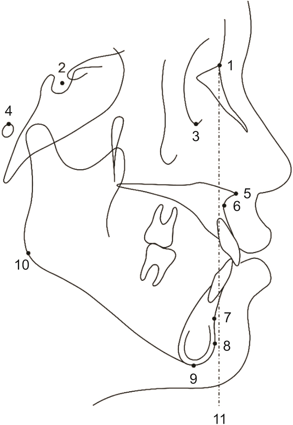
Figure 2 Sagittal landmarks and the vertical reference plane assessed on lateral cephalogram. 1, nasion; 2, sella; 3, orbitale; 4, porion; 5, anterior nasal spine; 6, A point; 7, B point; 8, pogonion; 9, menton; 10, gonion; and 11, nasion perpendicular plane: a line perpendicular to the Frankfort horizontal plane and passing through the nasion.
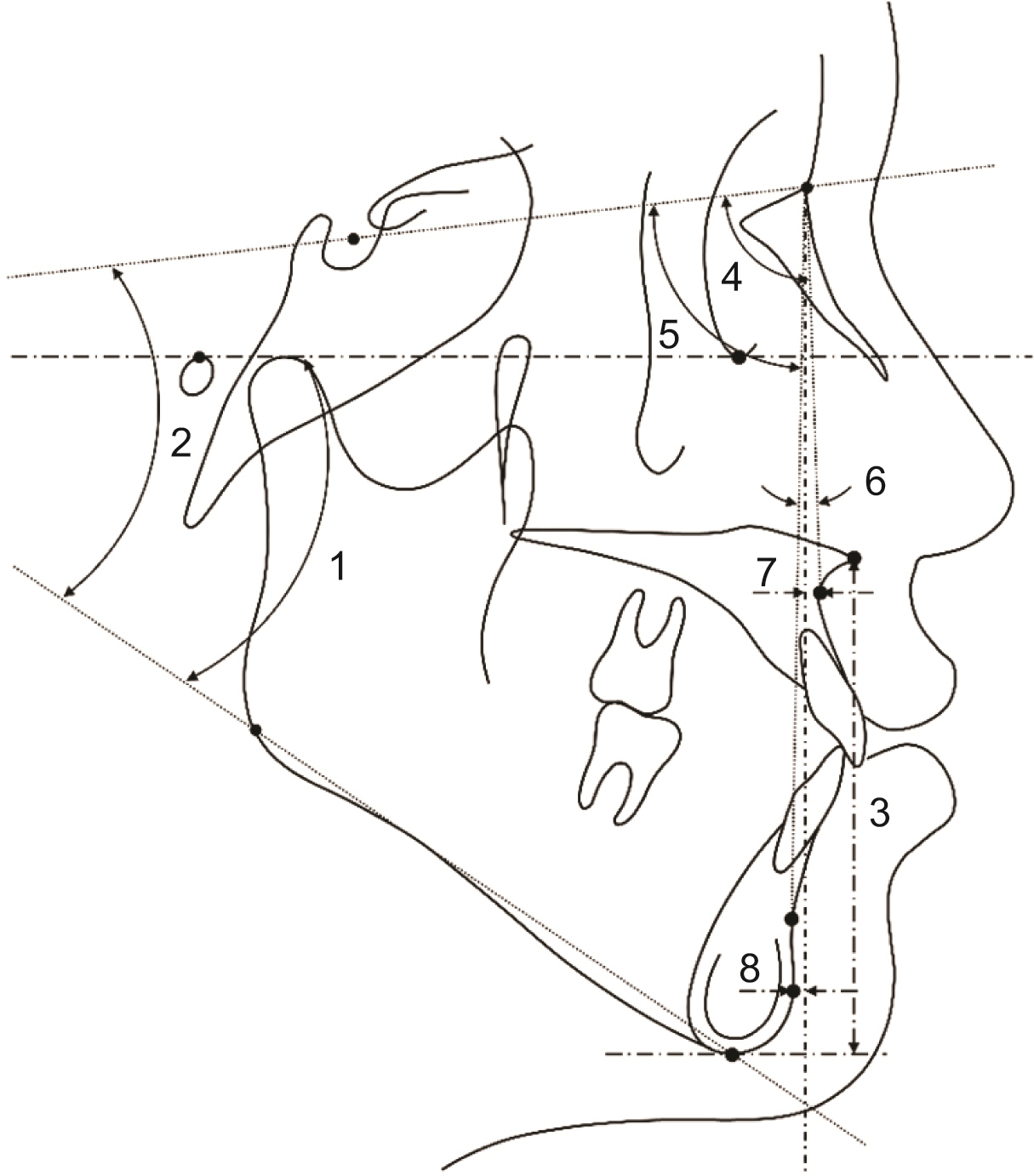
Figure 3 Sagittal skeletal variables assessed on lateral cephalogram. 1, Frankfort-mandibular plane angle (FMA); 2, sella-nasion to mandibular plane angle (SN-MP); 3, lower anterior facial height (LAFH, distance between the anterior nasal spine and the menton parallel to the nasion perpendicular); 4, sella-nasion-A point (SNA); 5, sella-nasion-B point (SNB); 6, A point-nasion-B point (ANB); 7, A point to nasion perpendicular (A to N perp); and 8, pogonion to nasion perpendicular (Pog to N perp). A to N perp, Pog to N perp, and LAFH are linear measurements, while the remaining variables are angular measurements.

Figure 4 Soft tissue variables assessed on frontal photograph. 1, interpupillary distance: the distance between the left and right pupils; 2, alar width: the distance between the left and right alars; 3, nose length: the distance between the midpoint of the pupils and subnasale; 4, upper lip length: the distance between the subnasale and stomion; and 5, lip chin length: the distance between stomion and menton. Vertical measurements including the nose length, upper lip length, and lip chin length were measured as the distances parallel to the vertical bisector of the pupils.
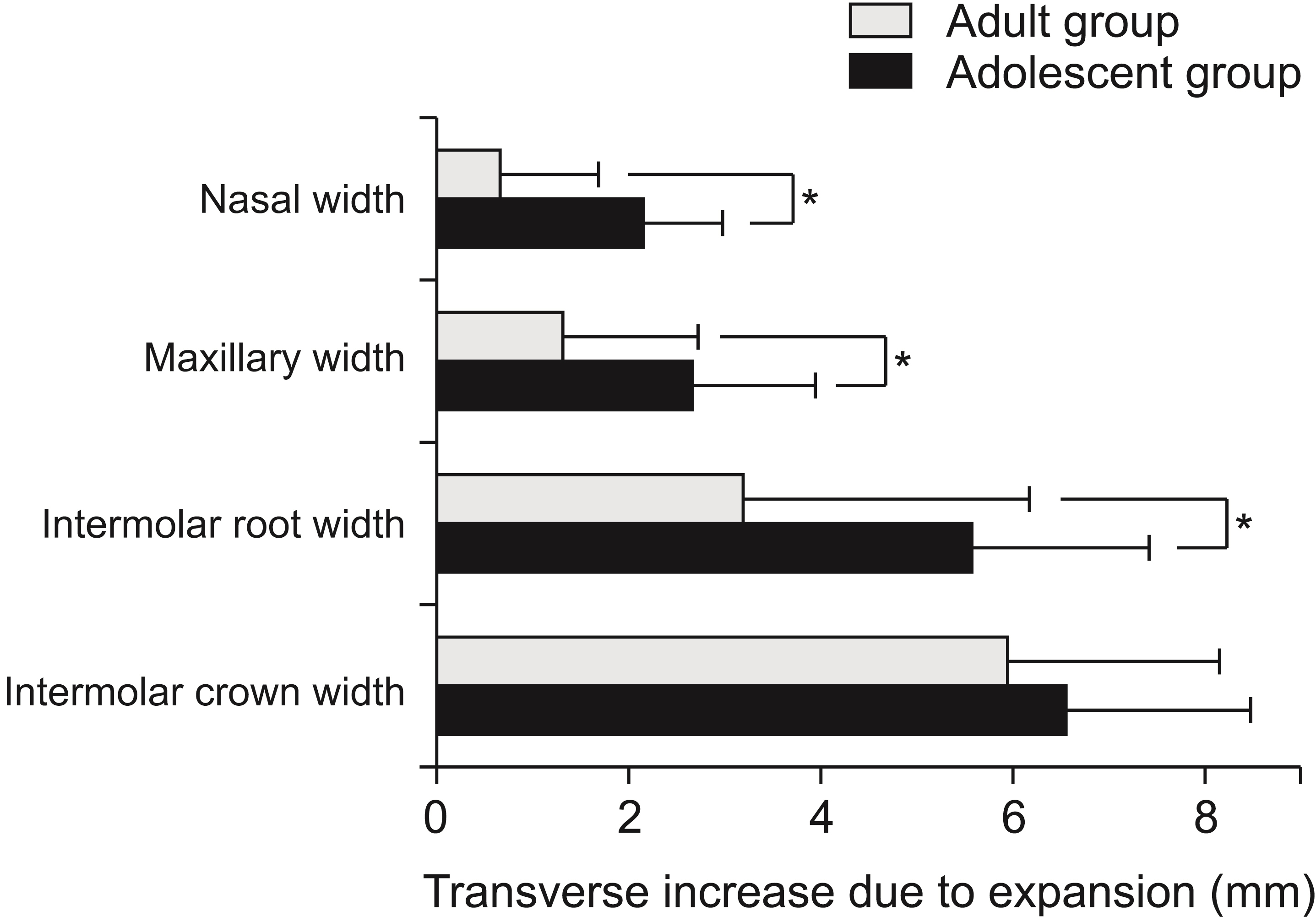
Figure 5 Differences in the transverse dentoskeletal variable changes between the groups after rapid maxillary expansion. *Statistically significant difference (p < 0.05).
Reference
-
References
1. Chung CH, Font B. 2004; Skeletal and dental changes in the sagittal, vertical, and transverse dimensions after rapid palatal expansion. Am J Orthod Dentofacial Orthop. 126:569–75. DOI: 10.1016/j.ajodo.2003.10.035. PMID: 15520689.
Article2. Liu S, Xu T, Zou W. 2015; Effects of rapid maxillary expansion on the midpalatal suture: a systematic review. Eur J Orthod. 37:651–5. DOI: 10.1093/ejo/cju100. PMID: 25700989.
Article3. Del Santo M Jr, Guerrero CA, Buschang PH, English JD, Samchukov ML, Bell WH. 2000; Long-term skeletal and dental effects of mandibular symphyseal distraction osteogenesis. Am J Orthod Dentofacial Orthop. 118:485–93. DOI: 10.1067/mod.2000.109887. PMID: 11094362.
Article4. Schuster G, Borel-Scherf I, Schopf PM. 2005; Frequency of and complications in the use of RPE appliances-- results of a survey in the Federal State of Hesse, Germany. J Orofac Orthop. 66:148–61. DOI: 10.1007/s00056-005-0431-6. PMID: 15827702.
Article5. Nanda R, Snodell SF, Bollu P. 2012; Transverse growth of maxilla and mandible. Semin Orthod. 18:100–17. DOI: 10.1053/j.sodo.2011.10.007.
Article6. Bishara SE, Staley RN. 1987; Maxillary expansion: clinical implications. Am J Orthod Dentofacial Orthop. 91:3–14. DOI: 10.1016/0889-5406(87)90202-2. PMID: 3541577.
Article7. Melsen B. 1975; Palatal growth studied on human autopsy material. A histologic microradiographic study. Am J Orthod. 68:42–54. DOI: 10.1016/0002-9416(75)90158-X. PMID: 1056143.8. Handelman CS, Wang L, BeGole EA, Haas AJ. 2000; Nonsurgical rapid maxillary expansion in adults: report on 47 cases using the Haas expander. Angle Orthod. 70:129–44. DOI: 10.1043/0003-3219(2000)070<0129:NRMEIA>2.0.CO;2. PMID: 10833001.9. Koudstaal MJ, Poort LJ, van der Wal KG, Wolvius EB, Prahl-Andersen B, Schulten AJ. 2005; Surgically assisted rapid maxillary expansion (SARME): a review of the literature. Int J Oral Maxillofac Surg. 34:709–14. DOI: 10.1016/j.ijom.2005.04.025. PMID: 15961279.
Article10. Ramieri GA, Spada MC, Austa M, Bianchi SD, Berrone S. 2005; Transverse maxillary distraction with a bone-anchored appliance: dento-periodontal effects and clinical and radiological results. Int J Oral Maxillofac Surg. 34:357–63. DOI: 10.1016/j.ijom.2004.10.011. PMID: 16053842.
Article11. Carlson C, Sung J, McComb RW, Machado AW, Moon W. 2016; Microimplant-assisted rapid palatal expansion appliance to orthopedically correct transverse maxillary deficiency in an adult. Am J Orthod Dentofacial Orthop. 149:716–28. DOI: 10.1016/j.ajodo.2015.04.043. PMID: 27131254.
Article12. Gunyuz Toklu M, Germec-Cakan D, Tozlu M. 2015; Periodontal, dentoalveolar, and skeletal effects of tooth-borne and tooth-bone-borne expansion appliances. Am J Orthod Dentofacial Orthop. 148:97–109. DOI: 10.1016/j.ajodo.2015.02.022. PMID: 26124033.
Article13. Kanomi R, Deguchi T, Kakuno E, Takano-Yamamoto T, Roberts WE. 2013; CBCT of skeletal changes following rapid maxillary expansion to increase arch-length with a development-dependent bonded or banded appliance. Angle Orthod. 83:851–7. DOI: 10.2319/082012-669.1. PMID: 23488528. PMCID: PMC8744536.
Article14. Sandikçioğlu M, Hazar S. 1997; Skeletal and dental changes after maxillary expansion in the mixed dentition. Am J Orthod Dentofacial Orthop. 111:321–7. DOI: 10.1016/S0889-5406(97)70191-4. PMID: 9082855.
Article15. Lione R, Franchi L, Cozza P. 2013; Does rapid maxillary expansion induce adverse effects in growing subjects? Angle Orthod. 83:172–82. DOI: 10.2319/041012-300.1. PMID: 22827478. PMCID: PMC8805530.
Article16. Berger JL, Pangrazio-Kulbersh V, Thomas BW, Kaczynski R. 1999; Photographic analysis of facial changes associated with maxillary expansion. Am J Orthod Dentofacial Orthop. 116:563–71. DOI: 10.1016/S0889-5406(99)70190-3. PMID: 10547518.
Article17. Pangrazio-Kulbersh V, Wine P, Haughey M, Pajtas B, Kaczynski R. 2012; Cone beam computed tomography evaluation of changes in the naso-maxillary complex associated with two types of maxillary expanders. Angle Orthod. 82:448–57. DOI: 10.2319/072211-464.1. PMID: 22032536. PMCID: PMC8865835.
Article18. Handelman CS. 1997; Nonsurgical rapid maxillary alveolar expansion in adults: a clinical evaluation. Angle Orthod. 67:291–305. discussion 306–8. DOI: 10.1043/0003-3219(1997)067<0291:NRMAEI>2.3.CO;2. PMID: 9267578.19. Northway WM, Meade JB Jr. 1997; Surgically assisted rapid maxillary expansion: a comparison of technique, response, and stability. Angle Orthod. 67:309–20. DOI: 10.1043/0003-3219(1997)067<0309:SARMEA>2.3.CO;2. PMID: 9267580.20. von Elm E, Altman DG, Egger M, Pocock SJ, Gøtzsche PC, Vandenbroucke JP. STROBE Initiative. 2014; The strengthening the reporting of observational studies in epidemiology (STROBE) statement: guidelines for reporting observational studies. Int J Surg. 12:1495–9. DOI: 10.1016/j.ijsu.2014.07.013. PMID: 25046131.
Article21. Kim JH, Yun S, Hwang SS, Shim JO, Chae HW, Lee YJ, et al. 2018; ; Committee for the Development of Growth Standards for Korean Children and Adolescents; Committee for School Health and Public Health Statistics, the Korean Pediatric Society; Division of Health and Nutrition Survey, Korea Centers for Disease Control and Prevention. The 2017 Korean National Growth Charts for children and adolescents: development, improvement, and prospects. Korean J Pediatr. 61:135–49. DOI: 10.3345/kjp.2018.61.5.135. PMID: 29853938. PMCID: PMC5976563.
Article22. Baccetti T, Franchi L, McNamara JA Jr. 2005; The cervical vertebral maturation (CVM) method for the assessment of optimal treatment timing in dentofacial orthopedics. Semin Orthod. 11:119–29. DOI: 10.1053/j.sodo.2005.04.005.
Article23. Faul F, Erdfelder E, Lang AG, Buchner A. 2007; G*Power 3: a flexible statistical power analysis program for the social, behavioral, and biomedical sciences. Behav Res Methods. 39:175–91. DOI: 10.3758/BF03193146. PMID: 17695343.
Article24. Baydas B, Yavuz I, Uslu H, Dagsuyu IM, Ceylan I. 2006; Nonsurgical rapid maxillary expansion effects on craniofacial structures in young adult females. A bone scintigraphy study. Angle Orthod. 76:759–67. DOI: 10.1043/0003-3219(2006)076[0759:NRMEEO]2.0.CO;2. PMID: 17029507.25. Lagravere MO, Major PW, Flores-Mir C. 2005; Long-term skeletal changes with rapid maxillary expansion: a systematic review. Angle Orthod. 75:1046–52. DOI: 10.1043/0003-3219(2005)75[1046:LSCWRM]2.0.CO;2. PMID: 16448254.26. Chang JY, McNamara JA Jr, Herberger TA. 1997; A longitudinal study of skeletal side effects induced by rapid maxillary expansion. Am J Orthod Dentofacial Orthop. 112:330–7. DOI: 10.1016/S0889-5406(97)70264-6. PMID: 9294364.
Article27. Habeeb M, Boucher N, Chung CH. 2013; Effects of rapid palatal expansion on the sagittal and vertical dimensions of the maxilla: a study on cephalograms derived from cone-beam computed tomography. Am J Orthod Dentofacial Orthop. 144:398–403. DOI: 10.1016/j.ajodo.2013.04.012. PMID: 23992812.
Article28. Kim YC, Kwon JG, Kim SC, Huh CH, Kim HJ, Oh TS, et al. 2018; Comparison of periorbital anthropometry between beauty pageant contestants and ordinary young women with Korean ethnicity: a three-dimensional photogrammetric analysis. Aesthetic Plast Surg. 42:479–90. DOI: 10.1007/s00266-017-1040-7. PMID: 29273931.
Article29. Tonello DL, Ladewig VM, Guedes FP, Ferreira Conti ACC, Almeida-Pedrin RR, Capelozza-Filho L. 2017; Midpalatal suture maturation in 11- to 15-year-olds: a cone-beam computed tomographic study. Am J Orthod Dentofacial Orthop. 152:42–8. DOI: 10.1016/j.ajodo.2016.11.028. PMID: 28651767.
Article30. Kapetanović A, Theodorou CI, Bergé SJ, Schols JGJH, Xi T. 2021; Efficacy of miniscrew-assisted rapid palatal expansion (MARPE) in late adolescents and adults: a systematic review and meta-analysis. Eur J Orthod. 43:313–23. DOI: 10.1093/ejo/cjab005. PMID: 33882127. PMCID: PMC8186837.
Article
- Full Text Links
-
- Actions
-
Cited
- CITED
-
- Close
- Share
-
- Similar articles
-
- Influence of changing various parameters in miniscrew-assisted rapid palatal expansion: A three-dimensional finite element analysis
- Effect of bone-borne maxillary skeletal expanders on cranial and circummaxillary sutures: A cone-beam computed tomography study
- Evaluation of condylar dimension and position following rapid maxillary expansion with toothor tooth-bone-borne appliances
- Surgically assisted rapid palatal expansion with tent screws and a custom-made palatal expander: a case report
- Total Knee Arthroplasty Using Tissue Expander In Patient with Poor Soft Tissue Coverage of Knee: A Case Report
