Gallic acid-mitochondria targeting sequence-H3R9 induces mitochondria-targeted cytoprotection
- Affiliations
-
- 1Department of Physiology, Cardiovascular and Metabolic Disease Center, Smart Marine Therapeutic Center, Inje University College of Medicine, Busan 47392, Korea
- 2Division of Applied Medicine, Research Institute for Korea Medicine, School of Korean Medicine, Pusan National University, Yangsan 50612, Korea
- 3Department of Biology and Clinical Pharmacology, R&D Center, Samyang Biopharmaceuticals Corporation, Seongnam 13488, Korea
- 4Department of Internal Medicine, Sanggye Paik Hospital, Cardiovascular and Metabolic Disease Center, Inje University, Seoul 01757, Korea
- KMID: 2524507
- DOI: http://doi.org/10.4196/kjpp.2022.26.1.15
Abstract
- The development of selective targeting of drug molecules towards the mitochondria is an important issue related to therapy efficacy. In this study, we report that gallic acid (GA)-mitochondria targeting sequence (MTS)-H3R9 exhibits a dual role as a mitochondria-targeting vehicle with antioxidant activity for disease therapy. In viability assays, GA-MTS-H3R9 showed a better rescue action compared to that of MTS-H3R9 . GA-MTS-H3R9 dramatically exhibited cell penetration and intercellular uptake compared to MTS and fit escape from lysosome release to the cytosol. We demonstrated the useful targeting of GA-MTS-H3R9 towards mitochondria in AC16 cells. Also, we observed that the antioxidant properties of mitochondrial-accrued GA-MTSH3R9 alleviated cell damage by reactive oxygen species production and disrupted mitochondrial membrane potential. GA-MTS-H3R9 showed a very increased cytoprotective effect against anticancer activity compared to that of MTS-H3R9 . We showed that GA-MTS-H3R9 can act as a vehicle for mitochondria-targeting and as a reagent for therapeutic applications intended for cardiovascular disease treatment.
Keyword
Figure
-

Fig. 1 Schematic description for the localization of the GA-MTS-H3R9 inside cells and its cytoprotective effect.

Fig. 2 Cell viability assay with GA-MTS-H3R9. Cell viability was determined using WST-1 and lactate dehydrogenase (LDH) assay. AC16 cells were exposed to different concentrations of GA, MTS, MTS-H3R6, MTS-H3R9, GA-MTS-H3R6, and GA-MTS-H3R9 up to 50 μM for 24 h. Panel (A) presents the results of the WST-1 assay and Panel (B) presents the results of LDH assay. GA, gallic acid; MTS, mitochondria targeting sequence. Data are indicted as the mean ± SD (n = 3).
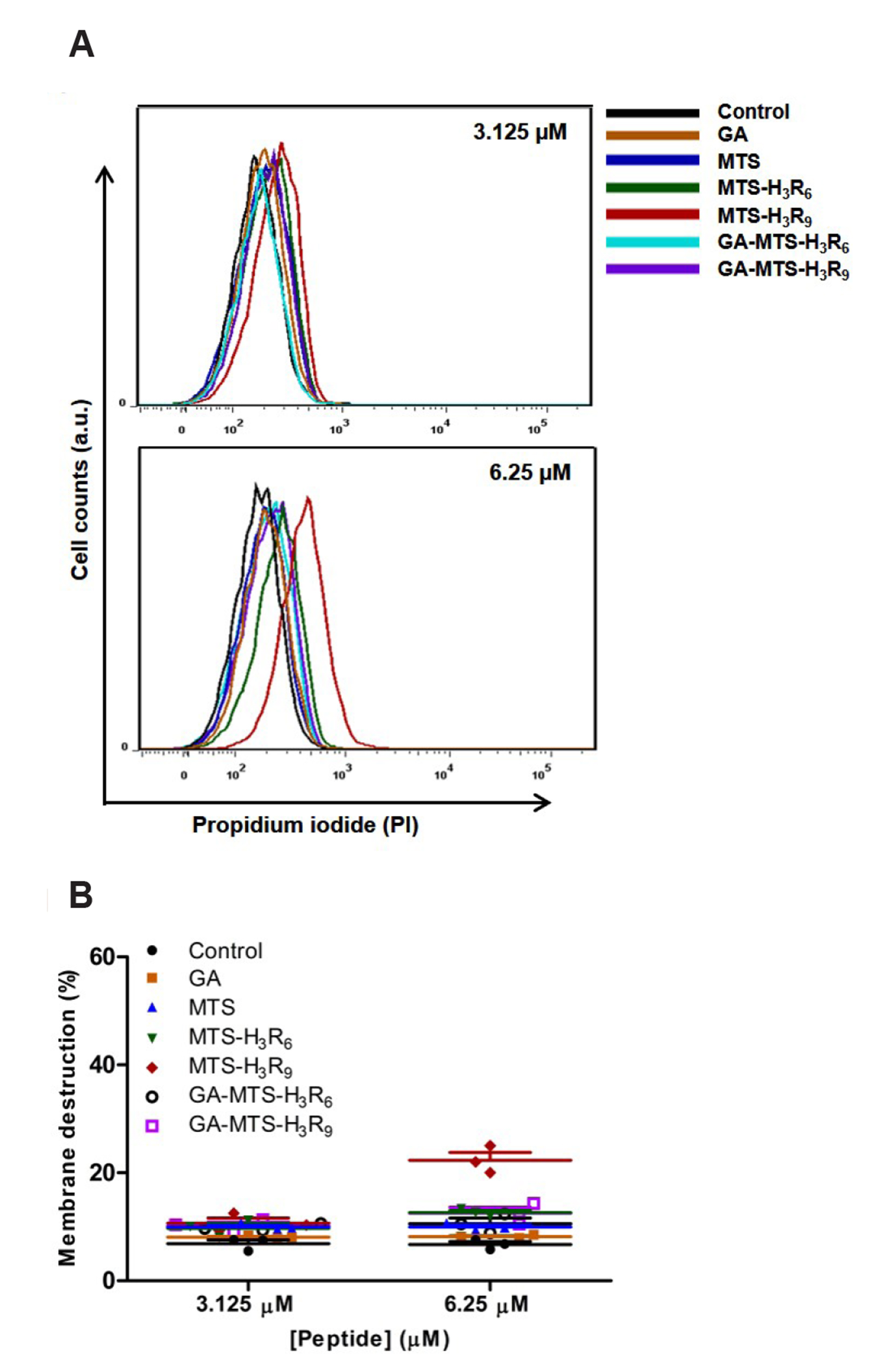
Fig. 3 Membrane internalization by GA-MTS-H3R9. AC16 cells were treated with 3.125 and 6.25 μM of GA, MTS, MTS-H3R6, MTS-H3R9, GA-MTS-H3R6, and GA-MTS-H3R9 for 16 h and were subsequently incubated with 5 μl of PI for 20 min. (A) PI internalization using flow cytometry analysis. (B) Quantitative graph showing each peptide from the PI fluorescence data shown in panel (A). GA, gallic acid; MTS, mitochondria targeting sequence; PI, propidium iodide.
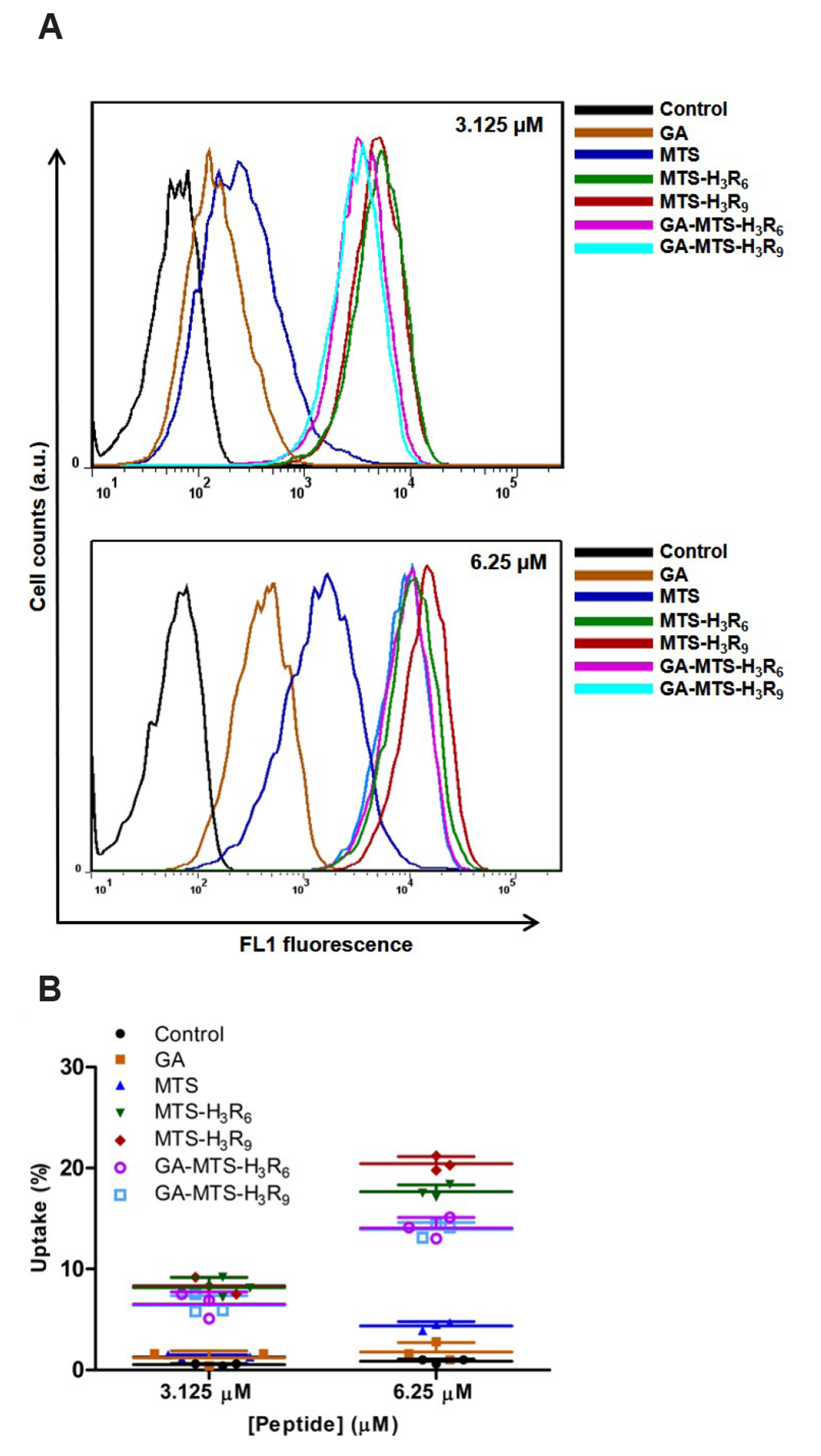
Fig. 4 Intracellular uptake of GA-MTS-H3R9. AC16 cells were treated with 3.125 and 6.25 μM of FITC-labeled GA, MTS, MTS-H3R6, MTS-H3R9, GA-MTS-H3R6, and GA-MTS-H3R9 for 16 h. (A) Cellular uptake of each FITC-labeled peptide using flow cytometry analysis. (B) Quantitative graph showing each peptide from uptake data depicted in panel (A). GA, gallic acid; MTS, mitochondria targeting sequence.
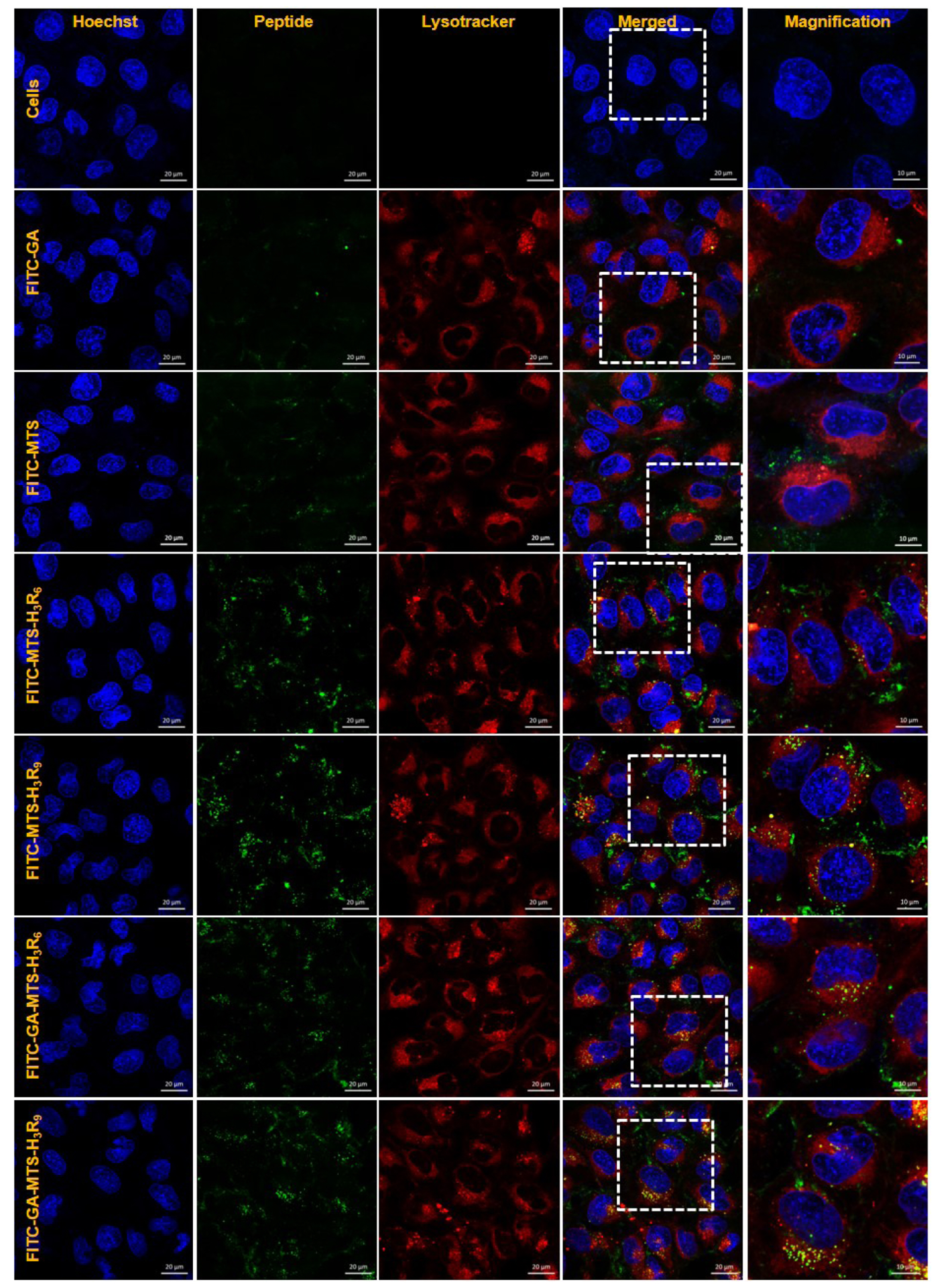
Fig. 5 Intracellular distribution of GA-MTS-H3R9. AC16 cells were treated with 1 μM of FITC-labeled GA, MTS, MTS-H3R6, MTS-H3R9, GA-MTS-H3R6, and GA-MTS-H3R9 for 16 h. The cells were stained with LysoTracker (red) for 20 min and counterstained with Hoechst stain (blue) for 10 min and imaged by confocal microscopy. Scale bar is 10 µm. GA, gallic acid; MTS, mitochondria targeting sequence.

Fig. 6 Intracellular mitochondria localization of GA-MTS-H3R9. AC16 cells were exposed to 1 μM of FITC-labeled GA, MTS, MTS-H3R6, MTS-H3R9, GA-MTS-H3R6, and GA-MTS-H3R9 for 24 h. The cells were stained with MitoTracker (red) for 20 min and counterstained with Hoechst stain (blue) and then imaged by confocal laser scanning microscopy. Scale bar is 20 µm. GA, gallic acid; MTS, mitochondria targeting sequence.

Fig. 7 ROS levels and mitochondrial membrane potential (MMP) with GA-MTS-H3R9. (A) AC16 cells were exposed to 6.25 μM of GA, MTS, MTS-H3R6, MTS-H3R9, GA-MTS-H3R6, and GA-MTS-H3R9 for 16 h. Intracellular ROS levels were assessed by GSH assay. (B) AC16 cells were treated using the same conditions as those panel (A). The cells were treated with 20 μM MitoSox for 16 h. Cells were imaged by confocal microscopy. Scale bar is 10 µm. (C) AC16 cells were treated using the same conditions as those in panel (A). The cells were added to 2 μM JC-1 and then incubated for 20 min. MMP was determined by flow cytometry analysis. Data are indicted as the mean ± SD (n = 3). ROS, reactive oxygen species; GA, gallic acid; GSH, glutathione; MTS, mitochondria targeting sequence; CCCP, carbonyl cyanide 3-chlorophenylhydrazone. **p < 0.01.

Fig. 8 Induction of cytoprotection by GA-MTS-H3R9. (A) Anticancer activity of each peptide. AC16 cells were exposed to 6.25 μM of GA, MTS, MTS-H3R6, MTS-H3R9, GA-MTS-H3R6, and GA-MTS-H3R9 for 24 h. Apoptosis levels for each group were measured by flow cytometry analysis after annexin V staining. Q1 indicates necrotic cells. Q2 indicates late apoptotic cells, Q3 indicates intact cells, and Q4 indicates early apoptotic cells. (B) Caspase-3 activity of each peptide. AC16 cells were treated using the same conditions as those in panel (A). Caspase-3 activity was assessed as described in Methods. Data are indicted as the mean ± SD (n = 3). GA, gallic acid; MTS, mitochondria targeting sequence. **p < 0.01.
Reference
-
1. Wolfram JA, Donahue JK. 2013; Gene therapy to treat cardiovascular disease. J Am Heart Assoc. 2:e000119. DOI: 10.1161/JAHA.113.000119. PMID: 23963752. PMCID: PMC3828796.
Article2. Leo CH, Jelinic M, Ng HH, Parry LJ, Tare M. 2019; Recent developments in relaxin mimetics as therapeutics for cardiovascular diseases. Curr Opin Pharmacol. 45:42–48. DOI: 10.1016/j.coph.2019.04.001. PMID: 31048209.
Article3. Rossignol P, Hernandez AF, Solomon SD, Zannad F. 2019; Heart failure drug treatment. Lancet. 393:1034–1044. DOI: 10.1016/S0140-6736(18)31808-7. PMID: 30860029.
Article4. Dvir T, Bauer M, Schroeder A, Tsui JH, Anderson DG, Langer R, Liao R, Kohane DS. 2011; Nanoparticles targeting the infarcted heart. Nano Lett. 11:4411–4414. DOI: 10.1021/nl2025882. PMID: 21899318. PMCID: PMC3192253.
Article5. Tan KX, Pan S, Jeevanandam J, Danquah MK. 2019; Cardiovascular therapies utilizing targeted delivery of nanomedicines and aptamers. Int J Pharm. 558:413–425. DOI: 10.1016/j.ijpharm.2019.01.023. PMID: 30660748.
Article6. Mahapatro A, Singh DK. 2011; Biodegradable nanoparticles are excellent vehicle for site directed in-vivo delivery of drugs and vaccines. J Nanobiotechnology. 9:55. DOI: 10.1186/1477-3155-9-55. PMID: 22123084. PMCID: PMC3238292.
Article7. Sanna V, Pala N, Sechi M. 2014; Targeted therapy using nanotechnology: focus on cancer. Int J Nanomedicine. 9:467–483. DOI: 10.2147/IJN.S36654. PMID: 24531078. PMCID: PMC3896284.8. Calzoni E, Cesaretti A, Polchi A, Di Michele A, Tancini B, Emiliani C. 2019; Biocompatible polymer nanoparticles for drug delivery applications in cancer and neurodegenerative disorder therapies. J Funct Biomater. 10:4. DOI: 10.3390/jfb10010004. PMID: 30626094. PMCID: PMC6463038.
Article9. Cosentino K, García-Sáez AJ. 2014; Mitochondrial alterations in apoptosis. Chem Phys Lipids. 181:62–75. DOI: 10.1016/j.chemphyslip.2014.04.001. PMID: 24732580.
Article10. Rin Jean S, Tulumello DV, Wisnovsky SP, Lei EK, Pereira MP, Kelley SO. 2014; Molecular vehicles for mitochondrial chemical biology and drug delivery. ACS Chem Biol. 9:323–333. DOI: 10.1021/cb400821p. PMID: 24410267.
Article11. Weissig V, Boddapati SV, Jabr L, D'Souza GG. 2007; Mitochondria-specific nanotechnology. Nanomedicine (Lond). 2:275–285. DOI: 10.2217/17435889.2.3.275. PMID: 17716174.
Article12. Lin R, Zhang P, Cheetham AG, Walston J, Abadir P, Cui H. 2015; Dual peptide conjugation strategy for improved cellular uptake and mitochondria targeting. Bioconjug Chem. 26:71–77. DOI: 10.1021/bc500408p. PMID: 25547808. PMCID: PMC4306504.
Article13. Klimpel A, Neundorf I. 2018; Bifunctional peptide hybrids targeting the matrix of mitochondria. J Control Release. 291:147–156. DOI: 10.1016/j.jconrel.2018.10.029. PMID: 30367921.
Article14. von Heijne G. 1986; Mitochondrial targeting sequences may form amphiphilic helices. EMBO J. 5:1335–1342. DOI: 10.1002/j.1460-2075.1986.tb04364.x. PMID: 3015599. PMCID: PMC1166945.
Article15. Yu GS, Han J, Ko KS, Choi JS. 2014; Cationic oligopeptide-conjugated mitochondria targeting sequence as a novel carrier system for mitochondria. Macromol Res. 22:42–46. DOI: 10.1007/s13233-014-2003-3.
Article16. Schmidt N, Mishra A, Lai GH, Wong GC. 2010; Arginine-rich cell-penetrating peptides. FEBS Lett. 584:1806–1813. DOI: 10.1016/j.febslet.2009.11.046. PMID: 19925791.
Article17. Bae Y, Joo C, Kim GY, Ko KS, Huh KM, Han J, Choi JS. 2019; Cationic oligopeptide-functionalized mitochondria targeting sequence show mitochondria targeting and anticancer activity. Macromol Res. 27:1071–1080. DOI: 10.1007/s13233-019-7153-x.
Article18. Xie M, Hu B, Wang Y, Zeng X. 2014; Grafting of gallic acid onto chitosan enhances antioxidant activities and alters rheological properties of the copolymer. J Agric Food Chem. 62:9128–9136. DOI: 10.1021/jf503207s. PMID: 25198516.
Article19. Perron NR, Brumaghim JL. 2009; A review of the antioxidant mechanisms of polyphenol compounds related to iron binding. Cell Biochem Biophys. 53:75–100. DOI: 10.1007/s12013-009-9043-x. PMID: 19184542.
Article20. Bae Y, Green ES, Kim GY, Song SJ, Mun JY, Lee S, Park JI, Park JS, Ko KS, Han J, Choi JS. 2016; Dipeptide-functionalized polyamidoamine dendrimer-mediated apoptin gene delivery facilitates apoptosis of human primary glioma cells. Int J Pharm. 515:186–200. DOI: 10.1016/j.ijpharm.2016.09.083. PMID: 27732896.
Article21. Bae Y, Jung MK, Lee S, Song SJ, Mun JY, Green ES, Han J, Ko KS, Choi JS. 2018; Dequalinium-based functional nanosomes show increased mitochondria targeting and anticancer effect. Eur J Pharm Biopharm. 124:104–115. DOI: 10.1016/j.ejpb.2017.12.013. PMID: 29305141.
Article22. AshaRani PV, Low Kah Mun G, Hande MP, Valiyaveettil S. 2009; Cytotoxicity and genotoxicity of silver nanoparticles in human cells. ACS Nano. 3:279–290. DOI: 10.1021/nn800596w. PMID: 19236062.
Article23. Holder AL, Goth-Goldstein R, Lucas D, Koshland CP. 2012; Particle-induced artifacts in the MTT and LDH viability assays. Chem Res Toxicol. 25:1885–1892. DOI: 10.1021/tx3001708. PMID: 22799765. PMCID: PMC3446248.
Article24. Yang Y, Xiang Y, Xu M. 2015; From red to green: the propidium iodide-permeable membrane of Shewanella decolorationis S12 is repairable. Sci Rep. 5:18583. DOI: 10.1038/srep18583. PMID: 26687136. PMCID: PMC4685271.
Article25. Xiang L, Xie G, Liu C, Zhou J, Chen J, Yu S, Li J, Pang X, Shi H, Liang H. 2013; Knock-down of glutaminase 2 expression decreases glutathione, NADH, and sensitizes cervical cancer to ionizing radiation. Biochim Biophys Acta. 1833:2996–3005. DOI: 10.1016/j.bbamcr.2013.08.003. PMID: 23954443.
Article26. Soumya RS, Vineetha VP, Salin Raj P, Raghu KG. 2014; Beneficial properties of selenium incorporated guar gum nanoparticles against ischemia/reperfusion in cardiomyoblasts (H9c2). Metallomics. 6:2134–2147. DOI: 10.1039/C4MT00241E. PMID: 25307064.
Article27. He H, Li DW, Yang LY, Fu L, Zhu XJ, Wong WK, Jiang FL, Liu Y. 2015; A novel bifunctional mitochondria-targeted anticancer agent with high selectivity for cancer cells. Sci Rep. 5:13543. DOI: 10.1038/srep13543. PMID: 26337336. PMCID: PMC4559806.
Article28. Perelman A, Wachtel C, Cohen M, Haupt S, Shapiro H, Tzur A. 2012; JC-1: alternative excitation wavelengths facilitate mitochondrial membrane potential cytometry. Cell Death Dis. 3:e430. DOI: 10.1038/cddis.2012.171. PMID: 23171850. PMCID: PMC3542606.
Article29. Sancho P, Galeano E, Nieto E, Delgado MD, García-Pérez AI. 2007; Dequalinium induces cell death in human leukemia cells by early mitochondrial alterations which enhance ROS production. Leuk Res. 31:969–978. DOI: 10.1016/j.leukres.2006.11.018. PMID: 17250890.
Article30. Madani F, Lindberg S, Langel U, Futaki S, Gräslund A. 2011; Mechanisms of cellular uptake of cell-penetrating peptides. J Biophys. 2011:414729. DOI: 10.1155/2011/414729. PMID: 21687343. PMCID: PMC3103903.
Article
- Full Text Links
-
- Actions
-
Cited
- CITED
-
- Close
- Share
-
- Similar articles
-
- The Interplay between Autophagy and Aging
- Understanding the Role of Heat Shock Protein Isoforms in Male Fertility, Aging and Apoptosis
- Ultrastructural Studies on Mitochondria of Preimplantaion Rabbit Embryos
- Myopathy, Drugs, and Mitochondria
- The Formation of Giant Mitochondria in the Liver Cells Induced by Hydrazine
