Int J Stem Cells.
2021 Aug;14(3):320-330. 10.15283/ijsc20186.
Galectin-3 Derived from HucMSC Exosomes Promoted Myocardial Fibroblast-to-Myofibroblast Differentiation Associated with β-catenin Upregulation
- Affiliations
-
- 1School of Medicine, Jiangsu University, Zhenjiang, China
- 2Department of Obstetrics, Affiliated Hospital of Jiangsu University, Zhenjiang, China
- 3Department of Clinical Laboratory, Zhenjiang Provincial Blood Center, Zhenjiang, China
- KMID: 2519374
- DOI: http://doi.org/10.15283/ijsc20186
Abstract
- Background and Objectives
Galectin-3 promotes fibroblast-to-myofibroblast differentiation and facilitates injury repair. Previous studies have shown that exosomes derived from human umbilical cord mesenchymal stem cells (hucMSC-ex) promote the differentiation of myocardial fibroblasts into myofibroblasts under inflammatory environment. Whether hucMSC-ex derived Galectin-3 (hucMSC-ex-Galectin-3) plays an important role in fibroblast-to-myofibroblast differentiation is the focus of this study.
Methods and Results
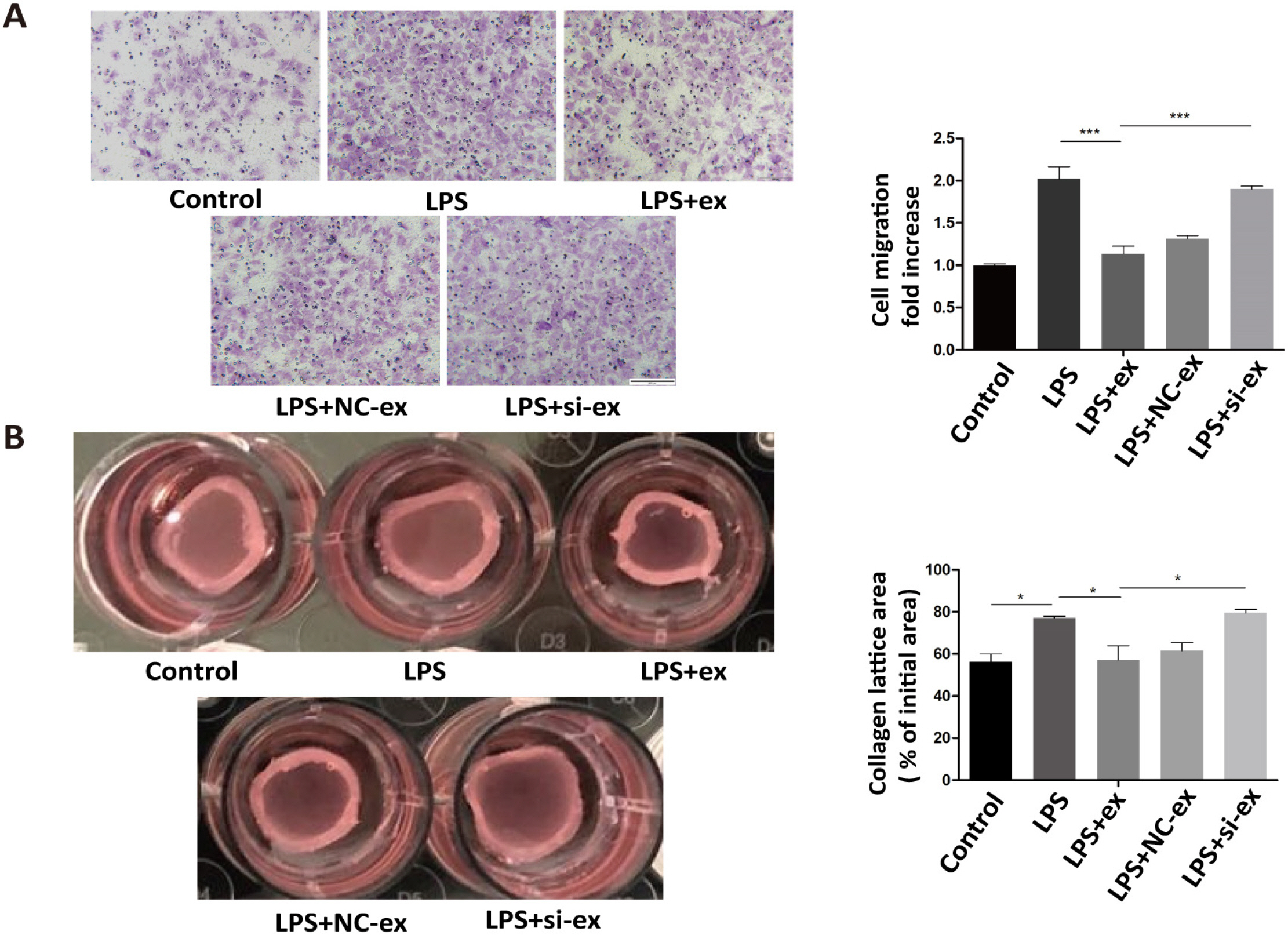
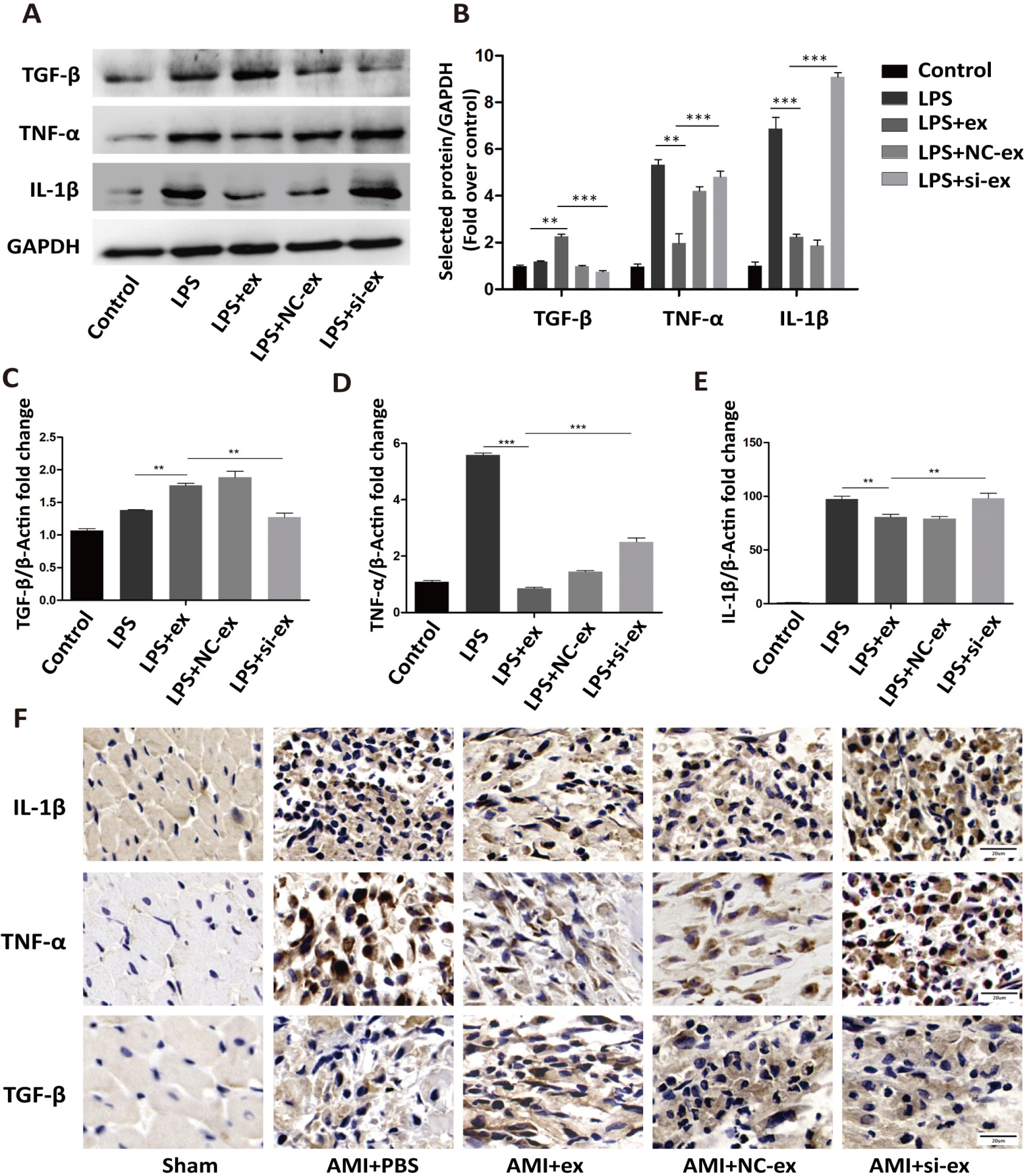
Galectin-3 was knocked-down by siRNA in hucMSCs, and then exosomes were extracted. Fibroblasts were treated with LPS, LPS+hucMSC-ex, LPS+negative control-siRNA-ex (NC-ex), or LPS+ Galectin-3-siRNA-ex (si-ex) in vitro. The coronary artery of the left anterior descending (LAD) branch was permanently ligated, followed by intramyocardial injection with phosphate buffered saline(PBS), hucMSC-ex, hucMSC-NC-ex, or hucMSC-si-ex in vivo. Western blot, RT-PCR, and immunohistochemistry were used to detect the expression of markers related to fibroblast-to-myofibroblast differentiation and inflammatory factors. Migration and contraction functions of fibroblasts were evaluated using Transwell migration and collagen contraction assays, respectively. β-catenin expression was detected by western blot and immunofluorescence. The results showed that hucMSC-ex increased the protein expression of myofibroblast markers, anti-inflammatory factors, and β-catenin. HucMSC-ex also reduced the migration and promoted the contractility of fibroblasts. However, hucMSC-si-ex did not show these activities.
Conclusions
HucMSC-ex-Galectin-3 promoted the differentiation of cardiac fibroblasts into myofibroblasts in an inflammatory environment, which was associated with increased β-catenin levels.
Figure
Reference
-
References
1. Fan D, Takawale A, Lee J, Kassiri Z. 2012; Cardiac fibroblasts, fibrosis and extracellular matrix remodeling in heart disease. Fibrogenesis Tissue Repair. 5:15. DOI: 10.1186/1755-1536-5-15. PMID: 22943504. PMCID: PMC3464725.
Article2. Tallquist MD, Molkentin JD. 2017; Redefining the identity of cardiac fibroblasts. Nat Rev Cardiol. 14:484–491. DOI: 10.1038/nrcardio.2017.57. PMID: 28436487. PMCID: PMC6329009.
Article3. Shin EY, Wang L, Zemskova M, Deppen J, Xu K, Strobel F, García AJ, Tirouvanziam R, Levit RD. 2018; Adenosine production by biomaterial-supported mesenchymal stromal cells reduces the innate inflammatory response in myocardial ischemia/reperfusion injury. J Am Heart Assoc. 7:e006949. DOI: 10.1161/JAHA.117.006949. PMID: 29331956. PMCID: PMC5850147.
Article4. Teng X, Chen L, Chen W, Yang J, Yang Z, Shen Z. 2015; Mesenchymal stem cell-derived exosomes improve the microenvironment of infarcted myocardium contributing to angiogenesis and anti-inflammation. Cell Physiol Biochem. 37:2415–2424. DOI: 10.1159/000438594. PMID: 26646808.
Article5. Shi Y, Yang Y, Guo Q, Gao Q, Ding Y, Wang H, Xu W, Yu B, Wang M, Zhao Y, Zhu W. 2019; Exosomes derived from human umbilical cord mesenchymal stem cells promote fibroblast-to-myofibroblast differentiation in inflammatory environments and benefit cardioprotective effects. Stem Cells Dev. 28:799–811. DOI: 10.1089/scd.2018.0242. PMID: 30896296.
Article6. Deng H, Sun C, Sun Y, Li H, Yang L, Wu D, Gao Q, Jiang X. 2018; Lipid, protein, and microRNA composition within mesenchymal stem cell-derived exosomes. Cell Reprogram. 20:178–186. DOI: 10.1089/cell.2017.0047. PMID: 29782191.
Article7. Hu K, Gu Y, Lou L, Liu L, Hu Y, Wang B, Luo Y, Shi J, Yu X, Huang H. 2015; Galectin-3 mediates bone marrow microenvironment-induced drug resistance in acute leukemia cells via Wnt/β-catenin signaling pathway. J Hematol Oncol. 8:1. DOI: 10.1186/s13045-014-0099-8. PMID: 25622682. PMCID: PMC4332970.
Article8. de Boer RA, Voors AA, Muntendam P, van Gilst WH, van Veldhuisen DJ. 2009; Galectin-3: a novel mediator of heart failure development and progression. Eur J Heart Fail. 11:811–817. DOI: 10.1093/eurjhf/hfp097. PMID: 19648160.
Article9. Li LC, Li J, Gao J. 2014; Functions of galectin-3 and its role in fibrotic diseases. J Pharmacol Exp Ther. 351:336–343. DOI: 10.1124/jpet.114.218370. PMID: 25194021.
Article10. Meijers WC, van der Velde AR, Pascual-Figal DA, de Boer RA. 2015; Galectin-3 and post-myocardial infarction cardiac remodeling. Eur J Pharmacol. 763(Pt A):115–121. DOI: 10.1016/j.ejphar.2015.06.025. PMID: 26101067.
Article11. González GE, Cassaglia P, Noli Truant S, Fernández MM, Wilensky L, Volberg V, Malchiodi EL, Morales C, Gelpi RJ. 2014; Galectin-3 is essential for early wound healing and ventricular remodeling after myocardial infarction in mice. Int J Cardiol. 176:1423–1425. DOI: 10.1016/j.ijcard.2014.08.011. PMID: 25150483.
Article12. Li M, Yuan Y, Guo K, Lao Y, Huang X, Feng L. 2020; Value of Galectin-3 in acute myocardial infarction. Am J Cardiovasc Drugs. 20:333–342. DOI: 10.1007/s40256-019-00387-9. PMID: 31784887.
Article13. Zhao X, Hua Y, Chen H, Yang H, Zhang T, Huang G, Fan H, Tan Z, Huang X, Liu B, Zhou Y. 2015; Aldehyde dehydrogenase-2 protects against myocardial infarction-related cardiac fibrosis through modulation of the Wnt/β-catenin signaling pathway. Ther Clin Risk Manag. 11:1371–1381. DOI: 10.2147/TCRM.S88297. PMID: 26392772. PMCID: PMC4574798.14. Ge Z, Li B, Zhou X, Yang Y, Zhang J. 2016; Basic fibroblast growth factor activates β-catenin/RhoA signaling in pulmonary fibroblasts with chronic obstructive pulmonary disease in rats. Mol Cell Biochem. 423:165–174. DOI: 10.1007/s11010-016-2834-7. PMID: 27734223.
Article15. Shimura T, Takenaka Y, Tsutsumi S, Hogan V, Kikuchi A, Raz A. 2004; Galectin-3, a novel binding partner of beta-catenin. Cancer Res. 64:6363–6367. DOI: 10.1158/0008-5472.CAN-04-1816. PMID: 15374939.16. Qiao C, Xu W, Zhu W, Hu J, Qian H, Yin Q, Jiang R, Yan Y, Mao F, Yang H, Wang X, Chen Y. 2008; Human mesenchymal stem cells isolated from the umbilical cord. Cell Biol Int. 32:8–15. DOI: 10.1016/j.cellbi.2007.08.002. PMID: 17904875.
Article17. Ma Y, Iyer RP, Jung M, Czubryt MP, Lindsey ML. 2017; Cardiac fibroblast activation post-myocardial infarction: current knowledge gaps. Trends Pharmacol Sci. 38:448–458. DOI: 10.1016/j.tips.2017.03.001. PMID: 28365093. PMCID: PMC5437868.
Article18. Nagpal V, Rai R, Place AT, Murphy SB, Verma SK, Ghosh AK, Vaughan DE. 2016; MiR-125b is critical for fibroblast-to-myofibroblast transition and cardiac fibrosis. Circulation. 133:291–301. DOI: 10.1161/CIRCULATIONAHA.115.018174. PMID: 26585673. PMCID: PMC5446084.
Article19. Nakaya M, Watari K, Tajima M, Nakaya T, Matsuda S, Ohara H, Nishihara H, Yamaguchi H, Hashimoto A, Nishida M, Nagasaka A, Horii Y, Ono H, Iribe G, Inoue R, Tsuda M, Inoue K, Tanaka A, Kuroda M, Nagata S, Kurose H. 2017; Cardiac myofibroblast engulfment of dead cells facilitates recovery after myocardial infarction. J Clin Invest. 127:383–401. DOI: 10.1172/JCI83822. PMID: 27918308. PMCID: PMC5199696.
Article20. Liu Y, Chen H, Zheng P, Zheng Y, Luo Q, Xie G, Ma Y, Shen L. 2017; ICG-001 suppresses growth of gastric cancer cells and reduces chemoresistance of cancer stem cell-like population. J Exp Clin Cancer Res. 36:125. DOI: 10.1186/s13046-017-0595-0. PMID: 28893318. PMCID: PMC5594604.
Article21. Sun XH, Wang X, Zhang Y, Hui J. 2019; Exosomes of bone-marrow stromal cells inhibit cardiomyocyte apoptosis under ischemic and hypoxic conditions via miR-486-5p targeting the PTEN/PI3K/AKT signaling pathway. Thromb Res. 177:23–32. DOI: 10.1016/j.thromres.2019.02.002. PMID: 30844685.
Article22. Xu R, Zhang F, Chai R, Zhou W, Hu M, Liu B, Chen X, Liu M, Xu Q, Liu N, Liu S. 2019; Exosomes derived from pro-inflammatory bone marrow-derived mesenchymal stem cells reduce inflammation and myocardial injury via mediating macrophage polarization. J Cell Mol Med. 23:7617–7631. DOI: 10.1111/jcmm.14635. PMID: 31557396. PMCID: PMC6815833.
Article23. Sun J, Shen H, Shao L, Teng X, Chen Y, Liu X, Yang Z, Shen Z. 2020; HIF-1α overexpression in mesenchymal stem cell-derived exosomes mediates cardioprotection in myocardial infarction by enhanced angiogenesis. Stem Cell Res Ther. 11:373. DOI: 10.1186/s13287-020-01881-7. PMID: 32859268. PMCID: PMC7455909.
Article24. Gu X, Li Y, Chen K, Wang X, Wang Z, Lian H, Lin Y, Rong X, Chu M, Lin J, Guo X. 2020; Exosomes derived from umbilical cord mesenchymal stem cells alleviate viral myocarditis through activating AMPK/mTOR-mediated autophagy flux pathway. J Cell Mol Med. 24:7515–7530. DOI: 10.1111/jcmm.15378. PMID: 32424968. PMCID: PMC7339183.
Article25. Wang XL, Zhao YY, Sun L, Shi Y, Li ZQ, Zhao XD, Xu CG, Ji HG, Wang M, Xu WR, Zhu W. 2018; Exosomes derived from human umbilical cord mesenchymal stem cells improve myocardial repair via upregulation of Smad7. Int J Mol Med. 41:3063–3072. DOI: 10.3892/ijmm.2018.3496. PMID: 29484378.
Article26. Sandanger Ø, Ranheim T, Vinge LE, Bliksøen M, Alfsnes K, Finsen AV, Dahl CP, Askevold ET, Florholmen G, Christensen G, Fitzgerald KA, Lien E, Valen G, Espevik T, Aukrust P, Yndestad A. 2013; The NLRP3 inflammasome is up-regulated in cardiac fibroblasts and mediates myocar-dial ischaemia-reperfusion injury. Cardiovasc Res. 99:164–174. DOI: 10.1093/cvr/cvt091. PMID: 23580606.
Article27. Shinde AV, Frangogiannis NG. 2014; Fibroblasts in myocardial infarction: a role in inflammation and repair. J Mol Cell Cardiol. 70:74–82. DOI: 10.1016/j.yjmcc.2013.11.015. PMID: 24321195. PMCID: PMC3995820.
Article28. Sharma UC, Pokharel S, van Brakel TJ, van Berlo JH, Cleutjens JP, Schroen B, André S, Crijns HJ, Gabius HJ, Maessen J, Pinto YM. 2004; Galectin-3 marks activated macrophages in failure-prone hypertrophied hearts and contributes to cardiac dysfunction. Circulation. 110:3121–3128. DOI: 10.1161/01.CIR.0000147181.65298.4D. PMID: 15520318.
Article29. Liu Y, Xie L, Wang D, Li D, Xu G, Wang L, Zhou H, Yu Y, Lin Z, Lu H. 2018; Galectin-3 and β-catenin are associated with a poor prognosis in serous epithelial ovarian cancer. Cancer Manag Res. 10:3963–3971. DOI: 10.2147/CMAR.S171146. PMID: 30310317. PMCID: PMC6165784.30. Song M, Pan Q, Yang J, He J, Zeng J, Cheng S, Huang Y, Zhou ZQ, Zhu Q, Yang C, Han Y, Tang Y, Chen H, Weng DS, Xia JC. 2020; Galectin-3 favours tumour metastasis via the activation of β-catenin signalling in hepatocellular carcinoma. Br J Cancer. 123:1521–1534. DOI: 10.1038/s41416-020-1022-4. PMID: 32801345. PMCID: PMC7653936.
Article31. Mackinnon AC, Gibbons MA, Farnworth SL, Leffler H, Nilsson UJ, Delaine T, Simpson AJ, Forbes SJ, Hirani N, Gauldie J, Sethi T. 2012; Regulation of transforming growth factor-β1-driven lung fibrosis by galectin-3. Am J Respir Crit Care Med. 185:537–546. DOI: 10.1164/rccm.201106-0965OC. PMID: 22095546. PMCID: PMC3410728.
Article
- Full Text Links
- Actions
-
Cited
- CITED
-
- Close
- Share
- Similar articles
-
- A Comprehensive Cancer-Associated MicroRNA Expression Profiling and Proteomic Analysis of Human Umbilical Cord Mesenchymal Stem Cell-Derived Exosomes
- PSC-MSC-Derived Exosomes Protect against Kidney Fibrosis In Vivo and In Vitro through the SIRT6/β-Catenin Signaling Pathway
- Exosomes Derived from Human Umbilical Cord Mesenchymal Stem Cells Regulate Macrophage Polarization to Attenuate Systemic Lupus Erythematosus-Associated Diffuse Alveolar Hemorrhage in Mice
- Tumor-Derived Exosomal Circular RNA Pinin Induces FGF13 Expression to Promote Colorectal Cancer Progression through miR-1225-5p
- Atractylochromene Is a Repressor of Wnt/beta-Catenin Signaling in Colon Cancer Cells

