White Opaque Substance, a New Optical Marker on Magnifying Endoscopy: Usefulness in Diagnosing Colorectal Epithelial Neoplasms
- Affiliations
-
- 1Department of Gastroenterology, Fukuoka University Chikushi Hospital, Fukuoka, Japan
- 2Department of Endoscopy, Fukuoka University Chikushi Hospital, Fukuoka, Japan
- 3Department of Pathology, Fukuoka University Chikushi Hospital, Fukuoka, Japan
- KMID: 2518861
- DOI: http://doi.org/10.5946/ce.2020.205
Abstract
- Background/Aims
A white substance that is opaque to endoscopic light is sometimes observed in the epithelium during narrowband imaging with magnifying endoscopy of gastric or colorectal epithelial neoplasms. This prospective observational study aimed to determine whether the morphology of the white opaque substance (WOS) allows differential diagnosis between colorectal adenoma and carcinoma.
Methods
A consecutive series of patients with colorectal adenomas or early carcinomas who underwent endoscopic resection or surgical excision were studied. The morphology of the WOS was determined based on endoscopic images before the histopathological diagnosis was performed. The primary outcome was the diagnostic performance of an irregular WOS as a marker of colorectal carcinoma.
Results
The study analyzed 125 lesions. A total of 33 lesions showed an irregular WOS, and 92 lesions showed a regular WOS. Among the 33 lesions found to show an irregular WOS, 30 were carcinomas. Among the 92 lesions showing a regular WOS, 79 were adenomas. With irregular WOS as a marker of carcinoma, the diagnostic accuracy was 87%, sensitivity was 91%, and specificity was 86%.
Conclusions
This study demonstrated the potential usefulness of the morphology of the WOS as a marker for the differential diagnosis between adenoma and carcinoma in cases of colorectal epithelial neoplasms.
Figure
-

Fig. 1. Endoscopic findings of white opaque substance (WOS)-positive and WOS-negative colorectal epithelial neoplasms. (A) Image of a WOS-negative colorectal epithelial neoplasm observed with white light. (B) Magnifying narrow-band imaging (NBI) endoscopic view of the square area. The WOS is absent, with the microvessels clearly visible. (C) Image of a WOS-positive colorectal epithelial neoplasm observed with white light. (D) Magnifying NBI endoscopic view of the square area. The WOS is present, with the microvessels practically obscured.
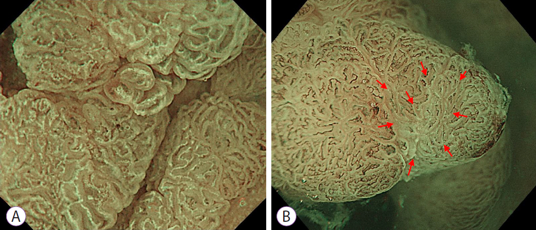
Fig. 2. Determination of whether a lesion is white opaque substance (WOS)-positive. (A) The morphology of the WOS can be visualized. The WOS is present in no less than half of the area when visualized under maximal magnification. (B) The morphology of the WOS cannot be evaluated. Although the WOS is seen in the areas identified by red arrows when visualized under maximal magnification, it is present in less than half of the overall area.
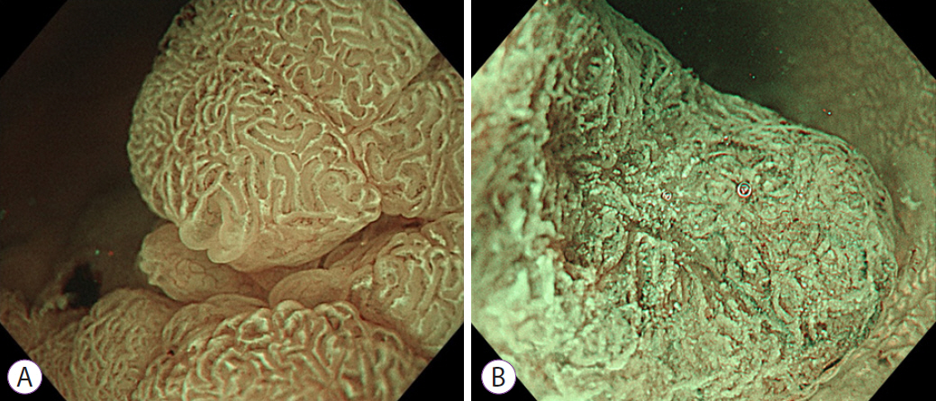
Fig. 3. Morphological findings of the white opaque substance (WOS). (A) Regular WOS: morphology of the WOS shows a well-organized and symmetrical distribution of a regular reticular pattern. (B) Irregular WOS: morphology of the WOS shows a disorganized and asymmetrical distribution of an irregular speckled pattern.
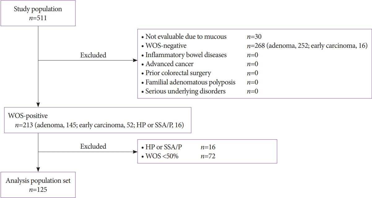
Fig. 4. Participant flow. HP, hyperplastic polyp; SSA/P, sessile serrated adenoma/polyp; WOS, white opaque substance.
Reference
-
1. Yao K, Anagnostopoulos GK, Ragunath K. Magnifying endoscopy for diagnosing and delineating early gastric cancer. Endoscopy. 2009; 41:462–467.
Article2. Sano Y, Ikematsu H, Fu KI, et al. Meshed capillary vessels by use of narrow-band imaging for differential diagnosis of small colorectal polyps. Gastrointest Endosc. 2009; 69:278–283.
Article3. Kanao H, Tanaka S, Oka S, Hirata M, Yoshida S, Chayama K. Narrow-band imaging magnification predicts the histology and invasion depth of colorectal tumors. Gastrointest Endosc. 2009; 69(3 Pt 2):631–636.
Article4. Ezoe Y, Muto M, Uedo N, et al. Magnifying narrowband imaging is more accurate than conventional white-light imaging in diagnosis of gastric mucosal cancer. Gastroenterology. 2011; 141:2017–2025.e3.
Article5. Machida H, Sano Y, Hamamoto Y, et al. Narrow-band imaging in the diagnosis of colorectal mucosal lesions: a pilot study. Endoscopy. 2004; 36:1094–1098.
Article6. East JE, Suzuki N, Bassett P, et al. Narrow band imaging with magnification for the characterization of small and diminutive colonic polyps: pit pattern and vascular pattern intensity. Endoscopy. 2008; 40:811–817.
Article7. Sikka S, Ringold DA, Jonnalagadda S, Banerjee B. Comparison of white light and narrow band high definition images in predicting colon polyp histology, using standard colonoscopes without optical magnification. Endoscopy. 2008; 40:818–822.
Article8. Hewett DG, Kaltenbach T, Sano Y, et al. Validation of a simple classification system for endoscopic diagnosis of small colorectal polyps using narrow-band imaging. Gastroenterology. 2012; 143:599–607.e1.
Article9. Wani S, Rastogi A. Narrow-band imaging in the prediction of submucosal invasive colon cancer: how “NICE” is it? Gastrointest Endosc. 2013; 78:633–636.10. Singh R, Nordeen N, Mei SL, Kaffes A, Tam W, Saito Y. West meets East: preliminary results of narrow band imaging with optical magnification in the diagnosis of colorectal lesions: a multicenter Australian study using the modified Sano’s classification. Dig Endosc. 2011; 23 Suppl 1:126–130.
Article11. Yao K, Iwashita A, Tanabe H, et al. White opaque substance within superficial elevated gastric neoplasia as visualized by magnification endoscopy with narrow-band imaging: a new optical sign for differentiating between adenoma and carcinoma. Gastrointest Endosc. 2008; 68:574–580.
Article12. Kanemitsu T, Yao K, Nagahama T, et al. Extending magnifying NBI diagnosis of intestinal metaplasia in the stomach: the white opaque substance marker. Endoscopy. 2017; 49:529–535.
Article13. Yao K, Iwashita A, Nambu M, et al. Nature of white opaque substance in gastric epithelial neoplasia as visualized by magnifying endoscopy with narrow-band imaging. Dig Endosc. 2012; 24:419–425.
Article14. Ueo T, Yonemasu H, Yada N, et al. White opaque substance represents an intracytoplasmic accumulation of lipid droplets: immunohistochemical and immunoelectron microscopic investigation of 26 cases. Dig Endosc. 2013; 25:147–155.
Article15. Hisabe T, Yao K, Imamura K, et al. White opaque substance visualized using magnifying endoscopy with narrow-band imaging in colorectal epithelial neoplasms. Dig Dis Sci. 2014; 59:2544–2549.
Article16. Hisabe T, Yao K, Imamura K, et al. Novel endoscopic findings as visualized by magnifying endoscopy with narrow-band imaging: white opaque substance is present in colorectal hyperplastic polyps. Digestion. 2016; 93:127–131.
Article17. Imamura K, Yao K, Hisabe T, et al. The nature of the white opaque substance within colorectal neoplastic epithelium as visualized by magnifying endoscopy with narrow-band imaging. Endosc Int Open. 2016; 4:E1151–E1157.
Article18. The Paris endoscopic classification of superficial neoplastic lesions: esophagus, stomach, and colon: November 30 to December 1, 2002. Gastrointest Endosc. 2003; 58:S3–S43.19. Schlemper RJ, Kato Y, Stolte M. Diagnostic criteria for gastrointestinal carcinomas in Japan and Western countries: proposal for a new classification system of gastrointestinal epithelial neoplasia. J Gastroenterol Hepatol. 2000; 15 Suppl:G49–G57.
Article20. Kawasaki K, Kurahara K, Yanai S, et al. Significance of a white opaque substance under magnifying narrow-band imaging colonoscopy for the diagnosis of colorectal epithelial neoplasms. Gastrointest Endosc. 2015; 82:1097–1104.
Article21. Kudo S, Tamura S, Nakajima T, Yamano H, Kusaka H, Watanabe H. Diagnosis of colorectal tumorous lesions by magnifying endoscopy. Gastrointest Endosc. 1996; 44:8–14.
Article22. Tanaka S, Sano Y. Aim to unify the narrow band imaging (NBI) magnifying classification for colorectal tumors: current status in Japan from a summary of the consensus symposium in the 79th annual meeting of the Japan Gastroenterological Endoscopy Society. Dig Endosc. 2011; 23 Suppl 1:131–139.
Article23. Sano Y, Tanaka S, Kudo SE, et al. Narrow-band imaging (NBI) magnifying endoscopic classification of colorectal tumors proposed by the Japan NBI Expert Team. Dig Endosc. 2016; 28:526–533.
Article24. Ikematsu H, Matsuda T, Emura F, et al. Efficacy of capillary pattern type IIIA/IIIB by magnifying narrow band imaging for estimating depth of invasion of early colorectal neoplasms. BMC Gastroenterol. 2010; 10:33.
Article25. Wada Y, Kudo SE, Kashida H, et al. Diagnosis of colorectal lesions with the magnifying narrow-band imaging system. Gastrointest Endosc. 2009; 70:522–531.
Article26. Hisabe T, Yao K, Beppu T, et al. Validity of the usefulness of microvascular architecture and microsurface structure using magnifying endoscopy with narrow-band imaging in the colorectal neoplasm. Ann Gastroenterol. 2013; 26:45–51.
- Full Text Links
-
- Actions
-
Cited
- CITED
-
- Close
- Share
-
- Similar articles
-
- Clinical Usefulness of Magnifying Chromoendoscopy and Magnifying Narrow Band Imaging Endoscopy for Predicting the Submucosal Invasion of Early Colorectal Cancers
- What Have We Accomplished in Endoscopic Image Analysis for Atrophic Gastritis?
- The Usefulness of Pit Patterns of Colorectal Tumors and Magnifying Colonoscopy
- Application of artificial intelligence for diagnosis of early gastric cancer based on magnifying endoscopy with narrow-band imaging
- Usefulness of Narrow-Band Imaging in Endoscopic Submucosal Dissection of the Stomach
