Endovascular Treatment of Intracranial Aneurysms Using the Novel Low Profile Visualized Intraluminal Support EVO Stent: Multicenter Early Feasibility Experience
- Foo M
 1
1 - Maingard J2,3
- Hall J1,4
- Ren Y1
- Mitreski G1
- Slater LA2,5
- Chandra R2,5
- Chong W2,6,7
- Jhamb A1,4
- Russell J8
- Kok HK3,9
- Brooks M1,4
- Asadi H1,2,3,10
- Affiliations
-
- 1Interventional Neuroradiology Service, Department of Radiology, Austin Health, Heidelberg, VIC, Australia
- 2Interventional Neuroradiology Service, Department of Radiology, Monash Health, Clayton, VIC, Australia
- 3School of Medicine, Deakin University, Waurn Ponds, VIC, Australia
- 4Interventional Neuroradiology Service, Department of Radiology, St Vincent’s Hospital Melbourne, Fitzroy, VIC, Australia
- 5Department of Imaging, Monash University, Clayton, VIC, Australia
- 6Faculty of Medicine and Health Sciences, Macquarie University, Macquarie Park, NSW, Australia
- 7Faculty of Medicine, Dentistry and Health Sciences, The University of Melbourne, Melbourne, VIC, Australia
- 8Neurosurgery Department, Austin Health, Heidelberg, VIC, Australia
- 9Interventional Radiology Service, Department of Radiology, Northern Health, Epping, VIC, Australia
- 10Florey Institute of Neurosciences and Mental Health, The University of Melbourne, Parkville, VIC, Australia
- KMID: 2517737
- DOI: http://doi.org/10.5469/neuroint.2021.00199
Abstract
- Purpose
Low-profile, self-expandable stents have broadened therapeutic options available for definitive treatment of intracranial aneurysms. The novel Low-Profile Visualized Intraluminal Support (LVIS) EVO stent extends upon the success of its predecessor, the LVIS Jr stent, aiming to enable higher visibility and greater opening ability within a self-expandable and fully retrievable microstent system. In this study, we aim to report the early safety and feasibility experience with the LVIS EVO stent.
Materials and Methods
A multicenter, retrospective, observational study was conducted on patients who had intracranial aneurysms treated with the LVIS EVO stent across 3 Australian neurovascular centers between February 2020 and September 2020. Short-term technical and clinical outcomes were evaluated.
Results
A total of 22 LVIS EVO stents were successfully implanted to treat 15 aneurysms (3 ruptured, 12 unruptured) in 15 patients. Aneurysms ranged from 2 mm to 35 mm in dome height. The LVIS EVO stent was used for stent-assisted coiling in 11 patients and flow diversion in 4 patients. There were no device-related procedural complications. There were 2 cases of peri-procedural symptomatic thromboembolic complications and no procedure-related mortality. At early radiological follow up, 10 patients had complete occlusion, 4 patients had small neck remnants, and 1 patient who was managed with flow diversion had a residual aneurysm.
Conclusion
Early experience with the LVIS EVO stent demonstrated safety and feasibility for stent-assisted coiling as well as flow diversion for intracranial aneurysms. In this heterogeneous cohort, including ruptured, complex, and large aneurysms, all cases were technically successful.
Keyword
Figure
-
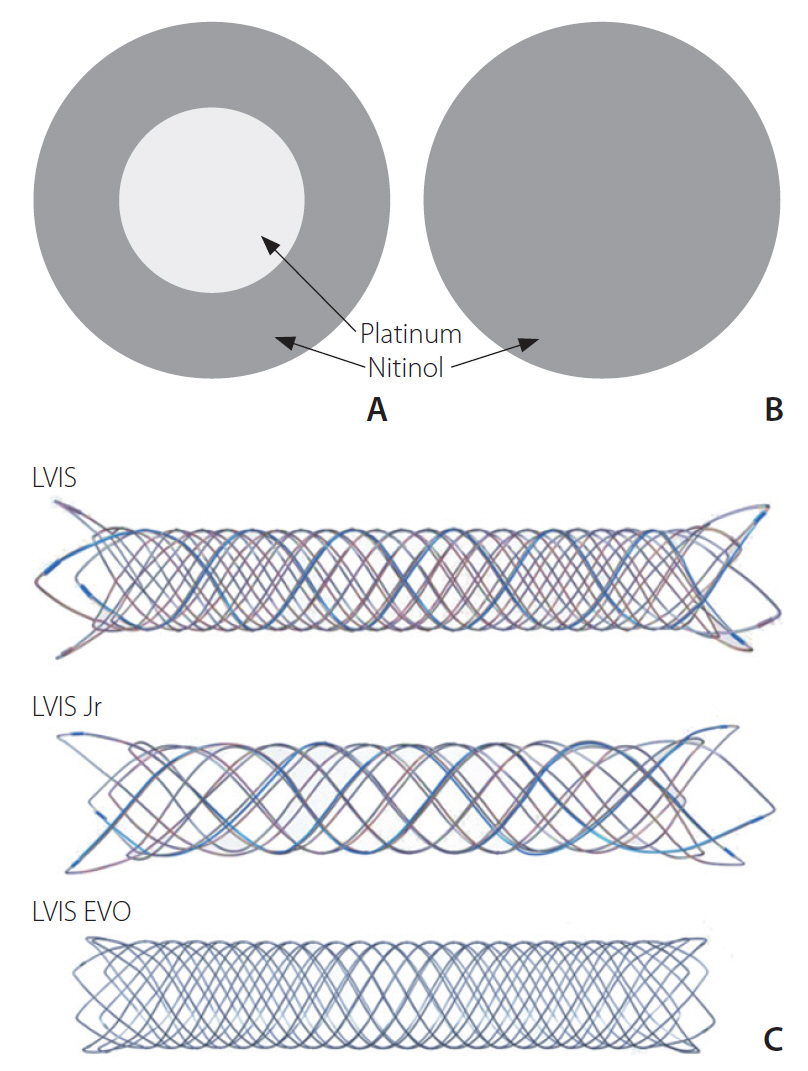
Fig. 1. Drawn-filled tube wire technology of the Low-Profile Visualized Intraluminal Support (LVIS) EVO device (A), allowing radio-opacity of the entire length of the stent body, as compared to the pure nitinol wires used in LVIS and LVIS Jr (B). (C) Visual comparison between LVIS, LVIS Jr, and LVIS EVO stents.
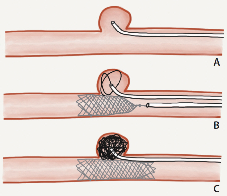
Fig. 2. Semi-jailing double microcatheter technique. (A) The first microcatheter is positioned with its tip in the aneurysm sac. (B) A second microcatheter is used to partially deploy the stent across the aneurysm neck, securing the first microcatheter between the stent and the vessel wall. (C) The stent is fully deployed after dense coil packing.
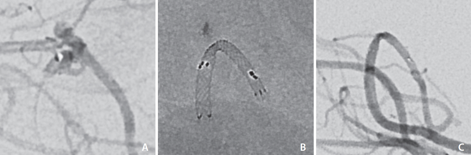
Fig. 3. Case 9. (A) Microcatheter tip near the base of a small aneurysm arising from a tortuous right P3 segment. (B) The Low-Profile Visualized Intraluminal Support (LVIS) EVO stent was easily and successfully deployed as a flow-diverting stent within an Acclino flex plus stent, achieving significant slowing of inflow into the aneurysmal sac on final angiographic run (C).
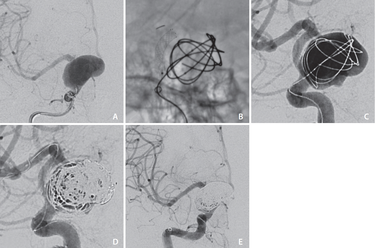
Fig. 4. Low-Profile Visualized Intraluminal Support (LVIS) EVO-assisted coiling of an unruptured right supraclinoid internal carotid artery (ICA) aneurysm. (A) Frontal projection DSA with contrast injection in the right cavernous ICA. (B) LVIS EVO device amid deployment, with radiopaque delivery tip visible within the right MCA and 4 radiopaque markers at the distal end of the implant. Note undetached coils within the aneurysm sac to help with stability of the jailed microcatheter. (C, D) Progressive packing of the aneurysm with coils. (E) Final DSA run demonstrating a subtle degree of contrast opacification along the aneurysmal wall.
Reference
-
1. Phan K, Huo YR, Jia F, Phan S, Rao PJ, Mobbs RJ, et al. Meta-analysis of stent-assisted coiling versus coiling-only for the treatment of intracranial aneurysms. J Clin Neurosci. 2016; 31:15–22.
Article2. Aydin K, Arat A, Sencer S, Barburoglu M, Men S. Stent-assisted coiling of wide-neck intracranial aneurysms using low-profile LEO baby stents: initial and midterm results. AJNR Am J Neuroradiol. 2015; 36:1934–1941.
Article3. Fiorella D, Boulos A, Turk AS, Siddiqui AH, Arthur AS, Diaz O, LVIS investigators, et al. The safety and effectiveness of the LVIS stent system for the treatment of wide-necked cerebral aneurysms: final results of the pivotal US LVIS trial. J Neurointerv Surg. 2019; 11:357–361.
Article4. Wang C, Tian Z, Liu J, Jing L, Paliwal N, Wang S, et al. Flow diverter effect of LVIS stent on cerebral aneurysm hemodynamics: a comparison with Enterprise stents and the Pipeline device. J Transl Med. 2016; 14:199.
Article5. Cagnazzo F, Cappucci M, Dargazanli C, Lefevre PH, Gascou G, Riquelme C, et al. Flow-diversion effect of LEO stents: aneurysm occlusion and flow remodeling of covered side branches and perforators. AJNR Am J Neuroradiol. 2018; 39:2057–2063.
Article6. Wang K, Huang Q, Hong B, Li Z, Fang X, Liu J. Correlation of aneurysm occlusion with actual metal coverage at neck after implantation of flow-diverting stent in rabbit models. Neuroradiology. 2012; 54:607–613.
Article7. Mascitelli JR, Moyle H, Oermann EK, Polykarpou MF, Patel AA, Doshi AH, et al. An update to the Raymond-Roy Occlusion Classification of intracranial aneurysms treated with coil embolization. J Neurointerv Surg. 2015; 7:496–502.
Article8. O’kelly CJ, Krings T, Fiorella D, Marotta TR. A novel grading scale for the angiographic assessment of intracranial aneurysms treated using flow diverting stents. Interv Neuroradiol. 2010; 16:133–137.
Article9. Hendricks BK, Yoon JS, Yaeger K, Kellner CP, Mocco J, De Leacy RA, et al. Wide-neck aneurysms: systematic review of the neurosurgical literature with a focus on definition and clinical implications. [published online ahead of print Jun 14, 2019]. J Neurosurg. 2019.
Article10. Regelsberger J, Matschke J, Grzyska U, Ries T, Fiehler J, Köppen J, et al. Blister-like aneurysms--a diagnostic and therapeutic challenge. Neurosurg Rev. 2011; 34:409–416.
Article11. Sirakov A, Bhogal P, Möhlenbruch M, Sirakov S. Endovascular treatment of patients with intracranial aneurysms: feasibility and successful employment of a new low profile visible intraluminal support (LVIS) EVO stent. Neuroradiol J. 2020; 33:377–385.
Article12. Poncyljusz W, Kubiak K. Initial experience with LVIS EVO stents for the treatment of intracranial aneurysms. J Clin Med. 2020; 9:3966.
Article13. Pandey AS, Koebbe C, Rosenwasser RH, Veznedaroglu E. Endovascular coil embolization of ruptured and unruptured posterior circulation aneurysms: review of a 10-year experience. Neurosurgery. 2007; 60:626–636. discussion 636-637.14. Ferns SP, Sprengers ME, van Rooij WJ, Rinkel GJ, van Rijn JC, Bipat S, et al. Coiling of intracranial aneurysms: a systematic review on initial occlusion and reopening and retreatment rates. Stroke. 2009; 40:e523–e529.15. Vollherbst DF, Berlis A, Maurer C, Behrens L, Sirakov S, Sirakov A, et al. Periprocedural safety and feasibility of the new LVIS EVO device for stent-assisted coiling of intracranial aneurysms: an observational multicenter study. AJNR Am J Neuroradiol. 2021; 42:319–326.
Article
- Full Text Links
-
- Actions
-
Cited
- CITED
-
- Close
- Share
-
- Similar articles
-
- Clinical and Angiographic Outcomes of Endovascular Treatment for Acute Intracranial Vertebral Artery Dissecting Aneurysms Using Double-Overlapping Stents : Low-Profile Visualized Intraluminal Support within Enterprise Stents
- Endovascular treatment of intracranial aneurysms: Past and present
- Flow diversion via telescoping stent with Low-profile Visualized Intraluminal Support Junior for treatment of ruptured dissecting aneurysm located at proximal posterior inferior cerebellar artery
- Preliminary Experience of Lvis Blue in the Internal Carotid Artery for The Treatment Of Wide-Necked Intracranial Aneurysms
- Current Update on the Randomized Controlled Trials of Intracranial Aneurysms
