Flow diversion via telescoping stent with Low-profile Visualized Intraluminal Support Junior for treatment of ruptured dissecting aneurysm located at proximal posterior inferior cerebellar artery
- Affiliations
-
- 1Department of Neurosurgery, Inje University, Ilsan Paik Hospital, Neuroscience & Radiosurgery Hybrid Neurosurgery Research Center, Goyang, Korea
- 2Department of Neurosurgery, Uijeongbu Eulji Medical Center, Eulji University College of Medicine, Uijeongbu, Korea
- KMID: 2517026
- DOI: http://doi.org/10.7461/jcen.2021.E2020.08.003
Abstract
- Dissecting aneurysm involving the posterior inferior cerebellar artery (PICA) are challenging because of its nature and anatomic relationship to medulla and lower cranial nerve. We introduce a case of ruptured dissecting aneurysm located at the proximal PICA treated with telescoping stents for flow diversion and dissection healing. A 49 years old female visited to the emergency room for ruptured dissecting aneurysm at right proximal PICA. Telescoping stent was deployed along the right vertebral artery to PICA covering the dissecting aneurysm bleb using two Low-profile Visualized Intraluminal Support Jr (LVIS Jr) stents. Three months follow up angiography revealed a disappearance of aneurysm bleb and healing of dissection by parent artery remodeling. Telescoping stent with LVIS Jr may be an effective treatment for dissecting aneurysm with small diameter (<2 mm) parent artery. Convenient navigation and targeted telescoping stent for minimizing metal coverage at perforating arteries are an advantage for this method.
Figure
-
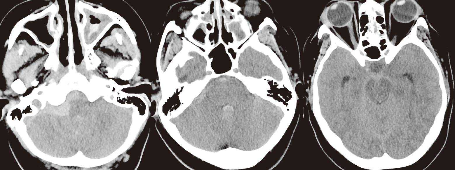
Fig. 1. Noncontrast brain computed tomography at the day of symptom onset. Subarachnoid hemorrhage was noted and dominant at right cerebello-medullary cistern. Intraventricle hemorrhage was also appeared at the fourth ventricle.
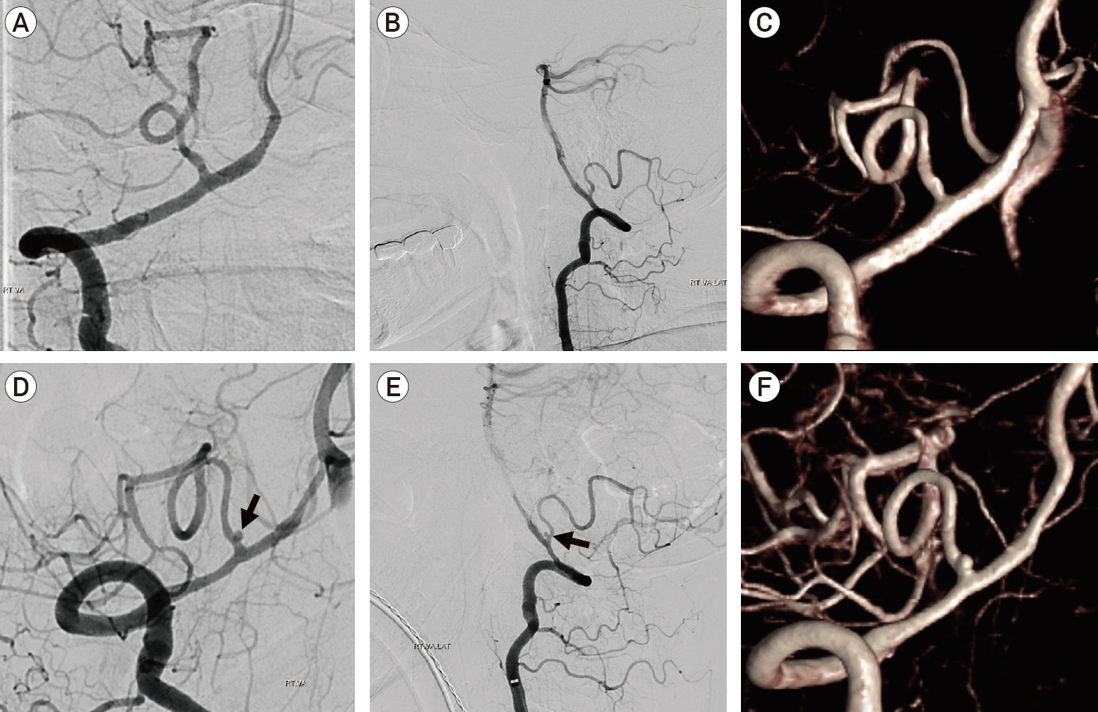
Fig. 2. Change of digital subtraction angiography between the day of subarachnoid hemorrhage (A, B, C) and follow up image which was taken four days later (D, E, F). Small dissection aneurysm bleb (black arrow) was noticed on the follow up image located at the anterior medullary segment of right posterior inferior cerebellar artery.
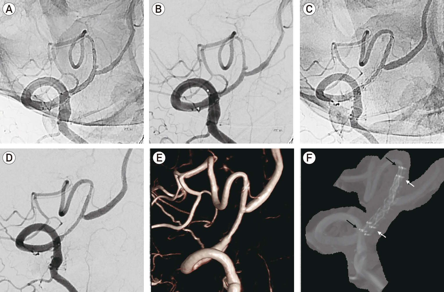
Fig. 3. (A, B) Right vertebral arteriography after the deployment of telescoping stent with LVIS Jr along the right VA and posterior inferior cerebellar artery. (C, D) Three months follow up cerebral angiography after the telescoping stent insertion. (E, F) Three months follow up digital subtraction and MIP image showing disappearance of dissection aneurysm bleb and healing of dissection (black arrow: proximal and distal tip of LVIS Jr 2.5×23 mm, white arrow: proximal and distal tip of LVIS Jr 2.5×17 mm). LVIS Jr, Low-profile Visualized Intraluminal Support Jr; VA, vertebral artery; MIP, maximum intensity projection.
Reference
-
1. Akmangit I, Aydin K, Sencer S, Topcuoglu OM, Topcuoglu ED, Daglioglu E, et al. Dual stenting using low-profile LEO baby stents for the endovascular management of challenging intracranial aneurysms. AJNR Am J Neuroradiol. 2015; Feb. 36(2):323–9.
Article2. Anil G, Sein L, Nga V, Teo K, Chou N, Yeo TT. Dissecting distal cerebellar artery aneurysms: options beyond a parent vessel sacrifice. Neurosurg Rev. 2020; Apr. 43(2):771–80.
Article3. Behme D, Weber A, Kowoll A, Berlis A, Burke TH, Weber W. Low-profile Visualized Intraluminal Support device (LVIS Jr) as a novel tool in the treatment of wide-necked intracranial aneurysms: initial experience in 32 cases. J Neurointerv Surg. 2015; Apr. 7(4):281–5.
Article4. Bhogal P, Chudyk J, Bleise C, Lylyk I, Henkes H, Lylyk P. The use of flow diverters to treat aneurysms of the posterior inferior cerebellar artery: Report of three cases. Interv Neuroradiol. 2018; Oct. 24(5):489–98.
Article5. Chung J, Suh SH, Hong CK, Joo JY, Lim YC, Shin YS, et al. Preliminary experience with self-expanding closed-cell stent placement in small arteries less than 2 mm in diameter for the treatment of intracranial aneurysms. J Neurosurg. 2015; Jun. 122(6):1503–10.
Article6. Lewis SB, Chang DJ, Peace DA, Lafrentz PJ, Day AL. Distal posterior inferior cerebellar artery aneurysms: clinical features and management. J Neurosurg. 2002; Oct. 97(4):756–66.
Article7. Martin AR, Cruz JP, O’Kelly C, Kelly M, Spears J, Marotta TR. Small pipes: preliminary experience with 3-mm or smaller pipeline flow-diverting stents for aneurysm repair prior to regulatory approval. AJNR Am J Neuroradiol. 2015; Mar. 36(3):557–61.
Article8. Mericle RA, Reig AS, Burry MV, Eskioglu E, Firment CS, Santra S. Endovascular surgery for proximal posterior inferior cerebellar artery aneurysms: an analysis of Glasgow Outcome Score by Hunt-Hess grades. Neurosurgery. 2006; Apr. 58(4):619–25. discussion 619-25.
Article9. Möhlenbruch MA, Kizilkilic O, Killer-Oberpfalzer M, Baltacioglu F, Islak C, Bendszus M, et al. Multicenter experience with FRED Jr flow re-direction endoluminal device for intracranial aneurysms in small arteries. AJNR Am J Neuroradiol. 2017; Oct. 38(10):1959–65.
Article10. Oğuz Ş, Dinc H. Treatment of posterior inferior cerebellar artery aneurysms using flow-diverter stents: A single-center experience. Interv Neuroradiol. 2019; Aug. 25(4):407–13.
Article11. Peluso JP, van Rooij WJ, Sluzewski M, Beute GN, Majoie CB. Posterior inferior cerebellar artery aneurysms: incidence, clinical presentation, and outcome of endovascular treatment. AJNR Am J Neuroradiol. 2008; Jan. 29(1):86–90.
Article12. Santillan A, Boddu S, Schwarz J, Lin N, Gobin YP, Knopman J, et al. LVIS Jr. stent for treatment of intracranial aneurysms with parent vessel diameter of 2.5 mm or less. Interv Neuroradiol. 2018; Jun. 24(3):246–53.13. Srinivasan VM, Ghali MGZ, Reznik OE, Cherian J, Mokin M, Dumont TM, et al. Flow diversion for the treatment of posterior inferior cerebellar artery aneurysms: a novel classification and strategies. J Neurointerv Surg. 2018; Jul. 10(7):663–8.
Article14. Williamson RW, Wilson DA, Abla AA, McDougall CG, Nakaji P, Albuquerque FC, et al. Clinical characteristics and long-term outcomes in patients with ruptured posterior inferior cerebellar artery aneurysms: a comparative analysis. J Neurosurg. 2015; Aug. 123(2):441–5.
Article
- Full Text Links
-
- Actions
-
Cited
- CITED
-
- Close
- Share
-
- Similar articles
-
- Clinical and Angiographic Outcomes of Endovascular Treatment for Acute Intracranial Vertebral Artery Dissecting Aneurysms Using Double-Overlapping Stents : Low-Profile Visualized Intraluminal Support within Enterprise Stents
- Stent-Jack Technique for Ruptured Vertebral Artery Dissecting Aneurysm Involving the Origin of Posterior Inferior Cerebellar Artery
- Coil Embolization of Ruptured Proximal Posterior Inferior Cerebellar Artery Aneurysm with Contralateral Retrograde Approach for LVIS Jr. Intraluminal Support Deployment
- In Situ Intersegmental Anastomosis within a Single Artery for Treatment of an Aneurysm at the Posterior Inferior Cerebellar Artery: Closing Omega Bypass
- Dissecting Aneurysm Associated with a Double Origin of the Posterior Inferior Cerebellar Artery Causing Subarachnoid Hemorrhage
