Coil Embolization of Ruptured Proximal Posterior Inferior Cerebellar Artery Aneurysm with Contralateral Retrograde Approach for LVIS Jr. Intraluminal Support Deployment
- Affiliations
-
- 1Department of Neurosurgery, St. Vincent's Hospital, College of Medicine, The Catholic University of Korea, Suwon, Korea. jaehoonsung@gmail.com
- KMID: 2461922
- DOI: http://doi.org/10.7461/jcen.2018.20.4.235
Abstract
- The safety and feasibility of simple coil embolization and stent deployment for the treatment of posterior inferior cerebellar artery (PICA) aneurysms, as well as their radiologic and clinical results, have not been adequately understood. Especially, if dissecting aneurysm of proximal PICA is associated with small caliber PICA and stenosis of ipsilateral vertebral artery orifice (VAO), endovascular coiling with saving of PICA is not always easy. This 64-year-old man presented with subarachnoid hemorrhage due to a ruptured dissecting aneurysm of left proximal PICA. The aneurysm was irregularly fusiform in nature with a shallow PICA orifice (1.4 mm) and narrow caliber (0.9-1.5 mm). Moreover, the ipsilateral VAO showed severe stenosis (1.8 mm). We performed bifemoral puncture and chose additional route from right vertebral artery to left vertebrobasilar junction for retrograde approach and deployment of LVIS Jr. intraluminal support at proximal PICA. And then, the antegrade approach and coiling of aneurysm was done. Despite of transient thrombus of PICA, the aneurysm was successfully secured with preservation of whole PICA course. For preservation of narrow PICA with ipsilateral VAO stenosis, the contralateral approach and deployment of LVIS Jr. intraluminal support may be considered.
MeSH Terms
Figure
-

Fig. 1 (A) The initial brain computed tomography scan shows acute subarachnoid hemorrhage on basal cistern with intraventricular hemorrhage. (B, C) The left vertebral angiography shows a left PICA dissecting fusiform aneurysm, measuring 3.95 mm (width) × 3.12 mm (height) × 4.75 mm (length). (D, E) The 3D volume rendering image shows shallow PICA orifice (1.3 mm) and narrow PICA just proximal and distal of aneurysm, giving rise to the PICA from the sac. (F) The left subclavian artery angiography shows severe stenosis of left vertebral artery orifice, about 1.8 mm (white arrow). PICA = posterior inferior cerebellar artery.
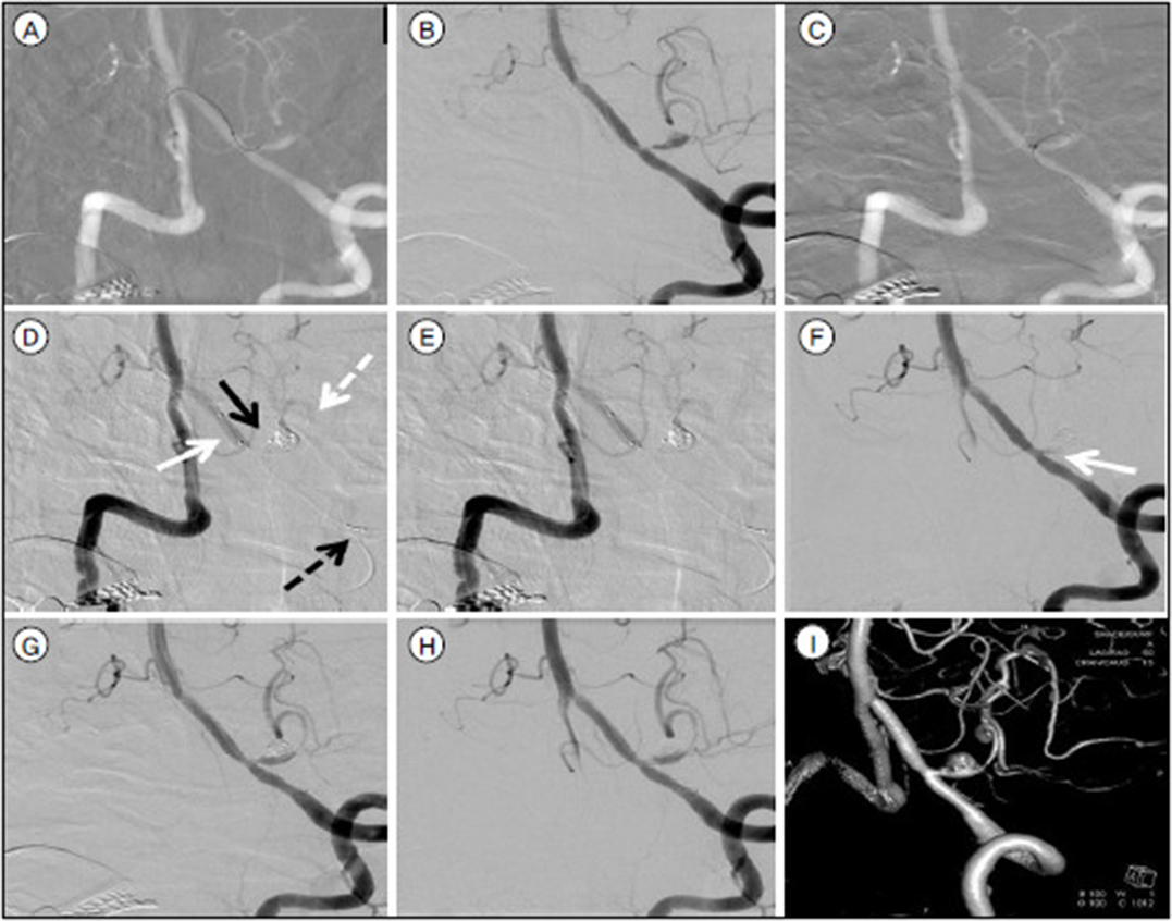
Fig. 2 The both vertebral angiography roadmap, via bifemoral puncture, enables Excelsior microcatheter from right VA to navigate left PICA, crossing aneurysm sac (A). The left VA angiography shows far distal, stable lodging of Excelsior microcatheter for future LVIS Jr. deployment (B). The both vertebral angiography roadmap shows another Excelsior microcatheter from left VA moving to fusiform aneurysm sac (C). The right VA angiography shows successful deployment of LVIS Jr intraluminal support from left VA, crossing aneurysm sac, to far distal PICA with preserved patency of PICA through contralateral VA approach (white arrow: proximal tip of deployed LVIS Jr., white dot arrow: distal tip of deployed LVIS Jr.) and left VA approach with microcatheter for detechable coil (black dot arrow: proximal tip of ipsilateral approached microcatheter for coil embolization, black arrow: distal tip of ipsilateral approached microcatheter for coil embolization) (D). Angiography shows near complete occlusion of aneurysm with preserved patency of PICA (E). After deployment of final coil, the left VA angiography revealed total thrombosis occlusion of left PICA (white arrow) (F). After reinsertion of right side microcatheter into proximal portion of PICA (through LVIS Jr. support), total 1 mg of tirofiban is injected. Rapid restroration of PICA flow is evident (G). Final angiography with 3D volume rendering image shows complete recanalization of left PICA with near obliteration of fusiform aneurysm (H, I). VA = vertebral artery; PICA = posterior inferior cerebellar artery.
Reference
-
1. Al-khayat H, Al-Khayat H, Beshay J, Manner D, White J. Vertebral artery-posteroinferior cerebellar artery aneurysms: clinical and lower cranial nerve outcomes in 52 patients. Neurosurgery. 2005; 56(1):2–10.
Article2. Cross DT 3rd, Moran CJ, Derdeyn CP, Mazumdar A, Rivet D, Chicoine MM. Neuroform stent deployment for treatment of a basilar tip aneurysm via a posterior communicating artery route. AJNR Am J Neuroradiol. 2005; Nov-Dec. 26(10):2578–2581.3. Jeon SI, Kwon BJ, Seo DH, Kang HI, Park SC, Choe IS. Bilateral approach for stent-assisted coiling of posterior inferior cerebellar artery aneurysms - two cases. J Cerebrovasc Endovasc Neurosurg. 2012; 09. 14(3):223–227.
Article4. Kim MJ, Chung J, Kim SL, Roh HG, Kwon BJ, Kim BS, et al. Stenting from the vertebral artery to the posterior inferior cerebellar artery. AJNR Am J Neuroradiol. 2012; 02. 33(2):348–352.
Article5. Lv X, Jiang C, Li Y, Wu Z. Clinical outcomes of ruptured and unruptured vertebral artery-posterior inferior cerebellar artery complex dissecting aneurysms after endovascular embolization. AJNR Am J Neuroradiol. 2010; 08. 31(7):1232–1235.
Article6. Mazur MD, Kilburg C, Wang V, Taussky P. Pipeline embolization device for the treatment of vertebral artery aneurysms: the fate of covered branch vessels. J Neurointerv Surg. 2016; 10. 8(10):1041–1047.
Article7. Peluso JP, van Rooij WJ, Sluzewski M, Beute GN, Majoie CB. Posterior inferior cerebellar artery aneurysms: incidence, clinical presentation, and outcome of endovascular treatment. AJNR Am J Neuroradiol. 2008; 01. 29(1):86–90.
Article8. Roh HG, Chun YI, Choi JW, Cho J, Moon WJ, Solander S. Retrograde stent placement for coil embolization of a wide-necked posterior inferior cerebellar artery aneurysm. Korean J Radiol. 2012; Jul-Aug. 13(4):510–514.
Article9. Song HH, Won YD, Kim YJ, Kim BS. The endovascular management of saccular posterior inferior cerebellar artery aneurysms. Korean J Radiol. 2008; Sep-Oct. 9(5):396–400.
Article10. Suh SH, Kim BM, Chung TS, Kim DI, Kim DJ, Hong CK, et al. Reconstructive endovascular treatment of intracranial fusiform aneurysms: a 1-stage procedure with stent and balloon. AJNR Am J Neuroradiol. 2010; 01. 31(1):155–160.
Article
- Full Text Links
-
- Actions
-
Cited
- CITED
-
- Close
- Share
-
- Similar articles
-
- Technical Consideration for Coiling of Ruptured Proximal Posterior Inferior Cerebellar Artery Aneurysm
- Flow diversion via telescoping stent with Low-profile Visualized Intraluminal Support Junior for treatment of ruptured dissecting aneurysm located at proximal posterior inferior cerebellar artery
- Persistent Trigeminal Artery with a Cerebellar Branch and Trigeminal-Cavernous Fistula from Ruptured Aneurysm: Transarterial Coil Embolization
- Retrograde Stent Placement for Coil Embolization of a Wide-Necked Posterior Inferior Cerebellar Artery Aneurysm
- Coil Embolization of Ruptured Thrombosed Distal Superior Cerebellar Artery Aneurysm: A Case Report
