Relationship of the fabella with the origins of the plantaris and gastrocnemius lateral head muscles in late-term fetuses: a histological study
- Affiliations
-
- 1Department of Anatomy, Wuxi School of Medicine, Jiangnan University, Wuxi, Jiangsu, China
- 2Department of Anatomy, Jeonbuk National University Medical School, Jeonju, Korea
- 3Divison of Common Curriculum, Hokkaido Chitose College of Rehabilitation, Chitose, Japan
- 4Divison of Rehabilitation, Hitsujigaoka Hospital of Orthopedics, Sapporo, Japan
- 5Division of Internal Medicine, Cupid Clinic, Iwamizawa, Hokkaido, Japan
- 6Emeritus Professor of Akita University School of Medicine, Akita, Japan
- 7Department of Anatomy and Embryology, School of Medicine, Complutense University, Madrid, Spain
- KMID: 2516911
- DOI: http://doi.org/10.5115/acb.20.326
Abstract
- Previous studies of midterm fetuses indicated that a cartilaginous fabella appeared to be embedded in the plantaris (PL), and was fused with the gastrocnemius lateral head (GL). We re-examined the topographical anatomy of the fabella or its analogue (a tight fibrous mass) originating in the GL and/or PL by evaluating histological sections of the unilateral knees of 15 late-term fetuses. Regardless of whether the cartilaginous fabella was present (6 fetuses) or absent (9 fetuses), the origins of the PL and GL muscles each had three parts. In each fetus, the fabella or its analogue was embedded in a thick common tendinous origin of the GL and PL. PL1 (whose origin is similar to that of the adult PL) originated from the femoral condyle immediately above the common tendon; PL2 originated from the posteromedial aspect of the fabella or its analogue; and PL3 originated from the inferior aspect of the fabella or its analogue. The muscle fibers of PL1, PL2, and PL3 joined to provide a thick plantaris. GL1 (which is adjacent to PL2) originated from the common tendon in the superior side of the fabella or its analogue and GL2 originated from the inferior side of the fabella or its analogue. GL1 and GL2 joined to provide a thick bundle, whereas GL3 (located far below the fabella or its analogue) originated from the posterior surface aponeurosis. Therefore, drastic reconstruction at these muscle origins was necessary during development. Due to the strong mechanical stress from the GL and the space-occupying effect of the muscle, we hypothesize that PL2 and PL3 are degraded or absorbed into the GL1 and GL2 during the postnatal period, so that the remaining PL1 was likely the remaining PL in adults.
Keyword
Figure
-

Fig. 1 Origins of the gastrocnemius lateral head (GL) and plantaris (PL) in a fetus (260 mm crown-rump length [CRL]) with a fabella. (A–C) show the topographical anatomy of bony elements and muscle attachments in the lateral half of the knee joint; (A) shows the most lateral site and (C) the most medial site. (A) shows the insertion of the PT to the femur (FE). (B) shows a common tendinous origin (star) of the GL and PL. (C) shows the fabella and anterior cruciate ligament. (D–I) show multiple muscle bundles at the origins of the GL and PL; (D) shows the most lateral site and (I) the most medial site. (D) corresponds to the square in (B), and (F) corresponds to the square in (C). (D, E) show the upper bundle of the PL (PL1) is adjacent to the posterior aspect of the common tendinous origin (stars). (F, G) show the fabella is surrounded by the PL origin, which has three muscle masses (PL1, PL2, PL3). (F) shows the upper muscle bundle of the GL (GL1, dotted oval) originates from the common tendon, and is adjacent to the inferior aspect of the PL1. (G) shows that after the GL1 combined with another muscle bundle originating from the fabella (GL2), the lateral head was separated from the PL, the lower most muscle bundle (GL3) originates from a surface aponeurosis (triangles in F) that extended from the GL2. (J) (a magnified view of the circle in F) shows the PL3 from the fabella. (A–C) are at low magnification (H&E, scale bar in A: 5 mm), (D–I) are at intermediate magnification (H&E, scale bar in D: 1 mm), and (J) is at high magnification (H&E, scale bar: 0.1 mm). See also Table 1. PA, patella; BF, biceps femoris; FI, fibula; T, tibia; ACL, anterior cruciate ligament; PM, popliteus muscle; PT, popliteus tendon.

Fig. 2 Origins of the gastrocnemius lateral head (GL) and plantaris (PL) in a fetus (328 mm crown-rump length [CRL]) with a fabella. (A–C) display the topographical anatomy of bony elements and muscle attachments in the lateral half of the knee joint; (A) shows the most lateral site and (C) the most medial site. (A) shows a common tendinous origin (star) of the GL and (B) shows the fabella. (D–H) show multiple muscle bundles at origins of these two muscles. (D, E, H) are magnified views of the squares in (A–C), respectively. (D) shows the most lateral site and (H) the most medial site. (D) shows that the PL1 attaches to the posterior aspect of the common tendinous origin (star). (E, F) show the origin of the GL1, which is distant from the PL1 and originates from the common tendon near fabella, but the GL3 originates from a surface aponeurosis (triangles). (I, J) show muscle fibers of the GL1 and PL3, and are higher magnifications of the circles in (E, F), respectively. (A–C) are at low magnification (H&E, scale bar in A: 5 mm), (D–H) are at intermediate magnification (H&E, scale bar in D: 1 mm), and (I, J) is at high magnification (H&E, scale bar: 0.1 mm). See also Table 1. BF, biceps femoris; FI, fibula; T, tibia; FE, femur; PM, popliteus muscle; PT, popliteus tendon; SO, soleus.

Fig. 3 Origins of the gastrocnemius lateral head (GL) and plantaris (PL) in a fetus (250 mm crown-rump length [CRL]) without a fabella. Panels show multiple muscle bundles at the origins of the GL and PL; (A) shows the most lateral site and (G) the most medial site. (D, E) show a fabella-like tight fibrous mass (green-dotted oval) instead of a fabella at the inferomedial end of a common tendinous origin (star) and (A–C) show this fibrous mass is surrounded by four muscle bundles (GL1, GL2, PL2, PL3). (E) shows a posterior surface aponeurosis (triangles) issuing from the lower muscle fibers of the lateral head (GL3). (G) shows the GL1 is separated from the PL1 and PL3 in the far medial side of the popliteus tendon, facing the joint cavity (PT). All panels are at the same magnification (H&E, scale bar in A: 1 mm). See also Table 1. BF, biceps femoris; FI, fibula; FE, femur; PM, popliteus muscle; PT, popliteus tendon; SO, soleus.

Fig. 4 Origins of the gastrocnemius lateral head (GL) and plantaris (PL) in a fetus (282 mm crown-rump length [CRL]) without a fabella. Panels show multiple muscle bundles at origins of the GL and PL; (A) shows the most lateral site and (G) the most medial site. (A) shows that the common tendinous origin of the GL and PL (dotted green line) is continuous to a fabella-like connective tissue mass (dotted green line in B, C) and these structures are surrounded by multiple muscle bundles (PL1, PL2, PL3, GL1, GL2). (E, F) show another origin of the PL3 appears in the medial side of the fabella-like mass. (F, G) shows the GL1 and GL2 fused to provide a thick muscle in this medial section. (D, E) show the GL3 originates from the surface aponeurosis (triangles). All panels are at the same magnification (H&E, scale bar in A: 1 mm). See also Table 1. BF, biceps femoris; FI, fibula; FE, femur; PM, popliteus muscle; PT, popliteus tendon.

Fig. 5 Lack of a common tendinous origin of the gastrocnemius lateral head (GL) and plantaris (PL) in a fetus (274 mm crown-rump length [CRL]) without a fabella. (A, B) show the topographical anatomy of bony elements and muscle attachments in the lateral half of the knee joint. The squares in (A, B) are at a higher magnification in (D, F), respectively. (C–J) show multiple muscle bundles at the origins of these two muscles; (C) is shows the most lateral site and (J) the most medial site. (E, F) show the three muscle bundles at the PL origin (PL1, PL2, and PL3) provide a belt on the immediate posterior side of the popliteus tendon. (G) shows that the GL1 attaches to the posterior aspect of the PL origins and the GL3 originates from a surface aponeurosis from the GL1 (triangles). (A, B) are at low magnification (H&E, scale bar in A: 5 mm) and (C–J) are at high magnification (H&E, scale bar in C: 1 mm). See also Table 1. PA, patella; BF, biceps femoris; FI, fibula; T, tibia; FE, femur; PM, popliteus muscle; PT, popliteus tendon; SO, soleus; LN, lymph node; TN, tibial nerve.
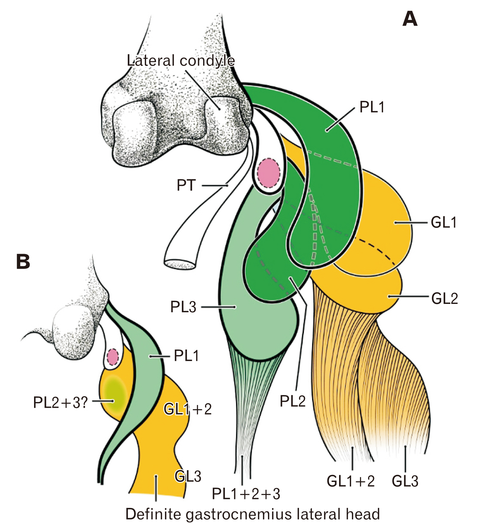
Fig. 6 Schematic representation of the gastrocnemius lateral head (GL) and plantaris (PL) origins in a late-term fetus, and hypothesized connections to adult morphology. The patella (or its analogue) is pink, the PL is green, and the GL is orange. (A) shows the topographical anatomy of the origins of the 3 GL bundles (GL1, GL2, and GL3) and the 3 PL bundles (PL1, PL2, and PL3). (B) shows the changes hypothesized to occur after birth, in which the PL1 remains, and the PL2 and PL3 degenerate or are absorbed into the gastrocnemius. PT, popliteus tendon.
Cited by 1 articles
-
Distally-extending muscle fibers across involved joints: study of long muscles and tendons of wrist and ankle in late-term fetuses and adult cadavers
Shaohe Wang, Shogo Hayashi, Zhe-Wu Jin, Ji Hyun Kim, Masahito Yamamoto, Gen Murakami, Shinichi Abe
Anat Cell Biol. 2023;56(1):46-53. doi: 10.5115/acb.22.133.
Reference
-
References
1. Jin ZW, Shibata S, Abe H, Jin Y, Li XW, Murakami G. 2017; A new insight into the fabella at knee: the foetal development and evolution. Folia Morphol (Warsz). 76:87–93. DOI: 10.5603/FM.a2016.0048. PMID: 27665955.
Article2. Rodríguez-Vázquez JF, Jin ZW, Zhao P, Murakami G, Li XW, Jin Y. 2018; Development of digastric muscles in human foetuses: a review and findings in the flexor digitorum superficialis muscle. Folia Morphol (Warsz). 77:362–70. DOI: 10.5603/FM.a2017.0083. PMID: 28868605.
Article3. Jin ZW, Jin Y, Yamamoto M, Abe H, Murakami G, Yan TF. 2016; Oblique cord (chorda obliqua) of the forearm and muscle-associated fibrous tissues at and around the elbow joint: a study of human foetal specimens. Folia Morphol (Warsz). 75:493–502. DOI: 10.5603/FM.a2016.0019. PMID: 27830875.
Article4. Shiraishi Y, Jin ZW, Mitomo K, Yamamoto M, Murakami G, Abe H, Wilting J, Abe S. 2018; Foetal development of the human gluteus maximus muscle with special reference to its fascial insertion. Folia Morphol (Warsz). 77:144–50. DOI: 10.5603/FM.a2017.0060. PMID: 28653302.
Article5. Minowa T, Murakami G, Kura H, Suzuki D, Han SH, Yamashita T. 2004; Does the fabella contribute to the reinforcement of the posterolateral corner of the knee by inducing the development of associated ligaments? J Orthop Sci. 9:59–65. DOI: 10.1007/s00776-003-0739-2. PMID: 14767706.
Article6. Nakamura T, Suzuki D, Murakami G, Cho BH, Fujimiya M, Kozuka N. 2011; Human fetal anatomy of the posterior semimembranosus complex at the knee with special reference to the gastrocnemio-semimembranosus bursa. Knee. 18:271–7. DOI: 10.1016/j.knee.2010.05.010. PMID: 20797867.
Article7. Milani-Comparetti A, Gidoni EA. 1967; Routine developmental examination in normal and retarded children. Dev Med Child Neurol. 9:631–8. DOI: 10.1111/j.1469-8749.1967.tb02335.x. PMID: 6066029.
Article8. Thelen E, Cooke DW. 1987; Relationship between newborn stepping and later walking: a new interpretation. Dev Med Child Neurol. 29:380–93. DOI: 10.1111/j.1469-8749.1987.tb02492.x. PMID: 3596074.
Article9. Okamoto T, Okamoto K, Andrew PD. 2003; Electromyographic developmental changes in one individual from newborn stepping to mature walking. Gait Posture. 17:18–27. DOI: 10.1016/S0966-6362(02)00049-8. PMID: 12535722.
Article10. Lambert HW. Tubbs RS, Shoja MM, Loukas M, editors. 2016. Leg muscles. Bergman's Comprehensive Encyclopedia of Human Anatomic Variation. John Wiley & Sons;Hoboken: p. 421–37. DOI: 10.1002/9781118430309.ch41. PMID: 27506436.11. Warburton NM, Yakovleff M, Malric A. 2012; Anatomical adaptations of the hind limb musculature of tree-kangaroos for arboreal locomotion (Marsupialia: macropodinae). Aust J Zool. 60:246–58. DOI: 10.1071/ZO12059.12. Doherty GP, Koike Y, Uhthoff HK, Lecompte M, Trudel G. 2006; Comparative anatomy of rabbit and human Achilles tendons with magnetic resonance and ultrasound imaging. Comp Med. 56:68–74. PMID: 16521862.13. Huisman ES, Andersson G, Scott A, Reno CR, Hart DA, Thornton GM. 2014; Regional molecular and cellular differences in the female rabbit Achilles tendon complex: potential implications for understanding responses to loading. J Anat. 224:538–47. DOI: 10.1111/joa.12169. PMID: 24571598. PMCID: PMC3981496.
Article14. Kawashima T, Takeishi H, Yoshitomi S, Ito M, Sasaki H. 2007; Anatomical study of the fabella, fabellar complex and its clinical implications. Surg Radiol Anat. 29:611–6. DOI: 10.1007/s00276-007-0259-4. PMID: 17882346.
Article15. Phukubye P, Oyedele O. 2011; The incidence and structure of the fabella in a South African cadaver sample. Clin Anat. 24:84–90. DOI: 10.1002/ca.21049. PMID: 20830786.
Article16. Zeng SX, Dong XL, Dang RS, Wu GS, Wang JF, Wang D, Huang HL, Guo XD. 2012; Anatomic study of fabella and its surrounding structures in a Chinese population. Surg Radiol Anat. 34:65–71. DOI: 10.1007/s00276-011-0828-4. PMID: 21626275.
Article17. Chew CP, Lee KH, Koh JS, Howe TS. 2014; Incidence and radiological characteristics of fabellae in an Asian population. Singapore Med J. 55:198–201. DOI: 10.11622/smedj.2014052. PMID: 24763835. PMCID: PMC4291947.
Article18. Benjamin M, Ralphs JR. 1998; Fibrocartilage in tendons and ligaments--an adaptation to compressive load. J Anat. 193(Pt 4):481–94. DOI: 10.1046/j.1469-7580.1998.19340481.x. PMID: 10029181. PMCID: PMC1467873.19. Milz S, Benjamin M, Putz R. 2005; Molecular parameters indicating adaptation to mechanical stress in fibrous connective tissue. Adv Anat Embryol Cell Biol. 178:1–71. PMID: 16080262.20. Eyal S, Blitz E, Shwartz Y, Akiyama H, Schweitzer R, Zelzer E. 2015; On the development of the patella. Development. 142:1831–9. DOI: 10.1242/dev.121970. PMID: 25926361.
Article
