Anat Cell Biol.
2024 Sep;57(3):459-462. 10.5115/acb.24.038.
A bilateral gastrocnemius tertius coexisting with a unilateral two-headed plantaris muscle
- Affiliations
-
- 1Department of Anatomy, School of Medicine, Faculty of Health Sciences, National and Kapodistrian University of Athens, Athens, Greece
- KMID: 2559765
- DOI: http://doi.org/10.5115/acb.24.038
Abstract
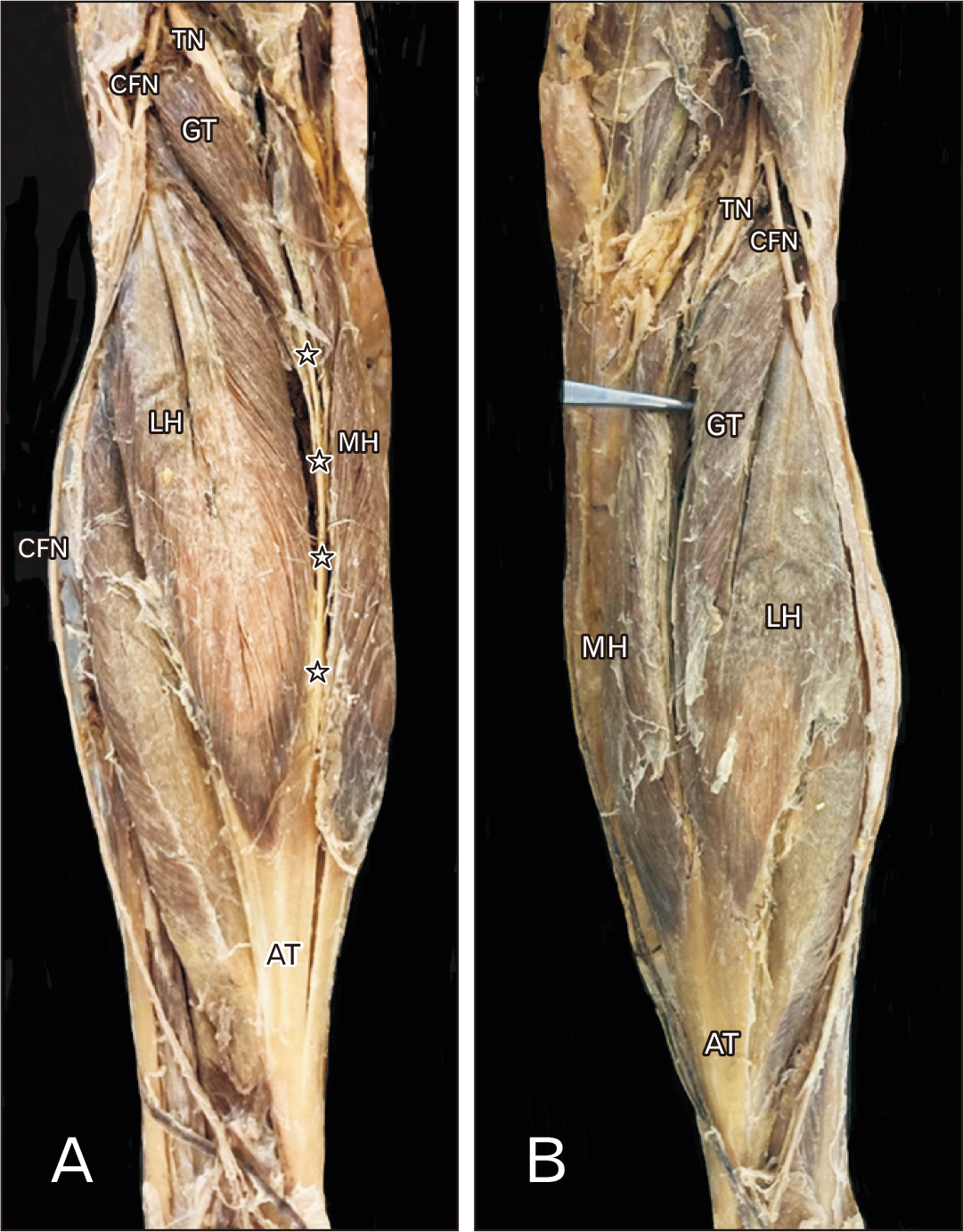
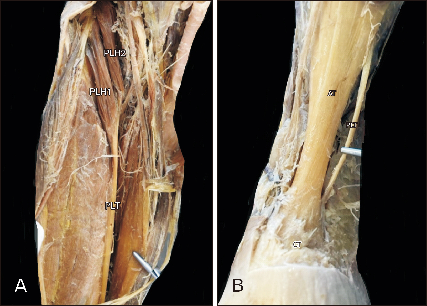
- The current cadaveric report aims to present a coexistence of two uncommon variants of the posterior leg compartment. The variations were detected, during classical dissection in an 84-year-old donated male cadaver. On the left lower limb, the gastrocnemius muscle was identified as having a third head that was attached to the lateral head. This variant is known as gastrocnemius tertius muscle and was bilaterally identified. The left-sided plantaris muscle had two distinct heads that fused into a common tendon that was inserted into the calcaneal tuberosity. Knowledge of these variants is important, due to their close relationship with the popliteal neurovascular bundle. Clinicians should be aware, to avoid pitfalls and take them into account in their differential diagnosis.
Keyword
Figure
Reference
-
References
1. Gonera B, Kurtys K, Paulsen F, Polguj M, LaPrade RF, Grzelecki D, Karauda P, Olewnik Ł. 2021; The plantaris muscle - anatomical curiosity or a structure with important clinical value? - A comprehensive review of the current literature. Ann Anat. 235:151681. DOI: 10.1016/j.aanat.2021.151681. PMID: 33561523.2. Bergman RA, Walker CW, el-Khour GY. 1995; The third head of gastrocnemius in CT images. Ann Anat. 177:291–4. DOI: 10.1016/S0940-9602(11)80205-0. PMID: 7598226.3. Bardeen CR. 1905; Studies of the development of the human skeleton. (A). The development of the lumbab, sacbal and coccygeal vertebwe. (B). The cubves and the pbopobtionate regional lengths of the spinal column during the first thbee months of embbyonic developnent. (C). The development of the skeleton of the posterior limb. Am J Anat. 4:265–302. DOI: 10.1002/aja.1000040302.4. Frey H. 1919; Musculus gastrocnemius tertius. Gegenbaurs Morph Jahrb. 50:517–30.5. Koplas MC, Grooff P, Piraino D, Recht M. 2009; Third head of the gastrocnemius: an MR imaging study based on 1,039 consecutive knee examinations. Skeletal Radiol. 38:349–54. DOI: 10.1007/s00256-008-0606-5. PMID: 19002457.6. Arefi IA, Kosco E, Werner E, Maxwell A, Fickert A, Frank PW. 2023; Bilateral gastrocnemius tertius muscles: cadaveric findings of a rare variant. Cureus. 15:e45316. DOI: 10.7759/cureus.45316. PMID: 37846245. PMCID: PMC10577022.7. Ishii T, Kawagishi K, Hayashi S, Yamada S, Yoshioka H, Matsuno Y, Mori Y, Kosaka J. 2021; A bilateral third head of the gastrocnemius which is morphologically similar to the plantaris. Surg Radiol Anat. 43:1095–8. DOI: 10.1007/s00276-020-02670-w. PMID: 33423145.8. Yildirim FB, Sarikcioglu L, Nakajima K. 2011; The co-existence of the gastrocnemius tertius and accessory soleus muscles. J Korean Med Sci. 26:1378–81. DOI: 10.3346/jkms.2011.26.10.1378. PMID: 22022193. PMCID: PMC3192352.9. Olewnik Ł, Kurtys K, Gonera B, Podgórski M, Sibiński M, Polguj M. 2020; Proposal for a new classification of plantaris muscle origin and its potential effect on the knee joint. Ann Anat. 231:151506. DOI: 10.1016/j.aanat.2020.151506. PMID: 32173563.10. Smędra A, Olewnik Ł, Łabętowicz P, Danowska-Klonowska D, Polguj M, Berent J. 2021; A bifurcated plantaris muscle: another confirmation of its high morphological variability? Another type of plantaris muscle. Folia Morphol (Warsz). 80:739–44. DOI: 10.5603/FM.a2020.0101. PMID: 32844386.11. Olewnik Ł, Zielinska N, Karauda P, Tubbs RS, Polguj M. 2020; A three-headed plantaris muscle: evidence that the plantaris is not a vestigial muscle? Surg Radiol Anat. 42:1189–93. DOI: 10.1007/s00276-020-02478-8. PMID: 32382814. PMCID: PMC7366563.12. Maślanka K, Zielinska N, Paulsen F, Niemiec M, Olewnik Ł. A three-headed plantaris muscle fused with Kaplan fibers: potential clinical significance. Folia Morphol (Warsz). 2023; Nov. 14. [Epub]. https://doi.org/10.5603/fm.95513. DOI: 10.5603/fm.95513. PMID: 37957936.13. Connell J. 1978; Popliteal vein entrapment. Br J Surg. 65:351. DOI: 10.1002/bjs.1800650518. PMID: 647204.14. Page PS, Paige S, Hanna A. 2021; Vascular entrapment neuropathy of the tibial nerve within the gastrocnemius muscle. Surg Neurol Int. 12:224. DOI: 10.25259/SNI_75_2021. PMID: 34221555. PMCID: PMC8247925.15. Sanchez JE, Conkling N, Labropoulos N. 2011; Compression syndromes of the popliteal neurovascular bundle due to Baker cyst. J Vasc Surg. 54:1821–9. DOI: 10.1016/j.jvs.2011.07.079. PMID: 21958564.
- Full Text Links
- Actions
-
Cited
- CITED
-
- Close
- Share
- Similar articles
-
- The Co-existence of the Gastrocnemius Tertius and Accessory Soleus Muscles
- Effects of Unilateral Sciatic Nerve Injury on Unaffected Hindlimb Muscles of Rats
- Intramuscular Baker's Cyst in Plantaris: A Case Report
- Variations for Incomplete Formation of the Plantaris Muscle: A Case Report
- Bicipital origin and the course of the plantaris muscle


