Enhancement of Wound Healing by Conditioned Medium of Adipose-Derived Stromal Cell with Photobiomodulation in Skin Wound
- Affiliations
-
- 1Cell Therapy Center, Ajou University School of Medicine, Suwon, Korea
- KMID: 2515997
- DOI: http://doi.org/10.15283/ijsc20175
Abstract
- Background and Objectives
The objective of this study was to investigate whether conditioned medium from photobiomodulation (PBM) irradiated adipose-derived stromal cell (ASC) spheroids prior to implanting could stimulate angiogenesis and tissue regeneration to improve functional recovery of skin tissue in an animal skin wound model.
Methods and Results
ASC were split and seeded on chitosan-coated 24 well plate at a density of 7.5×10 4 cells/cm 2 , and allowed to adhere at 37℃. Within 3 days of culture, ASC formed spheroids by PBM irradiation. Conditioned medium (CM) fractions were collected from the PBM-ASC to yield nor adipose-derived stromal cell spheroid (spheroid) and PBM-spheroid, respectively, centrifuged at 13,000 g at 4℃ for 10 min, and stored prior to use for ELISA, protein assay, or in vivo wound-healing assays. Phosphate-buffered saline, cultured CM from ASCs, PBM irradiation prior to implanting conditioned medium from ASC, cultured CM from ASC spheroid, and PBM–spheroid-CM (PSC) were transplanted into a wound bed in athymic mice to evaluate therapeutic effects of PSC in vivo. PSC enhanced wound closure in a skin injury model compared to PBS, CM, PBM–CM, and spheroid-CM. The density of vascular formations increased as a result of angiogenic factors released by the wound bed and enhanced tissue regeneration at the lesion site.
Conclusions
These results indicate that implant of PSC can significantly improve functional recovery compared to PBS, CM, PBM–CM, or spheroid-CM treatment. Implant of PSC may be an effective form of paracrine mediated therapy for treating a wound bed.
Keyword
Figure
-

Fig. 1 ASC formed spheroids by Low-Level light irradiation. (A) Brief overview of our experiment proce-dure. The light source used was LED (660 nm) designed to fit over a microplate (12.5×8.5 cm) for spheroid formation. ASC morphology on 24 well polystyrene plate at 72 h. Scale bar=500 μm. (B) Western blot analysis and quantification of hypoxia-induced survival factor as hypoxia-inducible factor (HIF)-1α in ASC cultured as spheroids, PBM.AS, and monolayers. (C) Enhanced secretion of angiogenic growth factors from PBM.AS in the wound bed. Angio-genesis-related protein analysis of PBM.AS (*, p<0.05, compared to the CM group, t-test, n=3 in each group). (D) ELISA measurement of spheroids cultured for 3 days. Con-centrations of VEGF are presented as pg-corrected for 104 cells (*, p< 0.05, compared with spheroid 6 J/cm2 group, t-test, n=3 in each group).

Fig. 2 Enhanced secretion of angiogenic growth factors in the wound bed. (A) Immunostaining was performed with anti-bFGF and anti-VEGF or anti-HGF antibody (red) at 14 days. The scale bar indicates 100 μm. (B) Western blot indicating the expression of bFGF, VEGF, and HGF at 14 days.
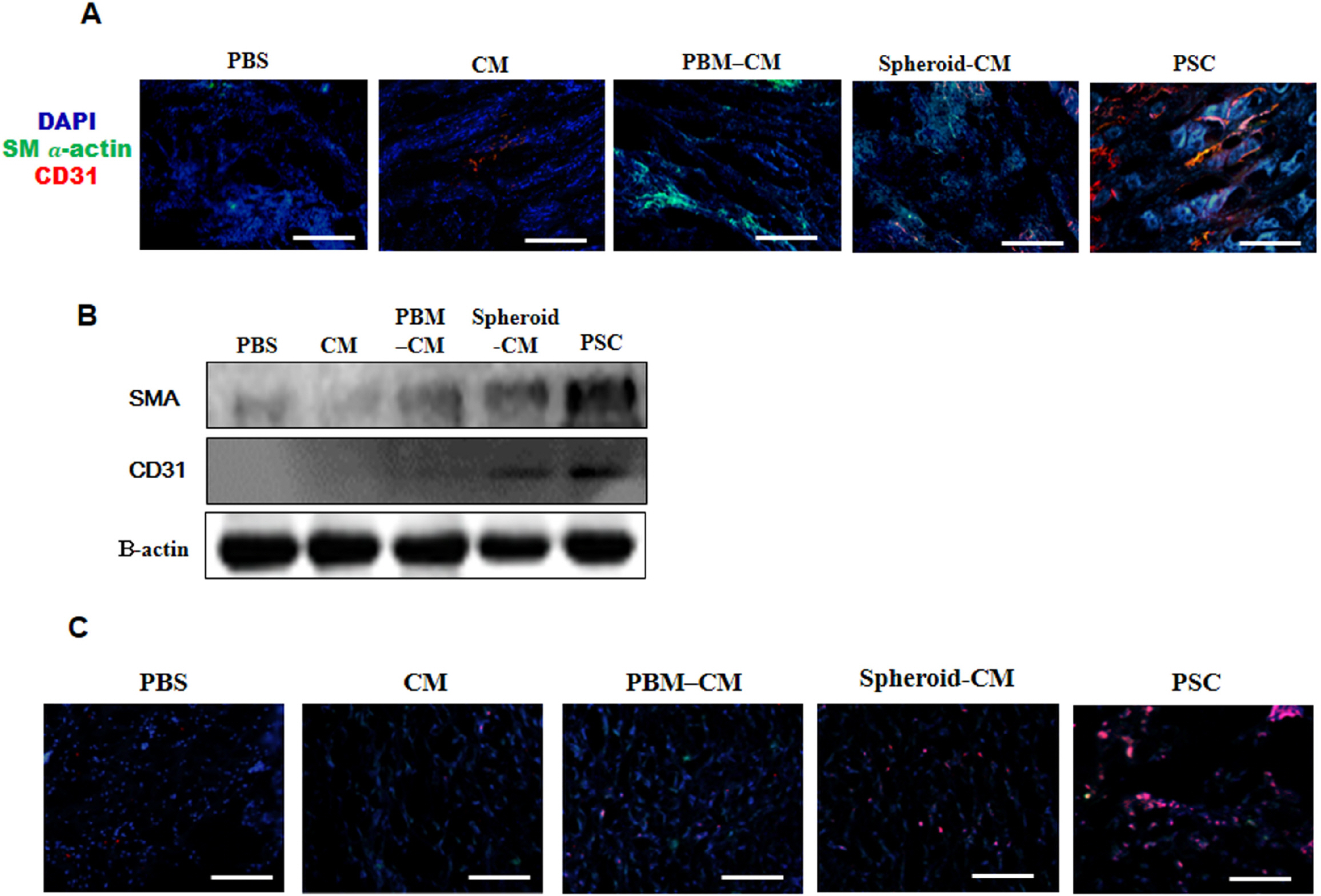
Fig. 3 Angiogenic efficacy in the wound bed. (A) Implants were removed on day 14 after implanted and stained with anti CD31 and αSMA antibodies. The scale bar indicates 200 μm. (B) Western blot showing the expression of CD31 and αSMA at 14 days. Beta-actin, also known as a “housekeeping” protein, is used as a loading control. (C) Immunofluorescence images showing cytokeratin-positive epithelial cells (red) at 14 days. The scale bar indicates 20 μm.

Fig. 4 Evaluation of wound closure. (A) The prepared excisional wound splinting model. Photographs of wounds. (B) Percentage of wound area was calculated using photographs of wounds at 1, 7, and 14 days. *p<0.05 versus the PSC group. (C) Histological analysis of the wound bed. Wounds were stained with H&E and Masson’s trichrome at 14 days. Wound edges are indicated with arrowheads. Closed arrows indicate skin appendages (hair follicles). The scale bar indicates 500 μm. (D) Regeneration of skin appendages was investigated by counting the number of skin appendages per wound section. (E) Histological scoring was performed using the criteria presented in Supplementary Table S3. *p<0.05 versus the PSC group.
Reference
-
References
1. Kyriakides TR, Maclauchlan S. 2009; The role of thrombospondins in wound healing, ischemia, and the foreign body reaction. J Cell Commun Signal. 3:215–225. DOI: 10.1007/s12079-009-0077-z. PMID: 19844806. PMCID: PMC2778594.
Article2. Metcalfe AD, Ferguson MW. 2007; Bioengineering skin using mechanisms of regeneration and repair. Biomaterials. 28:5100–5113. DOI: 10.1016/j.biomaterials.2007.07.031. PMID: 17688942.
Article3. Nie C, Yang D, Morris SF. 2009; Local delivery of adipose-derived stem cells via acellular dermal matrix as a scaffold: a new promising strategy to accelerate wound healing. Med Hypotheses. 72:679–682. DOI: 10.1016/j.mehy.2008.10.033. PMID: 19243892.
Article4. Heydarkhan-Hagvall S, Schenke-Layland K, Yang JQ, Heydarkhan S, Xu Y, Zuk PA, MacLellan WR, Beygui RE. 2008; Human adipose stem cells: a potential cell source for cardiovascular tissue engineering. Cells Tissues Organs. 187:263–274. DOI: 10.1159/000113407. PMID: 18196894.
Article5. Gimble J, Guilak F. 2003; Adipose-derived adult stem cells: isolation, characterization, and differentiation potential. Cyto-therapy. 5:362–369. DOI: 10.1080/14653240310003026. PMID: 14578098.
Article6. Joggerst SJ, Hatzopoulos AK. 2009; Stem cell therapy for cardiac repair: benefits and barriers. Expert Rev Mol Med. 11:e20. DOI: 10.1017/S1462399409001124. PMID: 19586557.
Article7. Don CW, Murry CE. 2013; Improving survival and efficacy of pluripotent stem cell-derived cardiac grafts. J Cell Mol Med. 17:1355–1362. DOI: 10.1111/jcmm.12147. PMID: 24118766. PMCID: PMC4049630.
Article8. Park IS, Chung PS, Ahn JC. 2017; Adipose-derived stem cell spheroid treated with low-level light irradiation accelerates spontaneous angiogenesis in mouse model of hindlimb ischemia. Cytotherapy. 19:1070–1078. DOI: 10.1016/j.jcyt.2017.06.005. PMID: 28739168.
Article9. Li P, Guo X. 2018; A review: therapeutic potential of adipose-derived stem cells in cutaneous wound healing and regeneration. Stem Cell Res Ther. 9:302. DOI: 10.1186/s13287-018-1044-5. PMID: 30409218. PMCID: PMC6225584.
Article10. Valcárcel M, Arteta B, Jaureguibeitia A, Lopategi A, Martínez I, Mendoza L, Muruzabal FJ, Salado C, Vidal-Va-naclocha F. 2008; Three-dimensional growth as multicellular spheroid activates the proangiogenic phenotype of colorectal carcinoma cells via LFA-1-dependent VEGF: implications on hepatic micrometastasis. J Transl Med. 6:57. DOI: 10.1186/1479-5876-6-57. PMID: 18844982. PMCID: PMC2579286.
Article11. Pastar I, Stojadinovic O, Yin NC, Ramirez H, Nusbaum AG, Sawaya A, Patel SB, Khalid L, Isseroff RR, Tomic-Canic M. 2014; Epithelialization in wound healing: a comprehensive review. Adv Wound Care (New Rochelle). 3:445–464. DOI: 10.1089/wound.2013.0473. PMID: 25032064. PMCID: PMC4086220.
Article12. Wu Y, Chen L, Scott PG, Tredget EE. 2007; Mesenchymal stem cells enhance wound healing through differentiation and angiogenesis. Stem Cells. 25:2648–2659. DOI: 10.1634/stemcells.2007-0226. PMID: 17615264.
Article13. Nie C, Yang D, Xu J, Si Z, Jin X, Zhang J. 2011; Locally administered adipose-derived stem cells accelerate wound healing through differentiation and vasculogenesis. Cell Transplant. 20:205–216. DOI: 10.3727/096368910X520065. PMID: 20719083.
Article14. Park IS, Rhie JW, Kim SH. 2014; A novel three-dimensional adipose-derived stem cell cluster for vascular regeneration in ischemic tissue. Cytotherapy. 16:508–522. DOI: 10.1016/j.jcyt.2013.08.011. PMID: 24210783.
Article15. Park IS, Kim SH, Jung Y, Rhie JW, Kim SH. 2013; Endothelial differentiation and vasculogenesis induced by three-dimensional adipose-derived stem cells. Anat Rec (Hoboken). 296:168–177. DOI: 10.1002/ar.22606. PMID: 23109231.
Article16. Pawitan JA. 2014; Prospect of stem cell conditioned medium in regenerative medicine. Biomed Res Int. 2014:965849. DOI: 10.1155/2014/965849. PMID: 25530971. PMCID: PMC4229962.
Article17. Kanji S, Das H. 2017; Advances of stem cell therapeutics in cutaneous wound healing and regeneration. Mediators Inflamm. 2017:5217967. DOI: 10.1155/2017/5217967. PMID: 29213192. PMCID: PMC5682068.
Article18. Chen P, Chen JZ, Shao CY, Li CY, Zhang YD, Lu WJ, Fu Y, Gu P, Fan X. 2015; Treatment with retinoic acid and lens epithelial cell-conditioned medium in vitro directed the differentiation of pluripotent stem cells towards corneal endothelial cell-like cells. Exp Ther Med. 9:351–360. DOI: 10.3892/etm.2014.2103. PMID: 25574197. PMCID: PMC4280952.
Article19. Choi K, Kang BJ, Kim H, Lee S, Bae S, Kweon OK, Kim WH. 2013; Low-level laser therapy promotes the osteogenic potential of adipose-derived mesenchymal stem cells seeded on an acellular dermal matrix. J Biomed Mater Res B Appl Biomater. 101:919–928. DOI: 10.1002/jbm.b.32897. PMID: 23529895.
Article20. Mvula B, Mathope T, Moore T, Abrahamse H. 2008; The effect of low level laser irradiation on adult human adipose derived stem cells. Lasers Med Sci. 23:277–282. DOI: 10.1007/s10103-007-0479-1. PMID: 17713825.
Article21. Mvula B, Moore TJ, Abrahamse H. 2010; Effect of low-level laser irradiation and epidermal growth factor on adult human adipose-derived stem cells. Lasers Med Sci. 25:33–39. DOI: 10.1007/s10103-008-0636-1. PMID: 19172344.
Article22. Wang X, Ge J, Tredget EE, Wu Y. 2013; The mouse excisional wound splinting model, including applications for stem cell transplantation. Nat Protoc. 8:302–309. DOI: 10.1038/nprot.2013.002. PMID: 23329003.
Article23. Kapur SK, Wang X, Shang H, Yun S, Li X, Feng G, Khurgel M, Katz AJ. 2012; Human adipose stem cells maintain proliferative, synthetic and multipotential properties when suspension cultured as self-assembling spheroids. Biofabri-cation. 4:025004. DOI: 10.1088/1758-5082/4/2/025004. PMID: 22522924. PMCID: PMC3401583.
Article24. Glicklis R, Merchuk JC, Cohen S. 2004; Modeling mass transfer in hepatocyte spheroids via cell viability, spheroid size, and hepatocellular functions. Biotechnol Bioeng. 86:672–680. DOI: 10.1002/bit.20086. PMID: 15137079.
Article25. Chen M, Przyborowski M, Berthiaume F. 2009; Stem cells for skin tissue engineering and wound healing. Crit Rev Biomed Eng. 37:399–421. DOI: 10.1615/CritRevBiomedEng.v37.i4-5.50. PMID: 20528733. PMCID: PMC3223487.
Article26. Santini MT, Rainaldi G, Indovina PL. 2000; Apoptosis, cell adhesion and the extracellular matrix in the three-dimensional growth of multicellular tumor spheroids. Crit Rev Oncol Hematol. 36:75–87. DOI: 10.1016/S1040-8428(00)00078-0. PMID: 11033298.
Article27. Ginani F, Soares DM, Barreto MP, Barboza CA. 2015; Effect of low-level laser therapy on mesenchymal stem cell proliferation: a systematic review. Lasers Med Sci. 30:2189–2194. DOI: 10.1007/s10103-015-1730-9. PMID: 25764448.
Article28. Hu WP, Wang JJ, Yu CL, Lan CC, Chen GS, Yu HS. 2007; Helium-neon laser irradiation stimulates cell proliferation through photostimulatory effects in mitochondria. J Invest Dermatol. 127:2048–2057. DOI: 10.1038/sj.jid.5700826. PMID: 17446900.
Article29. Bhang SH, Cho SW, La WG, Lee TJ, Yang HS, Sun AY, Baek SH, Rhie JW, Kim BS. 2011; Angiogenesis in ischemic tissue produced by spheroid grafting of human adipose-de-rived stromal cells. Biomaterials. 32:2734–2747. DOI: 10.1016/j.biomaterials.2010.12.035. PMID: 21262528.
Article30. Lee EJ, Park HW, Jeon HJ, Kim HS, Chang MS. 2013; Potentiated therapeutic angiogenesis by primed human mesenchymal stem cells in a mouse model of hindlimb ischemia. Regen Med. 8:283–293. DOI: 10.2217/rme.13.17. PMID: 23627823.
Article31. Alev C, Ii M, Asahara T. 2011; Endothelial progenitor cells: a novel tool for the therapy of ischemic diseases. Antioxid Redox Signal. 15:949–965. DOI: 10.1089/ars.2010.3872. PMID: 21254837.
Article
- Full Text Links
-
- Actions
-
Cited
- CITED
-
- Close
- Share
-
- Similar articles
-
- Effects of Adipose-derived Stromal Cells and of their Extract on Wound Healing in a Mouse Model
- Promising improvement in infected Wound Healing in Type two Diabetic rats by Combined effects of conditioned medium of human adipose‑derived stem cells plus Photobiomodulation
- Paracrine Effects of Adipose-Derived Stem Cells on Keratinocytes and Dermal Fibroblasts
- Clinical Application of Adipose Derived Stromal Cell Autograft for Wound Coverage
- The Effect of Curcumin and Human Adipose-derived Stromal Cells on Wound Healing of Lewis Rats
