J Korean Med Sci.
2010 May;25(5):746-751. 10.3346/jkms.2010.25.5.746.
Effects of Adipose-derived Stromal Cells and of their Extract on Wound Healing in a Mouse Model
- Affiliations
-
- 1Department of Plastic Surgery, College of Medicine, The Catholic University of Korea, Seoul, Korea. psyg@catholic.ac.kr
- KMID: 1713960
- DOI: http://doi.org/10.3346/jkms.2010.25.5.746
Abstract
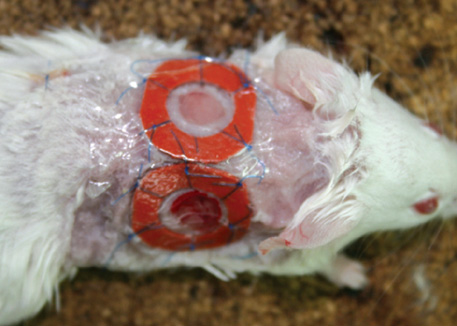
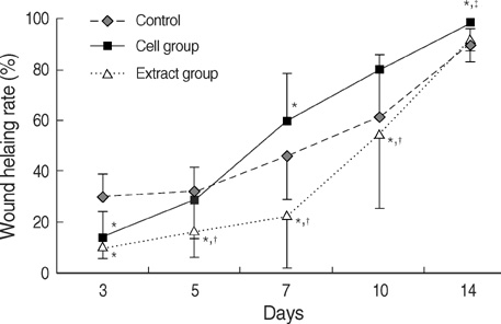
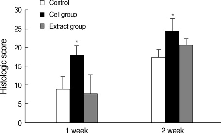
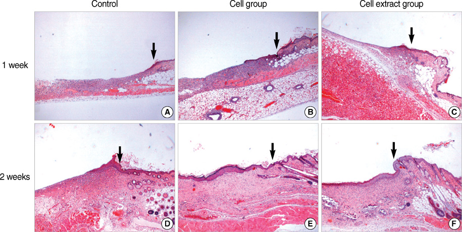
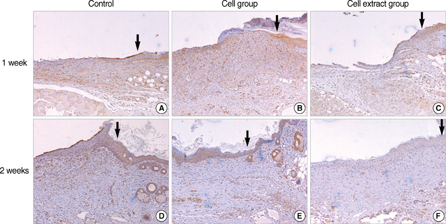
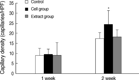
- In this study, the authors investigated the effects of adipose-derived stromal cells (ADSCs) and of their extract on wound healing. After creating wound healing splint model on the backs of mice, ADSCs and their extract were applied. Wound healing rates were calculated at 3, 5, 7, 10, and 14 days after the wounding, and tissues were harvested at 7 and 14 days for histological analysis. Wound healing rates were significantly higher at 7, 10, and 14 days in the cell group than in the control, but in the cell extract group wound healing rates were significantly decreased (P<0.05). Histological scores and capillary densities in the cell group were significantly higher at 2 weeks (P<0.05). In the cell group, thick inflammatory cell infiltration and many capillaries were observed at 1 week, and thick epithelium and numerous large capillaries were observed at 2 weeks. The present study suggests that ADSCs accelerate wound healing as known, and the effects of ADSCs on wound healing may be due to replacing insufficient cells by differentiation of ADSCs in the wound and secreting growth factors by differentiated cells, and not due to the effect of factors within ADSCs.
MeSH Terms
Figure
Reference
-
1. Martin P. Wound healing-aiming for perfect skin regeneration. Science. 1997. 276:75–81.
Article2. Falanga V. Wound healing and its impairment in the diabetic foot. Lancet. 2005. 366:1736–1743.
Article3. Zuk PA, Zhu M, Ashjian P, De Ugarte DA, Huang JI, Mizuno H, Alfonso ZC, Fraser JK, Benhaim P, Hedrick MH. Human adipose tissue is a source of multipotent stem cells. Mol Biol Cell. 2002. 13:4279–4295.
Article4. De Ugarte DA, Morizono K, Elbarbary A, Alfonso Z, Zuk PA, Zhu M, Dragoo JL, Ashjian P, Thomas B, Benhaim P, Chen I, Fraser J, Hedrick MH. Comparison of multi-lineage cells from human adipose tissue and bone marrow. Cells Tissues Organs. 2003. 174:101–109.
Article5. Izadpanah R, Trygg C, Patel B, Kriedt C, Dufour J, Gimble JM, Bunnell BA. Biologic properties of mesenchymal stem cells derived from bone marrow and adipose tissue. J Cell Biochem. 2006. 99:1285–1297.
Article6. Wu Y, Chen L, Scott PG, Tredget EE. Mesenchymal stem cells enhance wound healing through differentiation and angiogenesis. Stem Cells. 2007. 25:2648–2659.
Article7. Kim WS, Park BS, Sung JH, Yang JM, Park SB, Kwak SJ, Park JS. Wound healing effect of adipose-derived stem cells: a critical role of secretory factors on human dermal fibroblasts. J Dermatol Sci. 2007. 48:15–24.
Article8. Galiano RD, Michaels J, Dobryansky M, Levine JP, Gurtner GC. Quantitative and reproducible murine model of excisional wound healing. Wound Repair Regen. 2004. 12:485–492.
Article9. Jacobi J, Jang JJ, Sundram U, Dayoub H, Fajardo LF, Cooke JP. Nicotine accelerates angiogenesis and wound healing in genetically diabetic mice. Am J Pathol. 2002. 161:97–104.
Article10. Yoon YS, Murayama T, Gravereaux E, Tkebuchava T, Silver M, Curry C, Wecker A, Kirchmair R, Hu CS, Kearney M, Ashare A, Jackson DG, Kubo H, Isner JM, Losordo DW. Vegf-c gene therapy augments postnatal lymphangiogenesis and ameliorates secondary lymphedema. J Clin Invest. 2003. 111:717–725.
Article11. Badiavas EV, Abedi M, Butmarc J, Falanga V, Quesenberry P. Participation of bone marrow derived cells in cutaneous wound healing. J Cell Physiol. 2003. 196:245–250.
Article12. Nakagawa H, Akita S, Fukui M, Fujii T, Akino K. Human mesenchymal stem cells successfully improve skin-substitute wound healing. Br J Dermatol. 2005. 153:29–36.
Article13. Gharaee-Kermani M, Phan SH. Role of cytokines and cytokine therapy in wound healing and fibrotic diseases. Curr Pharm Des. 2001. 7:1083–1103.14. Gaustad KG, Boquest AC, Anderson BE, Gerdes AM, Collas P. Differentiation of human adipose tissue stem cells using extracts of rat cardiomyocytes. Biochem Biophys Res Commun. 2004. 314:420–427.
Article
- Full Text Links
- Actions
-
Cited
- CITED
-
- Close
- Share
- Similar articles
-
- The Effect of Curcumin and Human Adipose-derived Stromal Cells on Wound Healing of Lewis Rats
- Enhancement of Wound Healing by Conditioned Medium of Adipose-Derived Stromal Cell with Photobiomodulation in Skin Wound
- Clinical Application of Adipose Derived Stromal Cell Autograft for Wound Coverage
- Effects of Human Adipose-derived Stem Cells on Cutaneous Wound Healing in Nude Mice
- Effect of Allogenic Adipose-derived Stromal Cells on Wound Healing in BALB/c Mice

