Evaluation of strategic uprighting of the mandibular molars using an orthodontic miniplate and a nickel-titanium reverse curve arch wire: Preliminary cephalometric study
- Affiliations
-
- 1Department of Orthodontics, Graduate School, Kyung Hee University, Seoul, Korea
- 2Department of Plastic Surgery, Stanford University School of Medicine, Lucile Packard Children’s Hospital, Palo Alto, CA, USA
- KMID: 2515821
- DOI: http://doi.org/10.4041/kjod.2021.51.3.179
Abstract
Objective
To evaluate the overall treatment effects in terms of the amount of uprighting with changes in the sagittal and vertical positions of mandibular molars after applying an orthodontic miniplate with a nickel-titanium (NiTi) reverse curve arch wire (biocreative reverse curve [BRC] system).
Methods
A total of 30 female patients (mean age, 25.99 ± 8.96 years) were treated with the BRC system (mean BRC time, 10.3 ± 4.07 months). An I-shaped C-tube miniplate (Jin Biomed) was placed at the labial aspect for the alveolar bone of the mandibular incisors. A 0.017 × 0.025-inch NiTi reverse curve arch wire was engaged at the C-tube mini-plate anteriorly and the first and second premolars and molars posteriorly in the mandibular arch. Pre- and post-BRC lateral cephalograms were analyzed. A paired t-test was used to analyze the treatment effects of BRC.
Results
The mandibular second molars were intrusively uprighted successfully by the BRC system. Distal uprighting with a controlled vertical dimension was noted on the first molars when they remained engaged in the BRC and the distal ends of the arch wire were laid on the second molars. The mandibular first and second premolars showed a slight extrusion. The changes in the mandibular incisors were unremarkable, while the mandibular molar angulation improved significantly. The lower occlusal plane rotated counterclockwise (MP-LOP: 1.13° ± 2.60°).
Conclusions
The BRC system can provide very effective molar uprighting without compromising the position of the mandibular anterior teeth.
Figure
-

Figure 1 Schematic illustration of the biocreative reverse curve system. The C-tube miniplate is installed on the labial side of the anterior teeth in the symphysis area. The brackets are bonded on the mandibular posterior target teeth. A 0.017 × 0.025-inch reverse curve of Spee nickel-titanium arch wire is raised by the C-tube and the intrusive upright force would be induced to the molars. A, Frontal view. B, Lateral view. C, Occlusal view.
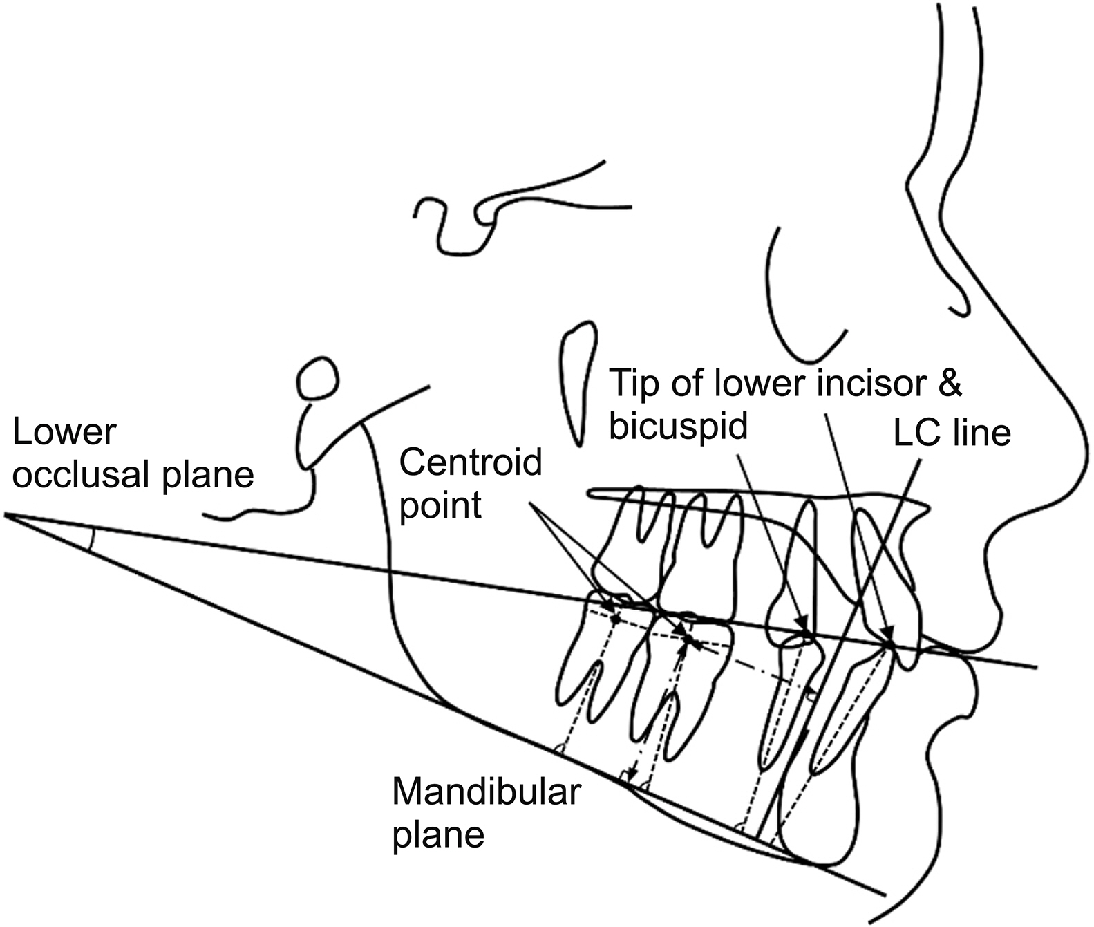
Figure 2 Reference points and lines used for cephalometric analysis. Dentoalveolar (containing occlusal plane) measurements. LC, mandibular lingual cortex.

Figure 3 Intraoral photographs of the case. A, Pretreatment. B, 6 months after biocreative reverse curve (BRC) procedure. C, 13 months after BRC procedure. D, Post-treatment (treatment periods: 21 months).

Figure 4 Lateral cephalograms and superimposed tracings of the case. A, Pretreatment. B, Right after biocreative reverse curve procedure. C, Post-treatment. D, Superimposition tracings from pretreatment (black) to posttreatment (gray).
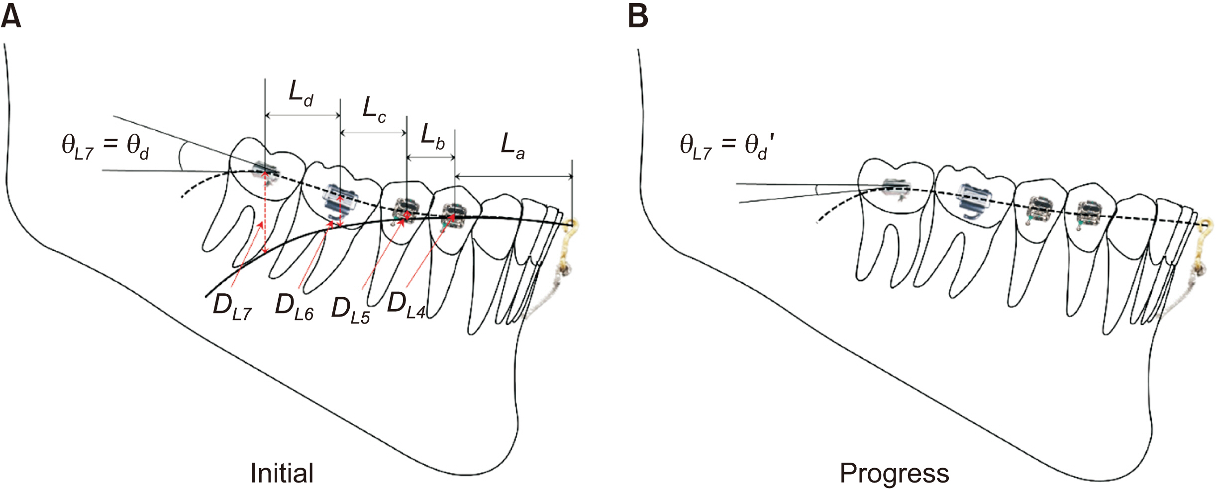
Figure 5 The progress of force-moment system change with the biocreative reverse curve system (non-equilibrium state). A, Initial state. B, Progress state.
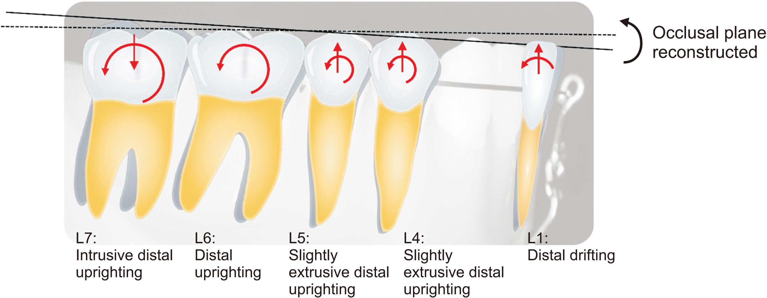
Figure 6 Overall treatment results of the biocreative reverse curve system (as referenced by the mean value in Table 2). L1, lower incisor (distalized: 0.48 mm, extruded: 0.44 mm); L4, first premolar (distalized: 0.94 mm, extruded: 0.75 mm, uprighted: 0.63°); L5, second premolar (distalized: 0.59 mm, extruded: 0.58 mm, uprighted: 1.46°); L6, first molar (distalized: 1.01 mm, uprighted: 7.29°); L7, second molar (distalized: 0.53 mm, intruded: 1.42 mm, uprighted: 10.83°).
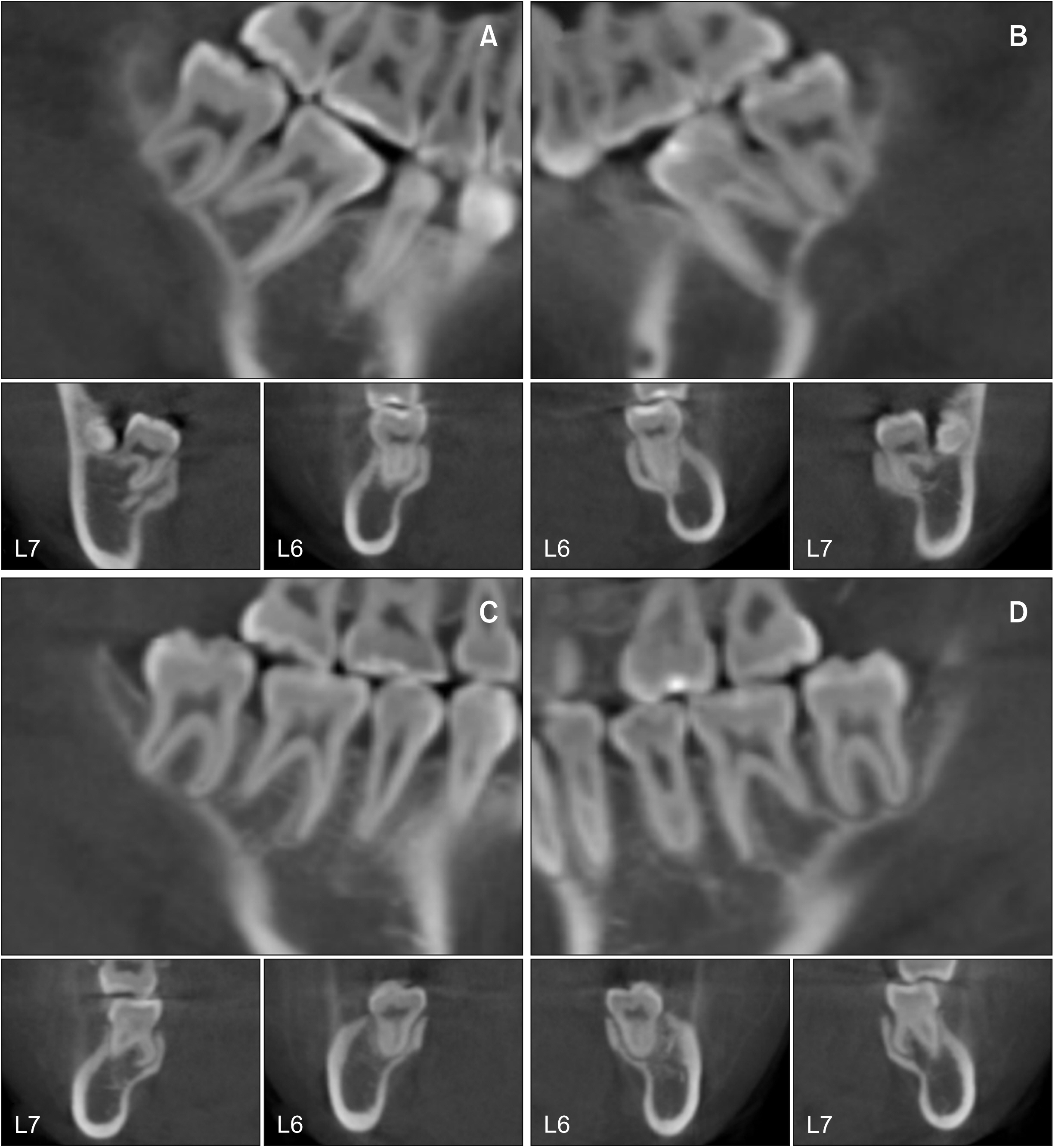
Figure 7 Three-dimensional tooth movement on cone-beam computed tomograms of the case. A, B, Initial state of the lower right and left molars, respectively (upper: sagittal, lower: frontal view). C, D, Final state of the lower right and left molars, respectively (upper: sagittal, lower: frontal view). L6, lower first molar; L7, lower second molar.
Cited by 1 articles
-
Root proximity of the anchoring miniscrews of orthodontic miniplates in the mandibular incisal area: Cone-beam computed tomographic analysis
Do-Min Jeong, Song Hee Oh, HyeRan Choo, Yong-Suk Choi, Seong-Hun Kim, Jin-Suk Lee, Eui-Hwan Hwang
Korean J Orthod. 2021;51(4):231-240. doi: 10.4041/kjod.2021.51.4.231.
Reference
-
1. Stern N, Revah A, Becker A. 1981; The tilted posterior tooth. Part I: etiology, syndrome, and prevention. J Prosthet Dent. 46:404–7. DOI: 10.1016/0022-3913(81)90447-9.
Article2. Mohlin B. 1983; Prevalence of mandibular dysfunction and relation between malocclusion and mandibular dysfunction in a group of women in Sweden. Eur J Orthod. 5:115–23. DOI: 10.1093/ejo/5.2.115. PMID: 6574917.
Article3. Roberts WW 3rd, Chacker FM, Burstone CJ. 1982; A segmental approach to mandibular molar uprighting. Am J Orthod. 81:177–84. DOI: 10.1016/0002-9416(82)90051-3. PMID: 6960706.
Article4. Simon RL. 1984; Rationale and practical technique for uprighting mesially inclined molars. J Prosthet Dent. 52:256–9. DOI: 10.1016/0022-3913(84)90107-0. PMID: 6381712.
Article5. Capelluto E, Lauweryns I. 1997; A simple technique for molar uprighting. J Clin Orthod. 31:119–25. PMID: 9511534.6. Shellhart WC, Oesterle LJ. 1999; Uprighting molars without extrusion. J Am Dent Assoc. 130:381–5. DOI: 10.14219/jada.archive.1999.0208. PMID: 10085661.
Article7. Zachrisson BU, Bantleon HP. 2005; Ask an expert: optimal mechanics for mandibular molar uprighting. World J Orthod. 6:80–7.8. Magkavali-Trikka P, Emmanouilidis G, Papadopoulos MA. 2018; Mandibular molar uprighting using orthodontic miniscrew implants: a systematic review. Prog Orthod. 19:1. DOI: 10.1186/s40510-017-0200-2. PMID: 29308540. PMCID: PMC5756736.
Article9. Ahn HW, Chung KR, Kang SM, Lin L, Nelson G, Kim SH. 2012; Correction of dental Class III with posterior open bite by simple biomechanics using an anterior C-tube miniplate. Korean J Orthod. 42:270–8. DOI: 10.4041/kjod.2012.42.5.270. PMID: 23173121. PMCID: PMC3495259.
Article10. Kim SH, Hwang YS, Ferreira A, Chung KR. 2009; Analysis of temporary skeletal anchorage devices used for en-masse retraction: a preliminary study. Am J Orthod Dentofacial Orthop. 136:268–76. DOI: 10.1016/j.ajodo.2007.08.023. PMID: 19651358.
Article11. Kojima K, Endo T, Shimooka S. 2009; Effects of maxillary second molar extraction on dentofacial morphology before and after anterior open-bite treatment: a cephalometric study. Odontology. 97:43–50. DOI: 10.1007/s10266-008-0093-0. PMID: 19184297.
Article12. Jee JH, Ahn HW, Seo KW, Kim SH, Kook YA, Chung KR, et al. 2014; En-masse retraction with a preformed nickel-titanium and stainless steel archwire assembly and temporary skeletal anchorage devices without posterior bonding. Korean J Orthod. 44:236–45. DOI: 10.4041/kjod.2014.44.5.236. PMID: 25309863. PMCID: PMC4192525.13. Ahn HW, Noh MK, Chung KR, Kim SH, Nelson G. 2020; Strategic molar uprighting using the biocreative reverse-curve technique. J Clin Orthod. 54:486–94. PMID: 32966268.14. Burstone CJ, Koenig HA. 1988; Creative wire bending--the force system from step and V bends. Am J Orthod Dentofacial Orthop. 93:59–67. DOI: 10.1016/0889-5406(88)90194-1. PMID: 3422122.
Article15. Lindauer SJ, Isaacson RJ. 1995; One-couple orthodontic appliance systems. Semin Orthod. 1:12–24. DOI: 10.1016/S1073-8746(95)80084-0. PMID: 8935039.
Article16. Burstone CJ, Koenig HA. 1974; Force systems from an ideal arch. Am J Orthod. 65:270–89. DOI: 10.1016/S0002-9416(74)90332-7. PMID: 4521361.
Article17. Smith RJ, Burstone CJ. 1984; Mechanics of tooth movement. Am J Orthod. 85:294–307. DOI: 10.1016/0002-9416(84)90187-8. PMID: 6585147.
Article18. Isaacson RJ, Lindauer SJ, Davidovitch M. 1995; The ground rules for arch wire design. Semin Orthod. 1:3–11. DOI: 10.1016/S1073-8746(95)80083-2. PMID: 8935038.
Article
- Full Text Links
-
- Actions
-
Cited
- CITED
-
- Close
- Share
-
- Similar articles
-
- Correction of dental Class III with posterior open bite by simple biomechanics using an anterior C-tube miniplate
- A photoelastic study of the effect of loops in arch wire and orthodontic elastics in relief of Curve of Spee
- A cone-beam computed tomography study on strategic uprighting of mandibular molars using a biocreative reverse curve system
- Biomechanical considerations for uprighting impacted mandibular molars
- Effects of heat treatment on the load-deflection properties of nickel-titanium wire
