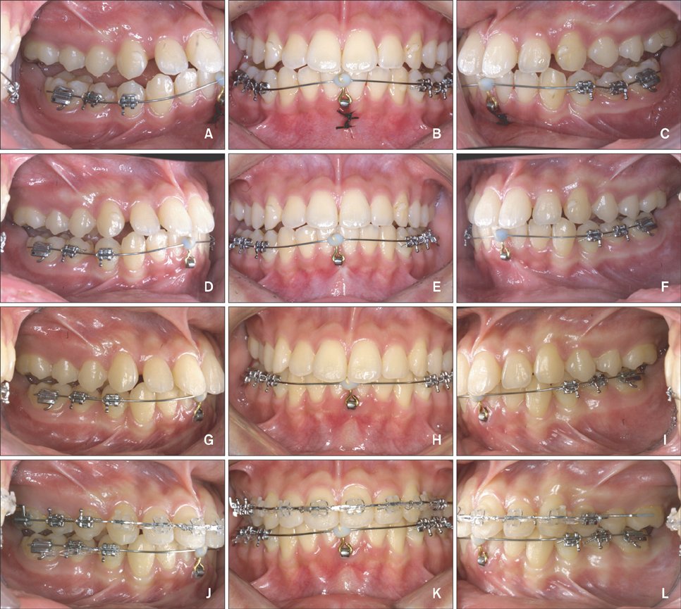
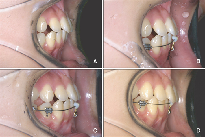
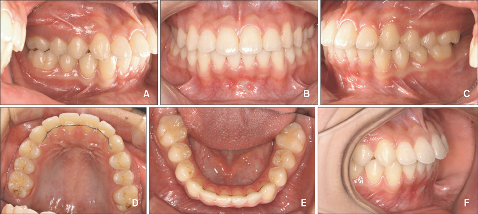
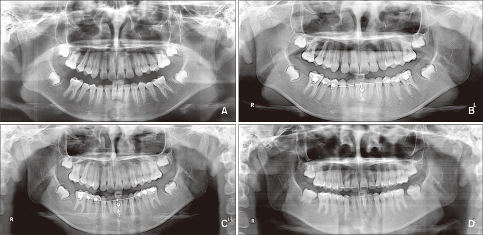
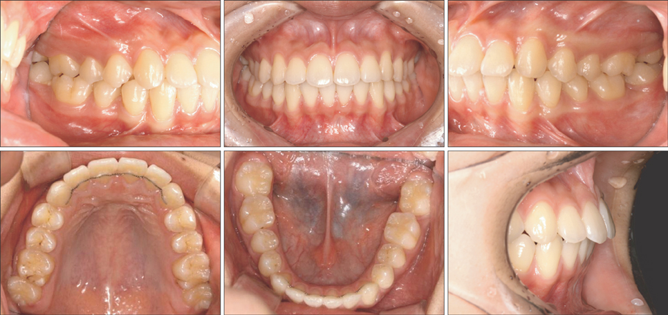
Correction of dental Class III with posterior open bite by simple biomechanics using an anterior C-tube miniplate
- Affiliations
-
- 1Department of Orthodontics, School of Dentistry, Kyung Hee University, Seoul, Korea. bravortho@khu.ac.kr
- 2Department of Orthodontics, School of Medicine, Ajou University, Suwon, Korea.
- 3Division of Orthodontics, University of California San Francisco, San Francisco, USA.
- KMID: 1435403
- DOI: http://doi.org/10.4041/kjod.2012.42.5.270
Abstract
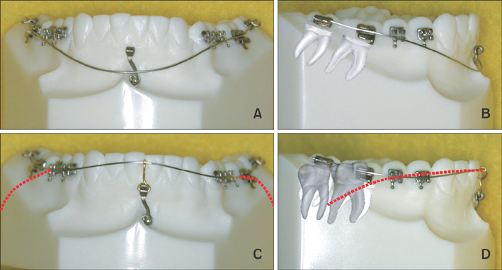
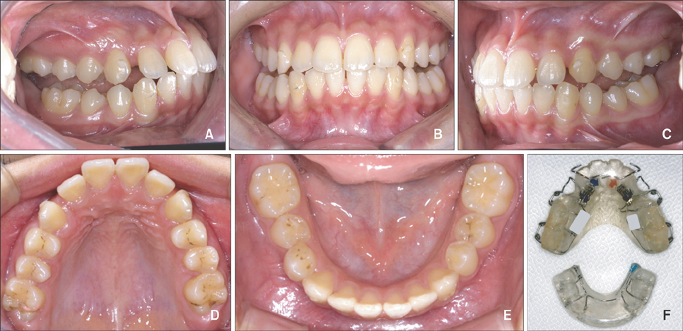
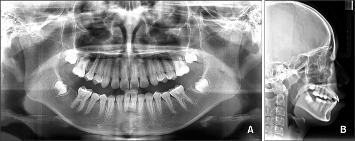
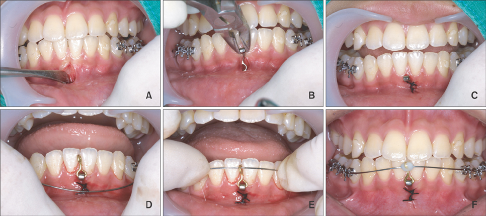
- In the correction of dental Class III molar relationship in skeletal Class II patients, uprighting of the mandibular posterior segments without opening the mandible is an important treatment objective. In the case reported herein, a C-tube miniplate fixed to the lower labial symphysis and connected with a nickel-titanium reverse-curved archwire provided effective uprighting of the lower molars, without the need of orthodontic appliances on the mandibular anteriors. Using this approach, an appropriate magnitude of force is exerted on the molars while avoiding any negative effect on the mandibular anteriors.
Keyword
Figure
Cited by 3 articles
-
En-masse retraction with a preformed nickel-titanium and stainless steel archwire assembly and temporary skeletal anchorage devices without posterior bonding
Jeong-Hyun Jee, Hyo-Won Ahn, Kyung-Won Seo, Seong-Hun Kim, Yoon-Ah Kook, Kyu-Rhim Chung, Gerald Nelson
Korean J Orthod. 2014;44(5):236-245. doi: 10.4041/kjod.2014.44.5.236.Evaluation of strategic uprighting of the mandibular molars using an orthodontic miniplate and a nickel-titanium reverse curve arch wire: Preliminary cephalometric study
Jae-Hyun Park, HyeRan Choo, Jin-Young Choi, Kyu-Rhim Chung, Seong-Hun Kim
Korean J Orthod. 2021;51(3):179-188. doi: 10.4041/kjod.2021.51.3.179.Root proximity of the anchoring miniscrews of orthodontic miniplates in the mandibular incisal area: Cone-beam computed tomographic analysis
Do-Min Jeong, Song Hee Oh, HyeRan Choo, Yong-Suk Choi, Seong-Hun Kim, Jin-Suk Lee, Eui-Hwan Hwang
Korean J Orthod. 2021;51(4):231-240. doi: 10.4041/kjod.2021.51.4.231.
Reference
-
1. Kim YS, Cha JY, Yu HS, Hwang CJ. Comparison of mandibular anterior alveolar bone thickness in different facial skeletal types. Korean J Orthod. 2010. 40:314–324.
Article2. Miyajima K, Iizuka T. Treatment mechanics in Class III open bite malocclusion with Tip Edge technique. Am J Orthod Dentofacial Orthop. 1996. 110:1–7.
Article3. Lee BR, Kang DK, Son WS, Park SB, Kim SS, Kim YI, et al. The relationship between condyle position, morphology and chin deviation in skeletal Class III patients with facial asymmetry using cone-beam CT. Korean J Orthod. 2011. 41:87–97.
Article4. Kim YH, Han UK, Lim DD, Serraon ML. Stability of anterior openbite correction with multiloop edgewise archwire therapy: A cephalometric follow-up study. Am J Orthod Dentofacial Orthop. 2000. 118:43–54.
Article5. Enacar A, Ugur T, Toroglu S. A method for correction of open bite. J Clin Orthod. 1996. 30:43–48.6. Park YC, Lee HA, Choi NC, Kim DH. Open bite correction by intrusion of posterior teeth with miniscrews. Angle Orthod. 2008. 78:699–710.
Article7. Moon CH, Lee JS, Lee HS, Choi JH. Non-surgical treatment and retention of open bite in adult patients with orthodontic mini-implants. Korean J Orthod. 2009. 39:402–419.
Article8. Kim MJ, Park SH, Kim HS, Mo SS, Sung SJ, Jang GW, et al. Effects of orthodontic mini-implant position in the dragon helix appliance on tooth displacement and stress distribution: a three-dimensional finite element analysis. Korean J Orthod. 2011. 41:191–199.
Article9. Deguchi T, Kurosaka H, Oikawa H, Kuroda S, Takahashi I, Yamashiro T, et al. Comparison of orthodontic treatment outcomes in adults with skeletal open bite between conventional edgewise treatment and implant-anchored orthodontics. Am J Orthod Dentofacial Orthop. 2011. 139:4 Suppl. S60–S68.
Article10. Kuroda S, Yamada K, Deguchi T, Hashimoto T, Kyung HM, Takano-Yamamoto T. Root proximity is a major factor for screw failure in orthodontic anchorage. Am J Orthod Dentofacial Orthop. 2007. 131:4 Suppl. S68–S73.
Article11. Asscherickx K, Vande Vannet B, Wehrbein H, Sabzevar MM. Success rate of miniscrews relative to their position to adjacent roots. Eur J Orthod. 2008. 30:330–335.
Article12. Park J, Cho HJ. Three-dimensional evaluation of interradicular spaces and cortical bone thickness for the placement and initial stability of microimplants in adults. Am J Orthod Dentofacial Orthop. 2009. 136:314.e1–314.e12.
Article13. Kim SH, Yoon HG, Choi YS, Hwang EH, Kook YA, Nelson G. Evaluation of interdental space of the maxillary posterior area for orthodontic mini-implants with cone-beam computed tomography. Am J Orthod Dentofacial Orthop. 2009. 135:635–641.
Article14. Chung KR, Kim SH, Kang YG, Nelson G. Orthodontic miniplate with tube as an efficient tool for borderline cases. Am J Orthod Dentofacial Orthop. 2011. 139:551–562.
Article15. Chang YI, Moon SC. Cephalometric evaluation of the anterior open bite treatment. Am J Orthod Dentofacial Orthop. 1999. 115:29–38.
Article16. Kim YH. Anterior openbite and its treatment with multiloop edgewise archwire. Angle Orthod. 1987. 57:290–321.17. Küçükkeleş N, Acar A, Demirkaya AA, Evrenol B, Enacar A. Cephalometric evaluation of open bite treatment with NiTi arch wires and anterior elastics. Am J Orthod Dentofacial Orthop. 1999. 116:555–562.
Article18. Janson G, Valarelli FP, Beltrão RT, de Freitas MR, Henriques JF. Stability of anterior open-bite extraction and nonextraction treatment in the permanent dentition. Am J Orthod Dentofacial Orthop. 2006. 129:768–774.
Article19. Korean Association of Orthodontists. Cephalometric norm of Korean adults with normal occlusion. 1998. Seoul: Ji-Sung Publishing Co.;589–595.
- Full Text Links
- Actions
-
Cited
- CITED
-
- Close
- Share
- Similar articles
-
- A cephalometric study on mesiodistal axial inclination of posterior teeth in open bite and deep bite
- A study on the vertical dysplasia in the skeletal class iii malocclusion
- Treatment of anterior open bites using nonextraction clear aligner therapy in adult patients
- Clincal analysis of skeletal stability after BSSRO for correction of skeletal class III malocclusion patients with anterir open bite
- A case report of unilateral posterior cross bite by the activator

