Anat Cell Biol.
2021 Mar;54(1):104-111. 10.5115/acb.20.228.
Anti-proliferative and apoptotic effects of hull-less pumpkin extract on human papillary thyroid carcinoma cell line
- Affiliations
-
- 1Cellular and Molecular Research Center, Faculty of Medicine, Guilan University of Medical Sciences, Rasht, Iran
- 2Department of Pharmaceutical Biotechnology, School of Pharmacy, Guilan University of Medical Sciences, Rasht, Iran
- 3Department of Endocrinology, Guilan University of Medical Sciences, Rasht, Iran
- 4Department of Anatomical Sciences, Faculty of Medicine, Baqiyatallah University of Medical Sciences, Tehran, Iran
- 5aqiyatallah Research Center for Gastroenterology and Liver Diseases (BRCGL), Baqiyatallah University of Medical Sciences, Tehran, Iran
- KMID: 2514590
- DOI: http://doi.org/10.5115/acb.20.228
Abstract
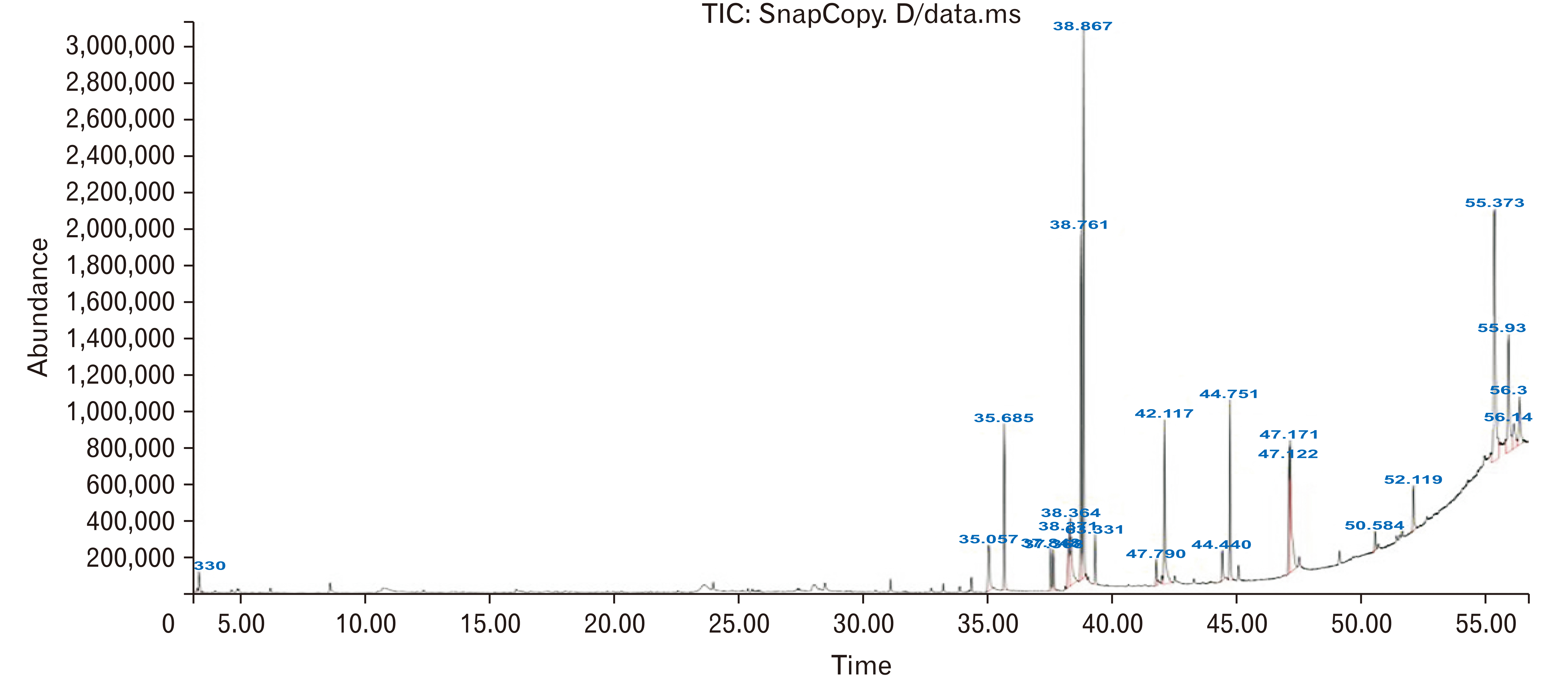
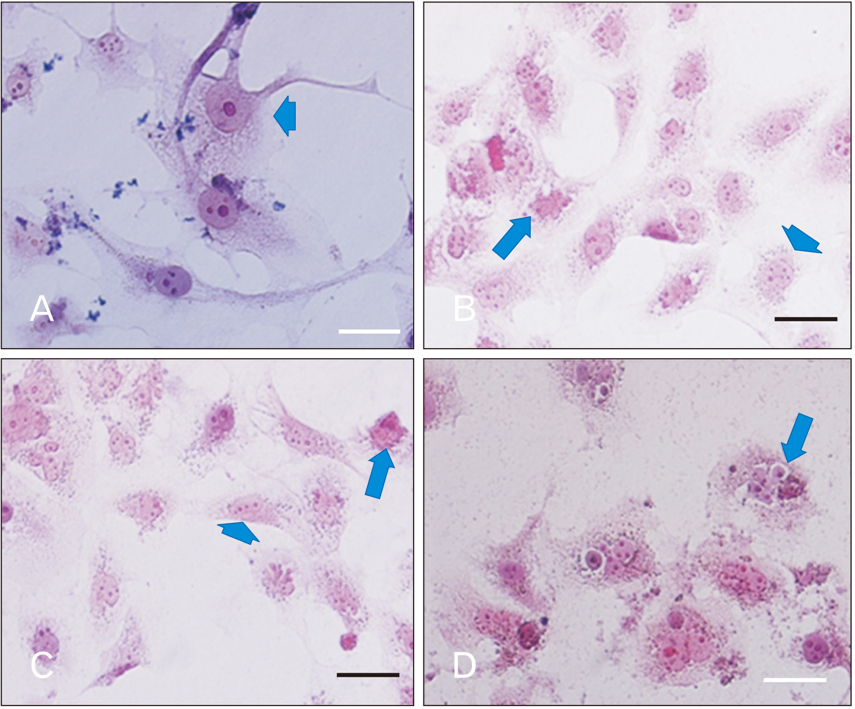
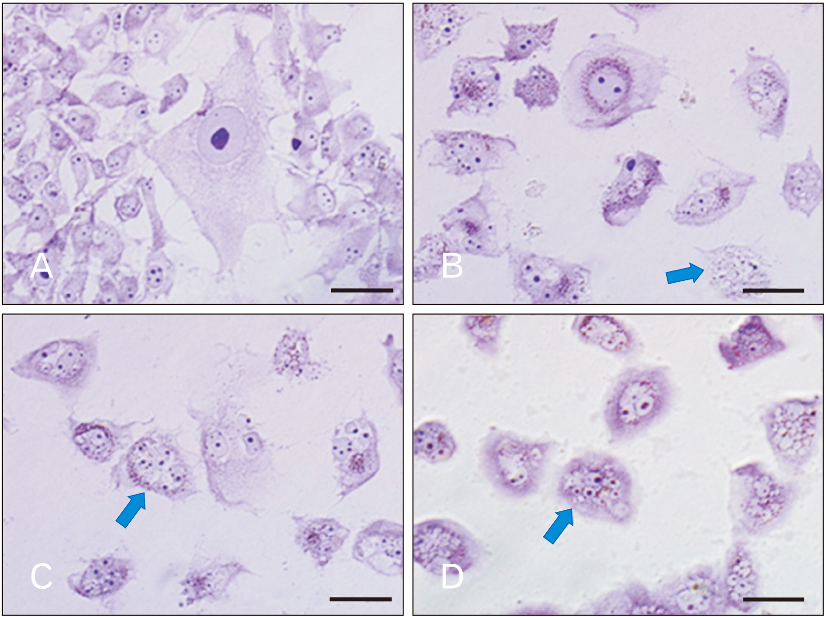
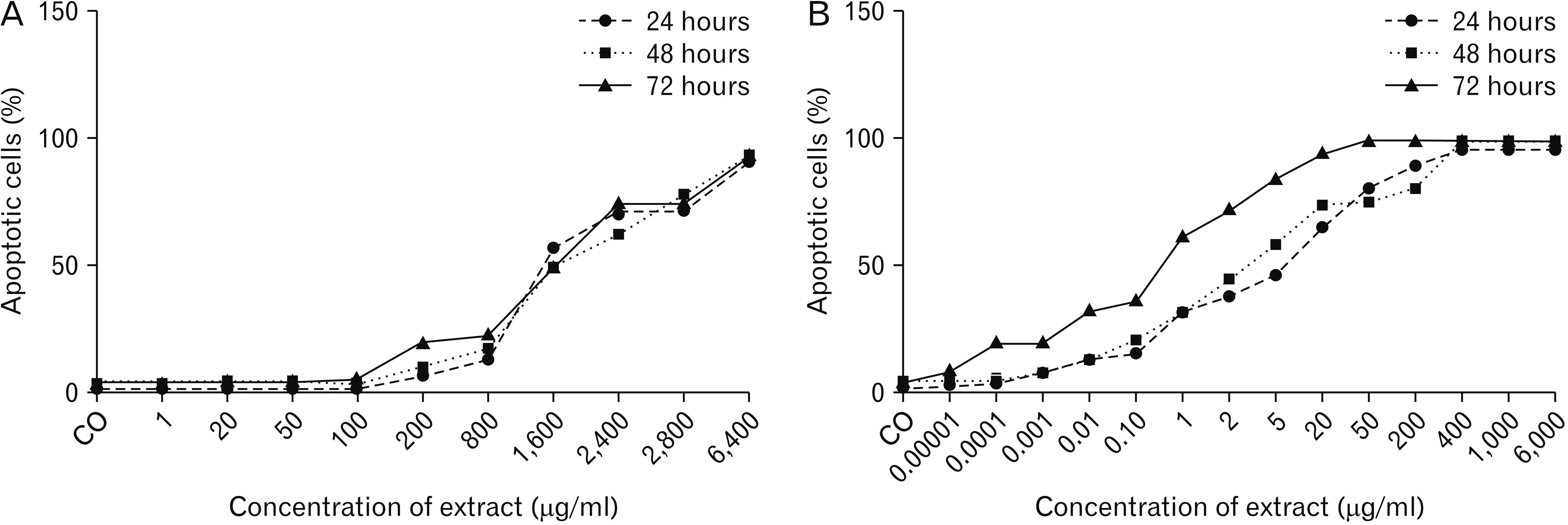
- Papillary thyroid carcinoma (PTC) is one of the most common cancers of the endocrine system. Previous studies have shown that the extract of hull-less pumpkin seed (HLPS) has a significant anti-cancer effect. The aim of this study was to evaluate the effect of this plant extract on the proliferation of PTC cells. In this study, an extract of this plant was prepared by soxhlet extraction method and analyzed by Gas Chromatography-Mass Spectrometry. The cytotoxicity of PTX and plant extract was investigated using the methylthiazol tetrazolium (MTT) method. For careful investigation of morphological alteration, we used hematoxylin and eosin and Giemsa stinging. Based on MTT assay test, the IC 50 value of paclitaxel (PTX) was significantly less than the hydro-alcoholic extract of HLPS at all of the incubation time. Our results of histological staining showed that HLPS and PTX induced significant morphological alteration in the PTC cultured cell that consistent with cell death. Comparing the groups treated by PTX or HLPS with control group showed significant differences. It seems that HLPS extract has an apparent effect on treatment of PTC, at least in laboratory condition, albeit for realistic decision about the effect of HLPS on PTC, more molecular investigations are necessary.
Figure
Reference
-
References
1. Halenka M, Fryšák Z. Halenka M, Fryšák Z, editors. 2017. Papillary thyroid carcinoma. Atlas of Thyroid Ultrasonography. Springer;Cham: p. 165–245. DOI: 10.1007/978-3-319-53759-7_15.
Article2. Valderrabano P, Simons S, Montilla-Soler J, Pal T, Zota V, Otto K, McIver B, Coppola D, Leon ME. Nasir A, Coppola D, editors. 2016. Medullary thyroid carcinoma. Neuroendocrine Tumors: Review of Pathology, Molecular and Therapeutic Advances. Springer;New York: p. 117–40. DOI: 10.1007/978-1-4939-3426-3_7.
Article3. Ganly I, Ibrahimpasic T, Rivera M, Nixon I, Palmer F, Patel SG, Tuttle RM, Shah JP, Ghossein R. 2014; Prognostic implications of papillary thyroid carcinoma with tall-cell features. Thyroid. 24:662–70. DOI: 10.1089/thy.2013.0503. PMID: 24262069.
Article4. Yardley DA. 2013; nab-Paclitaxel mechanisms of action and delivery. J Control Release. 170:365–72. DOI: 10.1016/j.jconrel.2013.05.041. PMID: 23770008.
Article5. Apaya MK, Chang MT, Shyur LF. 2016; Phytomedicine polypharmacology: cancer therapy through modulating the tumor microenvironment and oxylipin dynamics. Pharmacol Ther. 162:58–68. DOI: 10.1016/j.pharmthera.2016.03.001. PMID: 26969215.
Article6. Zubair H, Azim S, Ahmad A, Khan MA, Patel GK, Singh S, Singh AP. 2017; Cancer chemoprevention by phytochemicals: nature's healing touch. Molecules. 22:395. DOI: 10.3390/molecules22030395. PMID: 28273819. PMCID: PMC6155418.
Article7. Caili F, Huan S, Quanhong L. 2006; A review on pharmacological activities and utilization technologies of pumpkin. Plant Foods Hum Nutr. 61:73–80. DOI: 10.1007/s11130-006-0016-6. PMID: 16758316.
Article8. Yadav M, Jain S, Tomar R, Prasad GB, Yadav H. 2010; Medicinal and biological potential of pumpkin: an updated review. Nutr Res Rev. 23:184–90. DOI: 10.1017/S0954422410000107. PMID: 21110905.
Article9. Xie JM. 2004; Induced polarization effect of pumpkin protein on B16 cell. Fujian Med Univ Acta. 38:394–5.10. Cheong NE, Choi YO, Kim WY, Bae IS, Cho MJ, Hwang I, Kim JW, Lee SY. 1997; Purification and characterization of an antifungal PR-5 protein from pumpkin leaves. Mol Cells. 7:214–9. PMID: 9163735.11. Xanthopoulou MN, Nomikos T, Fragopoulou E, Antonopoulou S. 2009; Antioxidant and lipoxygenase inhibitory activities of pumpkin seed extracts. Food Res Int. 42:641–6. DOI: 10.1016/j.foodres.2009.02.003.
Article12. Jian L, Du CJ, Lee AH, Binns CW. 2005; Do dietary lycopene and other carotenoids protect against prostate cancer? Int J Cancer. 113:1010–4. DOI: 10.1002/ijc.20667. PMID: 15514967.
Article13. Pan HZ, Qiu XH, Li H, Jin J, Yu C, Zhao J. 2005; Effect of pumpkin extracts on tumor growth inhibition in S180-bearing mice. Pract Prev Med. 12:745–7.14. Richter D, Abarzua S, Chrobak M, Vrekoussis T, Weissenbacher T, Kuhn C, Schulze S, Kupka MS, Friese K, Briese V, Piechulla B, Makrigiannakis A, Jeschke U, Dian D. 2013; Effects of phytoestrogen extracts isolated from pumpkin seeds on estradiol production and ER/PR expression in breast cancer and trophoblast tumor cells. Nutr Cancer. 65:739–45. DOI: 10.1080/01635581.2013.797000. PMID: 23859042.
Article15. Medjakovic S, Hobiger S, Ardjomand-Woelkart K, Bucar F, Jungbauer A. 2016; Pumpkin seed extract: cell growth inhibition of hyperplastic and cancer cells, independent of steroid hormone receptors. Fitoterapia. 110:150–6. DOI: 10.1016/j.fitote.2016.03.010. PMID: 26976217.
Article16. Ghosh D, Biswas PK. 2016; Enzyme-aided extraction of carotenoids from pumpkin tissues. Indian Chem Eng. 58:1–11. DOI: 10.1080/00194506.2015.1046697.
Article17. Kurfurstova D, Bartkova J, Vrtel R, Mickova A, Burdova A, Majera D, Mistrik M, Kral M, Santer FR, Bouchal J, Bartek J. 2016; DNA damage signalling barrier, oxidative stress and treatment-relevant DNA repair factor alterations during progression of human prostate cancer. Mol Oncol. 10:879–94. DOI: 10.1016/j.molonc.2016.02.005. PMID: 26987799. PMCID: PMC5423169.
Article18. Poprac P, Jomova K, Simunkova M, Kollar V, Rhodes CJ, Valko M. 2017; Targeting free radicals in oxidative stress-related human diseases. Trends Pharmacol Sci. 38:592–607. DOI: 10.1016/j.tips.2017.04.005. PMID: 28551354.
Article19. Hecht F, Pessoa CF, Gentile LB, Rosenthal D, Carvalho DP, Fortunato RS. 2016; The role of oxidative stress on breast cancer development and therapy. Tumour Biol. 37:4281–91. DOI: 10.1007/s13277-016-4873-9. PMID: 26815507.
Article20. Gill JG, Piskounova E, Morrison SJ. 2016; Cancer, oxidative stress, and metastasis. Cold Spring Harb Symp Quant Biol. 81:163–75. DOI: 10.1101/sqb.2016.81.030791. PMID: 28082378.
Article21. Praveen KJ, Deepa M, Julius A, Nadiger HA. 2013; Study on thyroid status and oxidants in smokers and alcoholics. J Evol Med Dent Sci. 2:6982–7. DOI: 10.14260/jemds/1243.22. Asci A, Bulus D, Andiran N, Kocer-Gumusel B. Evaluation of the relation between thyroid dysfunction and oxidant/antioxidant status in obese children. Paper presented at: ESPE 2014. 2014 Sep 18-20; Dublin, Ireland. p. 82.23. Wang D, Feng JF, Zeng P, Yang YH, Luo J, Yang YW. 2011; Total oxidant/antioxidant status in sera of patients with thyroid cancers. Endocr Relat Cancer. 18:773–82. DOI: 10.1530/ERC-11-0230. PMID: 22002574. PMCID: PMC3230112.
Article24. Muzza M, Colombo C, Cirello V, Perrino M, Vicentini L, Fugazzola L. 2016; Oxidative stress and the subcellular localization of the telomerase reverse transcriptase (TERT) in papillary thyroid cancer. Mol Cell Endocrinol. 431:54–61. DOI: 10.1016/j.mce.2016.05.005. PMID: 27164443.
Article25. Tabur S, Aksoy ŞN, Korkmaz H, Ozkaya M, Aksoy N, Akarsu E. 2015; Investigation of the role of 8-OHdG and oxidative stress in papillary thyroid carcinoma. Tumour Biol. 36:2667–74. DOI: 10.1007/s13277-014-2889-6. PMID: 25434875.
Article26. Li-Weber M. 2013; Targeting apoptosis pathways in cancer by Chinese medicine. Cancer Lett. 332:304–12. DOI: 10.1016/j.canlet.2010.07.015. PMID: 20685036.
Article27. Saeedi Borujeni MJ, Hami J, Haghir H, Rastin M, Sazegar G. 2016; Evaluation of Bax and Bcl-2 proteins expression in the rat hippocampus due to childhood febrile seizure. Iran J Child Neurol. 10:53–60. PMID: 27057189. PMCID: PMC4815488.28. Shen W, Guan Y, Wang J, Hu Y, Tan Q, Song X, Jin Y, Liu Y, Zhang Y. 2016; A polysaccharide from pumpkin induces apoptosis of HepG2 cells by activation of mitochondrial pathway. Tumour Biol. 37:5239–45. DOI: 10.1007/s13277-015-4338-6. PMID: 26555544.
Article29. Bento AP, Gaulton A, Hersey A, Bellis LJ, Chambers J, Davies M, Krüger FA, Light Y, Mak L, McGlinchey S, Nowotka M, Papadatos G, Santos R, Overington JP. 2014; The ChEMBL bioactivity database: an update. Nucleic Acids Res. 42(Database issue):D1083–90. DOI: 10.1093/nar/gkt1031. PMID: 24214965. PMCID: PMC3965067.
Article30. Volpe DA, Hamed SS, Zhang LK. 2014; Use of different parameters and equations for calculation of IC50 values in efflux assays: potential sources of variability in IC50 determination. AAPS J. 16:172–80. DOI: 10.1208/s12248-013-9554-7. PMID: 24338112. PMCID: PMC3889528.31. Benbow SJ, Wozniak KM, Kulesh B, Savage A, Slusher BS, Littlefield BA, Jordan MA, Wilson L, Feinstein SC. 2017; Microtubule-targeting agents eribulin and paclitaxel differentially affect neuronal cell bodies in chemotherapy-induced peripheral neuropathy. Neurotox Res. 32:151–62. DOI: 10.1007/s12640-017-9729-6. PMID: 28391556. PMCID: PMC5474151.
Article32. Rouzier R, Rajan R, Wagner P, Hess KR, Gold DL, Stec J, Ayers M, Ross JS, Zhang P, Buchholz TA, Kuerer H, Green M, Arun B, Hortobagyi GN, Symmans WF, Pusztai L. 2005; Microtubule-associated protein tau: a marker of paclitaxel sensitivity in breast cancer. Proc Natl Acad Sci U S A. 102:8315–20. DOI: 10.1073/pnas.0408974102. PMID: 15914550. PMCID: PMC1149405.
Article
- Full Text Links
- Actions
-
Cited
- CITED
-
- Close
- Share
- Similar articles
-
- Concurrent Medullay and Papillary Carcinoma of the Thyroid
- Anti-tumor Activity of Saussurea laniceps against Pancreas Adenocarcinoma
- Mycelial Extract of Phellinus linteus Induces Cell Death in A549 Lung Cancer Cells and Elevation of Nitric Oxide in Raw 264.7 Macrophage Cells
- A Case of Mixed Follicular-Papillary Thyroid Carcinoma
- Immunohistochemical Study on the Proliferative Activity of Human Thyroid Tumors


