Long Non-coding RNA CASC15 Promotes Intrahepatic Cholangiocarcinoma Possibly through Inducing PRDX2/PI3K/AKT Axis
- Affiliations
-
- 1Department of Hepatobiliary and Pancreatic Surgery, The Second Affiliated Hospital, Zhejiang University School of Medicine, Hangzhou, China
- 2Key Laboratory of Precision Diagnosis and Treatment for Hepatobiliary and Pancreatic Tumor of Zhejiang Province, Hangzhou, China
- 3Research Center of Diagnosis and Treatment Technology for Hepatocellular Carcinoma of Zhejiang Province, Hangzhou, China
- 4Clinical Medicine Innovation Center of Precision Diagnosis and Treatment for Hepatobiliary and Pancreatic Disease of Zhejiang University, Hangzhou, China
- 5Clinical Research Center of Hepatobiliary and Pancreatic Diseases of Zhejiang Province, Hangzhou, China
- KMID: 2510660
- DOI: http://doi.org/10.4143/crt.2020.192
Abstract
- Purpose
Intrahepatic cholangiocarcinoma (ICC) is one of the most common liver primary tumors but its treatments are limited. Bioinformatics showed that the expression level of long non-coding RNA cancer-associated susceptibility 15 gene (CASC15) is correlated with ICC progression, but its functional mechanism remains unclear.
Materials and Methods
Tissues from ICC patients, tumor and adjacent tissue, were used for detection of the expression of CASC15. Clinical data were also collected for clinicopathologic and survival analysis. Short interfering RNA and lentiviral short hairpin RNA were used to knock down CASC15 and PRDX2 expression in ICC cell lines, for the analysis of changes of cell function and xenografts. RNA-pulldown and RNA immunoprecipitation assays were used to detect RNA-binding protein, PRDX2. Male nude mice were used for ICC xenografts, and livers were collected after 4 weeks for immunohistochemistry.
Results
CASC15 is highly expressed in ICC tissues and is related to higher TNM stage. Knockdown of CASC15 in ICC cells reduced cell proliferation, migration, invasiveness and increased apoptosis, and G1/S block. PRDX2 bound to CASC15. Knockdown of CASC15 decreased PRDX2 expression which was rescued by the inhibition of proteasome formation. Downregulation of PRDX2 resulted in G1/S block, reduced ICC cell invasion. Downregulation of CASC15 inhibited phosphoinositide 3-kinase (PI3K)/AKT/c-Myc pathway through downregulating of PRDX2 and overexpressed PRDX2 rescued the block. CASC15 knockout in ICC xenografts suppressed tumor development in vivo, decreased the expression of PRDX2 and Ki67 and inhibited PI3K/AKT pathway.
Conclusion
CASC15 promotes ICC possibly by targeting PRDX2 via the PI3K/AKT pathway, indicating poor prognosis and high degree of malignancy of ICC.
Figure
-

Fig. 1 Cancer-associated susceptibility 15 (CASC15) affected the prognosis of patients with intrahepatic cholangiocarcinoma (ICC). (A) Relative expression of long non-coding RNA (lncRNA)-CASC15 in tissues of 95 patients. (B) Survival curve analysis of the relationship between lncRNA-CANSC15 expression differences and survival time of patients. (C) Relative expression of ICC cell lines (CCLP, 9810, HuCCT1, and RBE). (D) The efficiency of knockdown of CASC15 in HuCCT1 and RBE cells. **p<0.01, ***p<0.001.
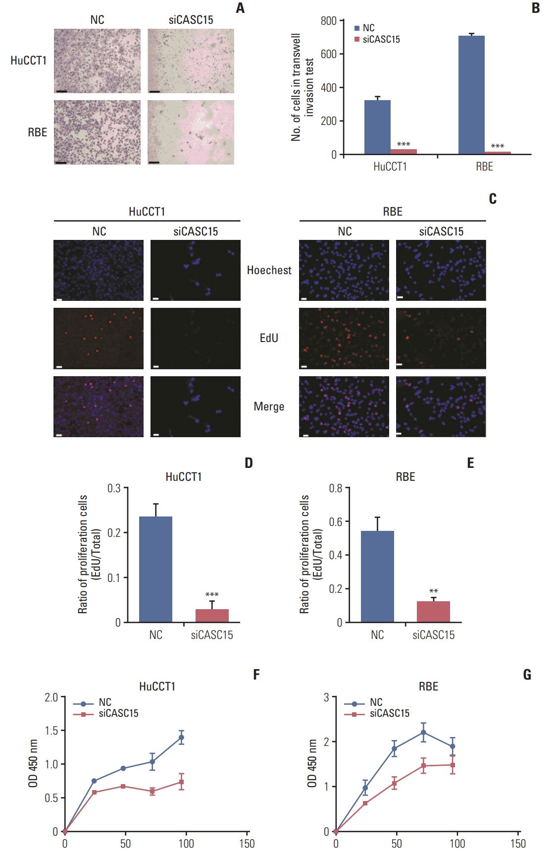
Fig. 2 The knockdown of cancer-associated susceptibility 15 (CASC15) expression inhibited cell invasion and proliferation. (A, B) Transwell invasion experiment and quantitative results. (C–E) EdU experiment showed that cell proliferation capacity was decreased in the siCASC15 group. Scale bars=100 μm. (F, G) Cell Counting Kit-8 (CCK-8) assay showed that the cell proliferation capacity decreased in the siCASC15 group. NC, negative control. **p<0.01, ***p<0.001.
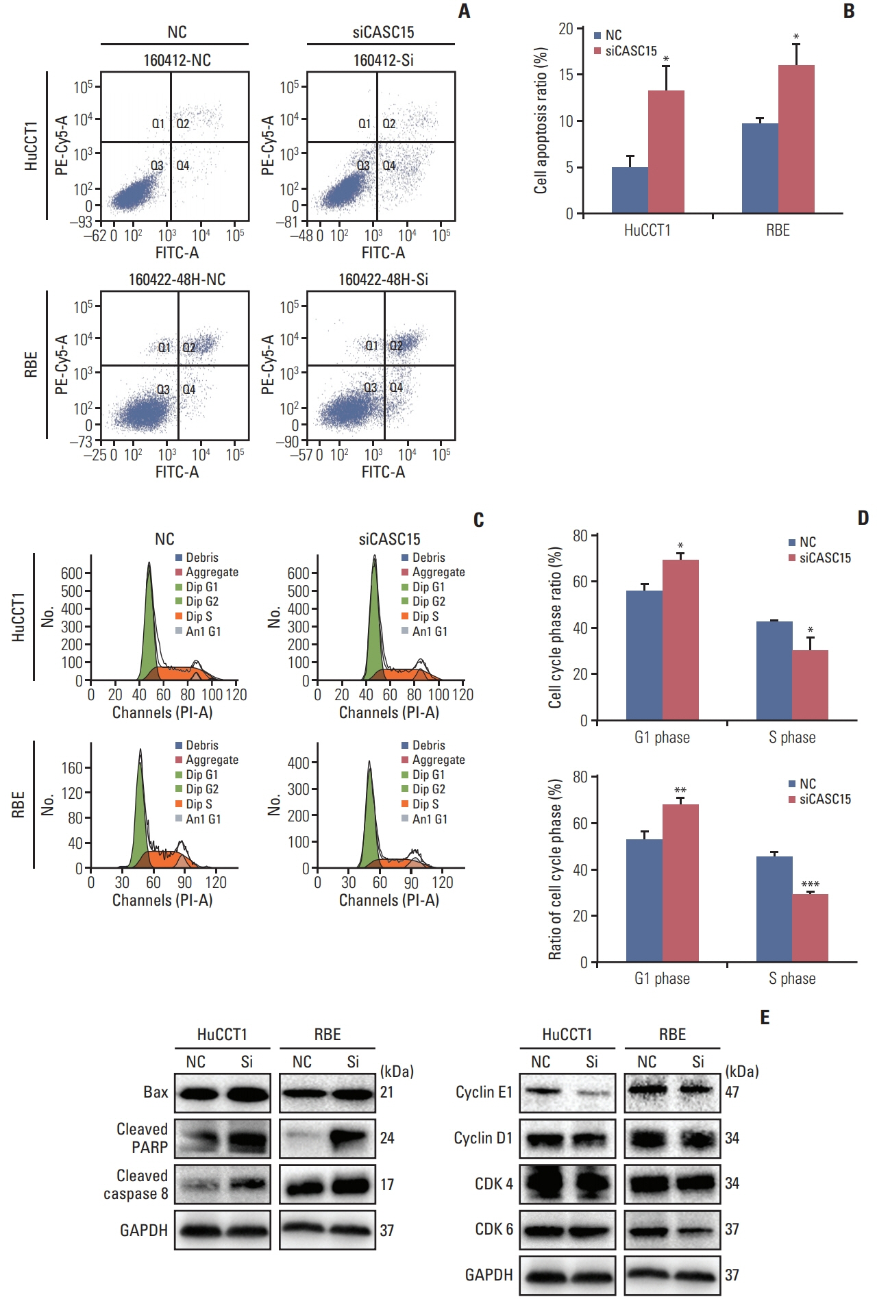
Fig. 3 The knockdown of cancer-associated susceptibility 15 (CASC15) expression promoted cell apoptosis and inhibited G1/S phase transformation. (A, B) Flow cytometry diagram of cell apoptosis and quantitative results. (C, D) Flow cytometry diagram of cell cycle and quantitative results. (E) Western blotting revealed that the expression of apoptosis and cell cycle-related proteins were changed in the siCASC15 group. GAPDH, glyceraldehyde 3-phosphate dehydrogenase; NC, negative control; PARP, poly(ADP-ribose) polymerase. *p<0.05, **p<0.01, ***p<0.001.
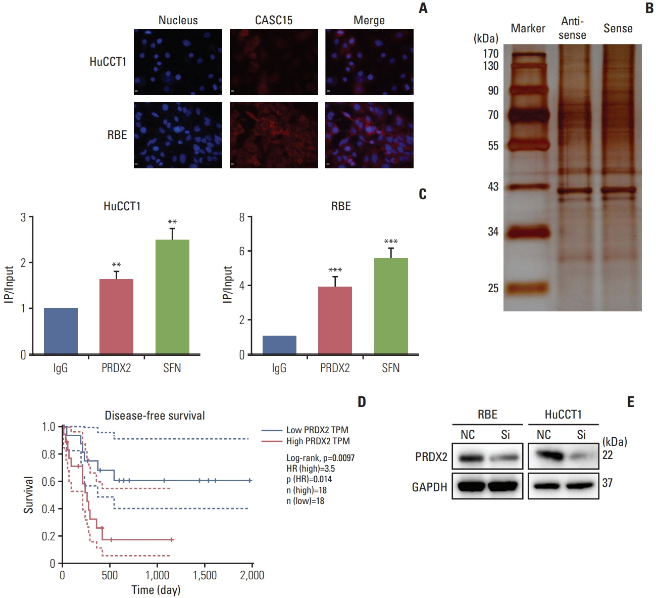
Fig. 4 Cancer-associated susceptibility 15 (CASC15) was associated with peroxiredoxin 2 (PRDX2) and regulated its expression. (A) Location of CASC15 in intrahepatic cholangiocarcinoma (ICC) cells. (B) RNA-protein complexes were resolved with sodium dodecyl sulfate polyacrylamide gel electrophoresis and visualized by silver staining, showing bands at 25–170 kDa. (C) RNA immunoprecipitation (IP)–quantitative polymerase chain reaction showed PRDX2 bound to CASC15. (D) Relationship of PRDX2 and disease-free survival of patients with ICC in The Cancer Genome Atlas database database. (E) Western blotting revealed that PRDX2 expression was decreased in the siCASC15 group. GAPDH, glyceraldehyde 3-phosphate dehydrogenase; NC, negative control. Scale bars=200 μm. **p<0.01, ***p<0.001.

Fig. 5 The knockdown of peroxiredoxin 2 (PRDX2) expression inhibited the invasion and G1/S phase transformation of intrahepatic cholangiocarcinoma (ICC) cells. (A) Efficiency of PRDX2 knockdown in RBE cells. (B) Flow cytometry diagram of cell cycle and quantitative results. (C) Transwell invasion experiment and quantitative results of RBE cells. Scale bars=100 μm. (D) Western blotting showed that the expression levels of cell cycle and endothelial mesenchymal transition (EMT)–related proteins were altered in the siPRDX2 group of RBE cells. (E) Western blotting showed that the phosphoinositide 3-kinase (PI3K)/AKT/c-Myc pathway was inhibited in the siPRDX2 group of RBE cells. GAPDH, glyceraldehyde 3-phosphate dehydrogenase; NC, negative control. *p<0.05 and **p<0.01.

Fig. 6 Cancer-associated susceptibility 15 (CASC15) affected phosphoinositide 3-kinase (PI3K)/AKT/c-Myc pathway through peroxiredoxin 2 (PRDX2) and CASC15 regulated PRDX2 by inhibiting ubiquitination. (A) Western blotting showed that the PI3K/AKT/c-Myc pathway was inhibited in the siCASC15 group. (B) Western blotting showed that the PI3K/AKT/c-Myc pathway was enhanced when PRDX2 was overexpressed in the siCASC15 group. (C, D) Expression of PRDX2 was increased and the PI3K/AKT pathway was activated when proteasome formation was inhibited (left panel). Relative expression of PRDX2 changed little in the siCASC15 group (right panel). (E, F) Flow cytometry diagram of cell apoptosis and quantitative results. 5-FU, 5-fluorouracil; GAPDH, glyceraldehyde 3-phosphate dehydrogenase; NC, negative control. **p<0.01.
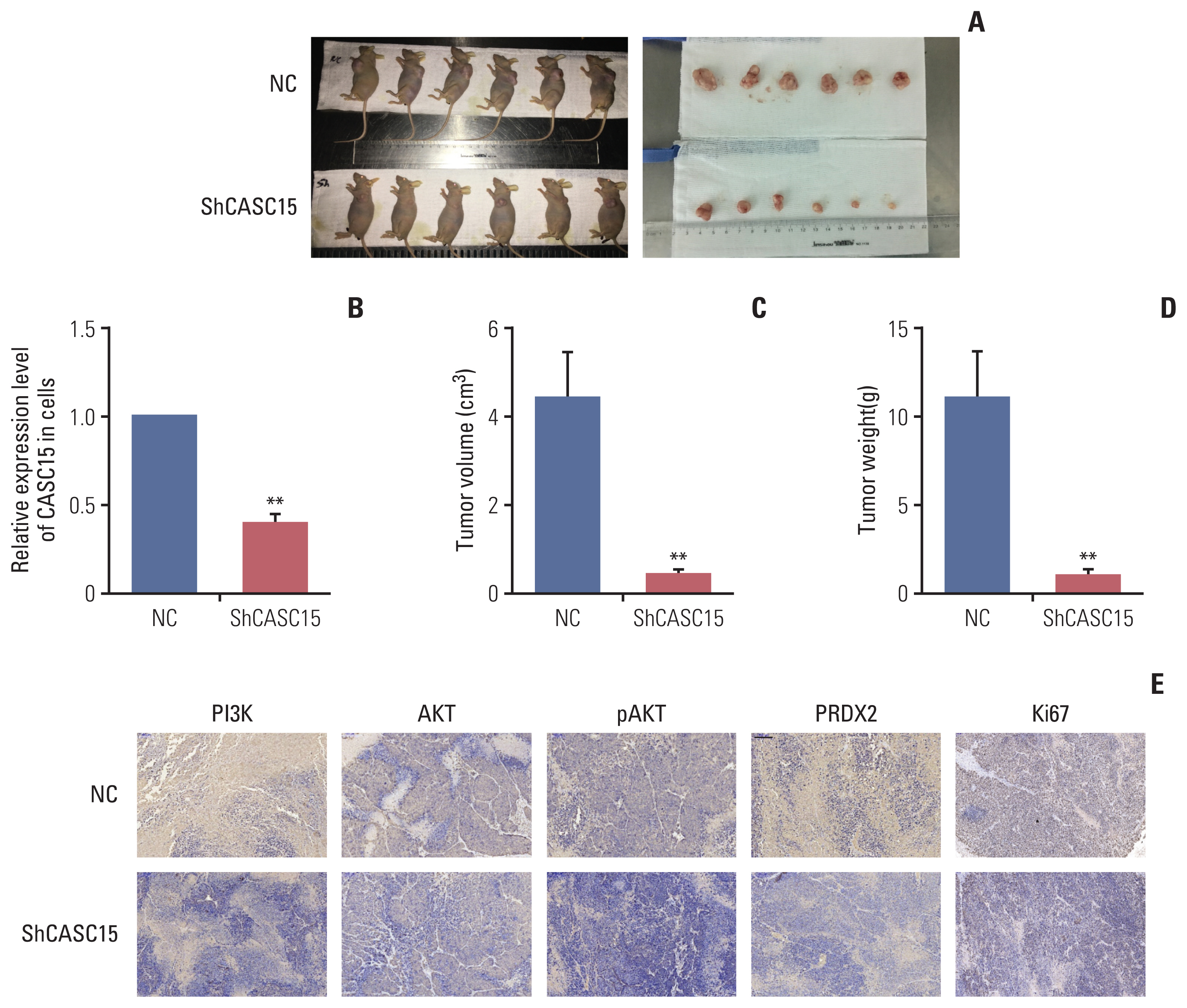
Fig. 7 The knockdown of cancer-associated susceptibility 15 (CASC15) in xenograft model inhibited the development of intrahepatic cholangiocarcinoma (ICC). (A–D) Knockdown of CASC15 in xenograft model decreased tumor size and weight. (E) Immunohistochemistry assay showed peroxiredoxin 2 (PRDX2), phosphoinositide 3-kinase (PI3K), AKT, pAKT, and Ki67 expression levels were decreased in the ShCASC15 group compared to in the control group. NC, negative control. Scale bar=100 μm. **p<0.01.

Fig. 8 Knockdown of cancer-associated susceptibility 15 (CASC-15) inhibits the phosphoinositide 3-kinase (PI3K)/AKT/c-Myc signal pathway possibly by binding to peroxiredoxin 2 (PRDX2) and promoting its degradation, resulting in the decrease of intrahepatic cholangiocarcinoma (ICC) development.
Reference
-
References
1. Mao K, Jiang W, Liu J, Wang J. Incidence of subsequent cholangiocarcinomas after another malignancy: trends in a population-based study. Medicine (Baltimore). 2015; 94:e596.2. Blechacz B, Gores GJ. Cholangiocarcinoma: advances in pathogenesis, diagnosis, and treatment. Hepatology. 2008; 48:308–21.
Article3. Buettner S, van Vugt JL, IJzermans JM, Groot Koerkamp B. Intrahepatic cholangiocarcinoma: current perspectives. Onco Targets Ther. 2017; 10:1131–42.
Article4. Anderson CD, Pinson CW, Berlin J, Chari RS. Diagnosis and treatment of cholangiocarcinoma. Oncologist. 2004; 9:43–57.
Article5. Manolio TA, Collins FS, Cox NJ, Goldstein DB, Hindorff LA, Hunter DJ, et al. Finding the missing heritability of complex diseases. Nature. 2009; 461:747–53.
Article6. Bertone P, Stolc V, Royce TE, Rozowsky JS, Urban AE, Zhu X, et al. Global identification of human transcribed sequences with genome tiling arrays. Science. 2004; 306:2242–6.
Article7. Zhang A, Zhao JC, Kim J, Fong KW, Yang YA, Chakravarti D, et al. LncRNA HOTAIR enhances the androgen-receptor-mediated transcriptional program and drives castration-resistant prostate cancer. Cell Rep. 2015; 13:209–21.
Article8. Gao S, Wang P, Hua Y, Xi H, Meng Z, Liu T, et al. ROR functions as a ceRNA to regulate Nanog expression by sponging miR-145 and predicts poor prognosis in pancreatic cancer. Oncotarget. 2016; 7:1608–18.
Article9. He T, Zhang L, Kong Y, Huang Y, Zhang Y, Zhou D, et al. Long non-coding RNA CASC15 is upregulated in hepatocellular carcinoma and facilitates hepatocarcinogenesis. Int J Oncol. 2017; 51:1722–30.
Article10. Merdrignac A, Angenard G, Allain C, Petitjean K, Bergeat D, Bellaud P, et al. A novel transforming growth factor beta-induced long noncoding RNA promotes an inflammatory microenvironment in human intrahepatic cholangiocarcinoma. Hepatol Commun. 2018; 2:254–69.
Article11. Jiang F, Ling X. The Advancement of long non-coding RNAs in cholangiocarcinoma development. J Cancer. 2019; 10:2407–14.
Article12. Bertoldi M. Human peroxiredoxins 1 and 2 and their interacting protein partners: through structure toward functions of biological complexes. Protein Pept Lett. 2016; 23:69–77.
Article13. Castaldo SA, Ajime T, Serrao G, Anastacio F, Rosa JT, Giacomantonio CA, et al. Annexin A2 regulates AKT upon H2O2-dependent signaling activation in cancer cells. Cancers (Basel). 2019; 11:492.
Article14. Jin Y, Yang Q, Liang L, Ding L, Liang Y, Zhang D, et al. Compound kushen injection suppresses human acute myeloid leukaemia by regulating the Prdxs/ROS/Trx1 signalling pathway. J Exp Clin Cancer Res. 2018; 37:277.
Article15. Zhang Y, Sun C, Xiao G, Shan H, Tang L, Yi Y, et al. S-nitrosylation of the peroxiredoxin-2 promotes S-nitrosoglutathione-mediated lung cancer cells apoptosis via AMPK-SIRT1 pathway. Cell Death Dis. 2019; 10:329.
Article16. Luthra S, Chandran U, Diergaarde B, Becich M, Lee AV, Neumann CA. Expression of reactive species related genes is associated with patient survival in luminal B breast cancer. Free Radic Biol Med. 2018; 120:170–80.
Article17. Gu C, Luo J, Lu X, Tang Y, Ma Y, Yun Y, et al. REV7 confers radioresistance of esophagus squamous cell carcinoma by recruiting PRDX2. Cancer Sci. 2019; 110:962–72.
Article18. Xu J, Zhang S, Wang R, Wu X, Zeng L, Fu Z. Knockdown of PRDX2 sensitizes colon cancer cells to 5-FU by suppressing the PI3K/AKT signaling pathway. Biosci Rep. 2017; 37:BSR20160447.
Article19. Xu D, Yang F, Yuan JH, Zhang L, Bi HS, Zhou CC, et al. Long noncoding RNAs associated with liver regeneration 1 accelerates hepatocyte proliferation during liver regeneration by activating Wnt/beta-catenin signaling. Hepatology. 2013; 58:739–51.20. Togami K, Yamaguchi K, Chono S, Tada H. Evaluation of permeability alteration and epithelial-mesenchymal transition induced by transforming growth factor-beta1 in A549, NCI-H441, and Calu-3 cells: development of an in vitro model of respiratory epithelial cells in idiopathic pulmonary fibrosis. J Pharmacol Toxicol Methods. 2017; 86:19–27.21. Richards EJ, Zhang G, Li ZP, Permuth-Wey J, Challa S, Li Y, et al. Long non-coding RNAs (LncRNA) regulated by transforming growth factor (TGF) beta: lncRNA-hit-mediated TGFbeta-induced epithelial to mesenchymal transition in mammary epithelia. J Biol Chem. 2015; 290:6857–67.
- Full Text Links
-
- Actions
-
Cited
- CITED
-
- Close
- Share
-
- Similar articles
-
- MicroRNA-21 promotes epithelial-mesenchymal transition and migration of human bronchial epithelial cells by targeting poly (ADP-ribose) polymerase-1 and activating PI3K/AKT signaling
- MLL4 Regulates the Progression of Non–Small-Cell Lung Cancer by Regulating the PI3K/AKT/SOX2 Axis
- CDK1 promotes the phosphorylation of KIFC1 to regulate the tumorgenicity of endometrial carcinoma
- Arctigenin Increases Hemeoxygenase-1 Gene Expression by Modulating PI3K/AKT Signaling Pathway in Rat Primary Astrocytes
- Long Non-Coding RNA LINC00525 Promotes the Stemness and Chemoresistance of Colorectal Cancer by Targeting miR-507/ELK3 Axis
