Human Placenta-Derived ECM Supports Tri-Lineage Differentiation of Human Induced Pluripotent Stem Cells
- Affiliations
-
- 1Institute of Regenerative Medicine, LifeNet Health, Virginia Beach, VA, USA
- KMID: 2508917
- DOI: http://doi.org/10.15283/ijsc20074
Abstract
- Human pluripotent stem cells (hPSCs) hold great promise for future applications in drug discovery and cell therapies. hPSC culture protocols require specific substrates and medium supplements to support cell expansion and lineage specific differentiation. The animal origin of these substrates is a severe limitation when considering the translation of hPSC derivatives to the clinic and in vitro disease modeling. The present study evaluates the use of a human placenta-derived extracellular matrix (ECM) hydrogel, HuGentraⓇ , to support tri-lineage differentiation of human induced pluripotent stem cells (hiPSCs). Lineage-specific embryoid bodies (EBs) were plated onto three separate matrices, and differentiation efficiency was evaluated based on morphology, protein, and gene expression. HuGentra was found to support the differentiation of hiPSCs to all three germ layers: ectodermal, mesodermal, and endodermal lineages. hiPSCs differentiated into neurons, cardiomyocytes, and hepatocytes on HuGentra had similar morphology, protein, and gene expression compared to differentiation on Matrigel or other cell preferred matrices. HuGentra can be considered as a suitable human substrate for hiPSC differentiation.
Keyword
Figure
-

Fig. 1 Characterization of hiPSC lines and differentiation schematic. (a) Three hiPSC lines derived from adult dermal fibroblasts, neonatal foreskin fibroblasts, and adult human osteoblasts display the typical hiPSC morphology and stain for Tra-1-81. Representative images were taken 10×. (b) Each stage of differentiation is displayed with the respective media supplements used to induce that stage of the differentiation as indicated by the numbering. Representative images are shown of the three lineages during each stage of the differentiations. Representative images were taken 4×, 10×, and 20×.
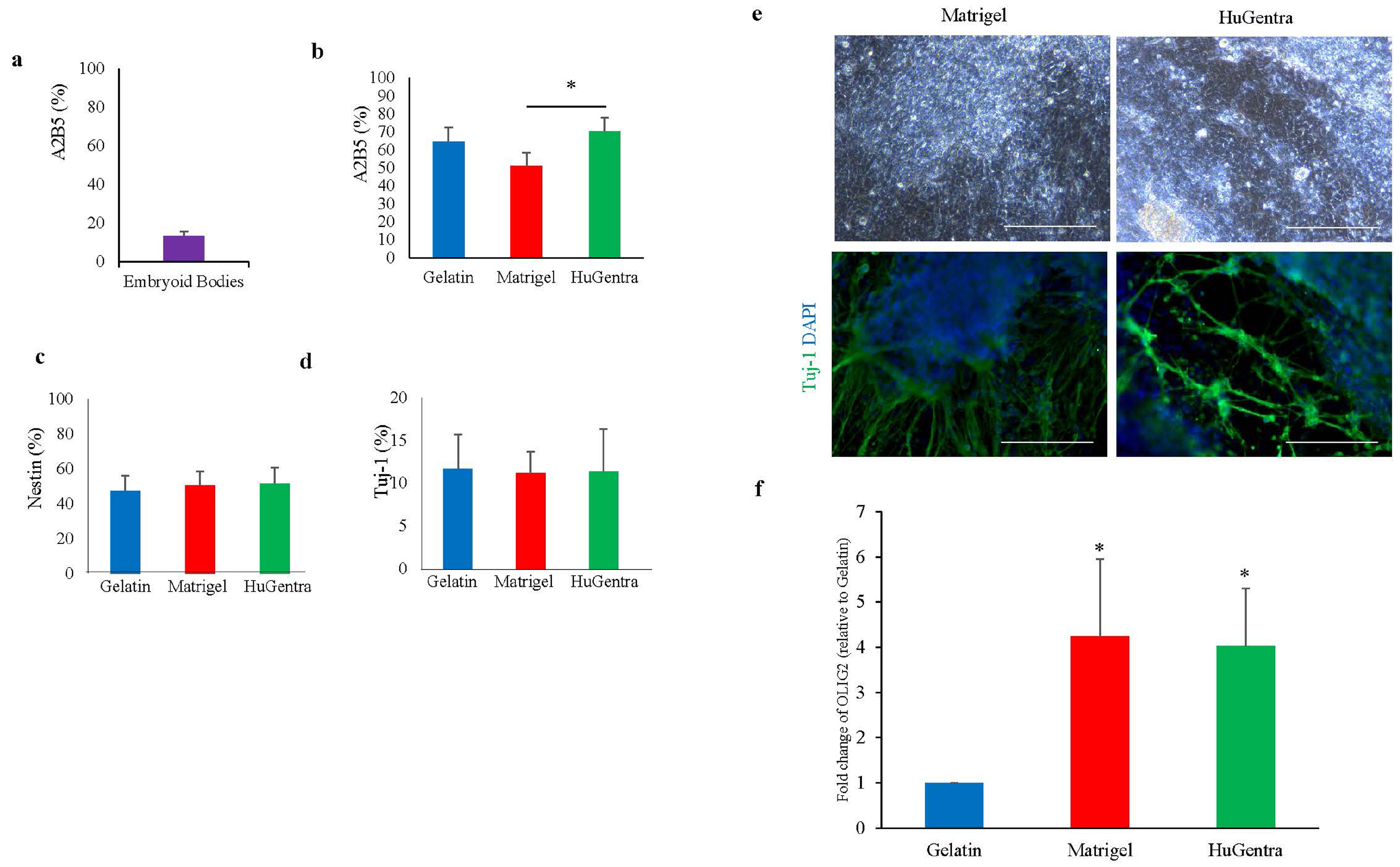
Fig. 2 Characterization of hiPSC-derived neurons on matrices. (a) The ectodermal lineage marker A2B5 was expressed at a frequency of 12%±2.3% by dissociated EBs. (b) A2B5 expression following differentiation on gelatin, Matrigel, or HuGentra. *p<0.05 to hiPSCs differentiated on HuGentra versus Matrigel. Flow analysis for neuronal markers. (c) NES and (d) Tuj-1. (e) ICC of Tuj-1 and representative morphological images. Representative images were taken 20×. (f) The gene expression of OLIG2 in Matrigel and HuGentra normalized to expression on gelatin. Error bars represent standard deviation. *p<0.05 to hiPSCs differentiated on gelatin. n≥3.
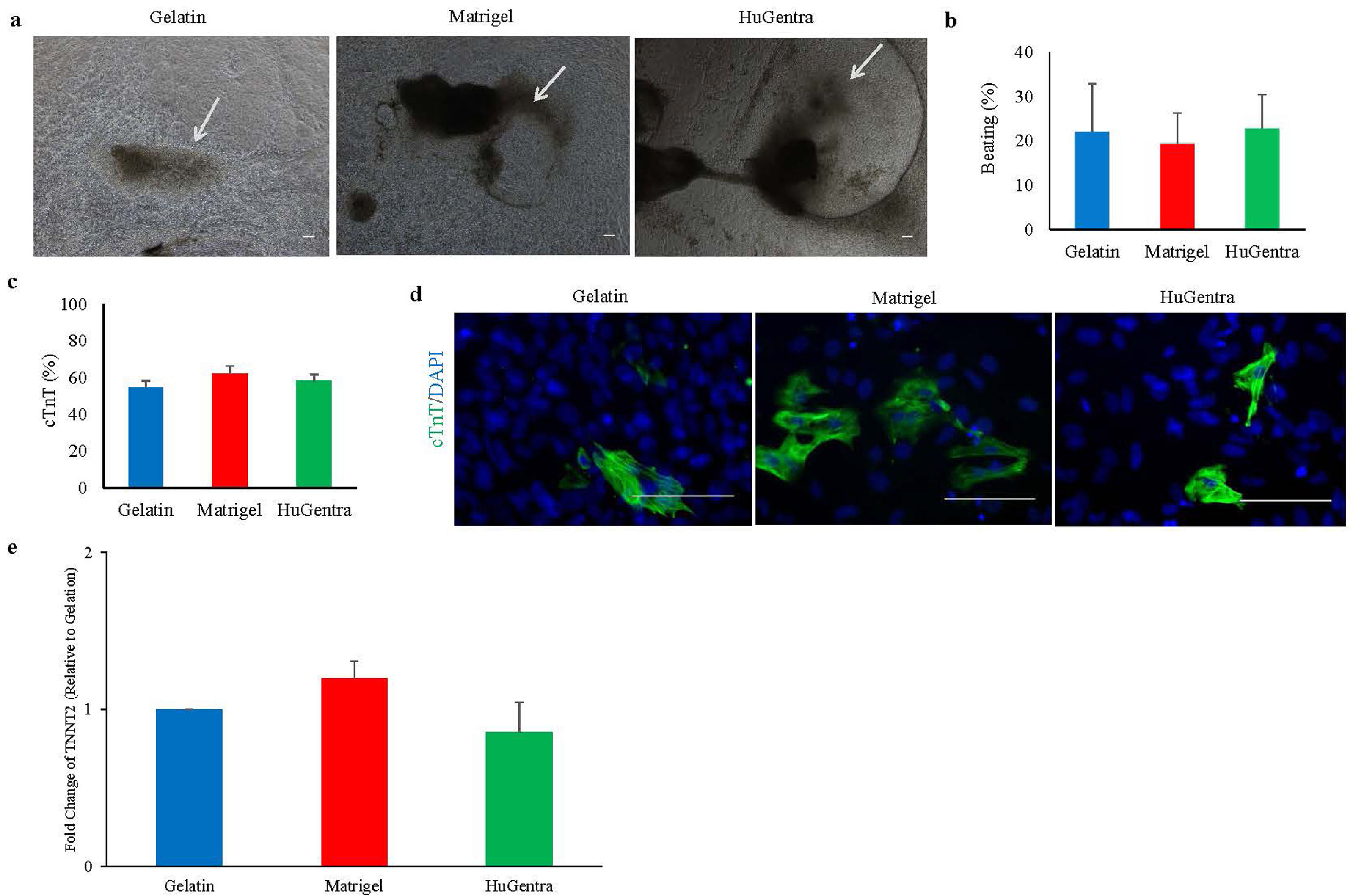
Fig. 3 Characterization of hiPSC-derived cardiomyocytes on matrices. (a) Following 14 days of differentiation, contracting colonies were found on all matrices as indicated by arrows. Representative images were taken 4×. (b) No significant differences in the frequency of the contracting colonies relative to the total attached colonies when quantitated on day 30. (c) After dissociation, the cells were assessed for expression cTnT via flow cytometry. The level of cTnT was similar between all three matrices. (d) cTnT expression was confirmed with ICC on the three matrices. Representative images were taken 20×. (e) Relative gene expression of the mature cardiac marker, TNNT2, was also shown and normalized to expression of gelatin. Error bars represent standard deviation. n≥3.
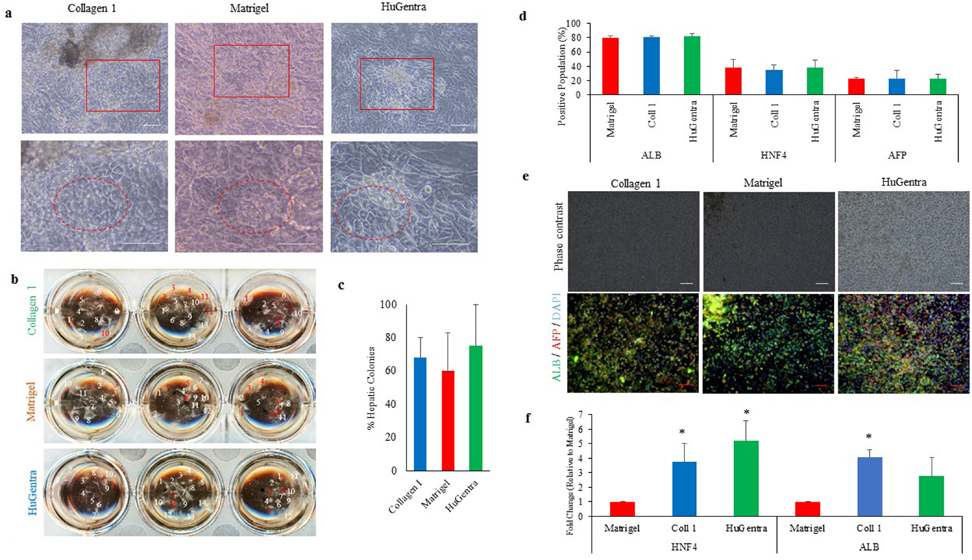
Fig. 4 Characterization of hiPSC-derived hepatocytes on matrices. (a) The frequency of the attached EBs and the resulting colonies were observed and recorded as containing the morphology of hepatic cells. Representative images were taken 10× (top row) and 20× (bottom row). (b) A representative plate with labeled hepatic colonies in white and non-hepatic colonies in red. (c) The percentage of EBs containing the characteristic morphology were calculated as a percentage of the total attached EBs. (d) Quantification of ALB, HNF4, and AFP on the dissociated cells via flow cytometry showed no significant differences in expression efficiencies across the three matrices. (e) ICC analysis for ALB and AFP was shown on differentiated hepatic like cells. Representative images were taken 10×. (f) Gene expression of HNF4 and ALB on Collagen I and HuGentra normalized to expression on Matrigel. Error bars represent standard deviation. *p<0.05 to hiPSCs differentiated on Matrigel. n≥3.
Reference
-
References
1. Kleinman HK, Martin GR. 2005; Matrigel: basement membrane matrix with biological activity. Semin Cancer Biol. 15:378–386. DOI: 10.1016/j.semcancer.2005.05.004. PMID: 15975825.
Article2. Del Álamo JC, Lemons D, Serrano R, Savchenko A, Cerignoli F, Bodmer R, Mercola M. 2016; High throughput physiological screening of iPSC-derived cardiomyocytes for drug development. Biochim Biophys Acta. 1863(7 Pt B):1717–1727. DOI: 10.1016/j.bbamcr.2016.03.003. PMID: 26952934. PMCID: PMC4885786.
Article3. Kondo Y, Iwao T, Nakamura K, Sasaki T, Takahashi S, Kamada N, Matsubara T, Gonzalez FJ, Akutsu H, Miyagawa Y, Okita H, Kiyokawa N, Toyoda M, Umezawa A, Nagata K, Matsunaga T, Ohmori S. 2014; An efficient method for differentiation of human induced pluripotent stem cells into hepatocyte-like cells retaining drug metabolizing activity. Drug Metab Pharmacokinet. 29:237–243. DOI: 10.2133/dmpk.DMPK-13-RG-104. PMID: 24334537. PMCID: PMC6594155.
Article4. Lee JB, Graham M, Collins TJ, Lee JH, Hong SH, Mcnicol AJ, Shapovalova Z, Bhatia M. 2015; Reversible lineage-specific priming of human embryonic stem cells can be exploited to optimize the yield of differentiated cells. Stem Cells. 33:1142–1152. DOI: 10.1002/stem.1952. PMID: 25639500. PMCID: PMC4413029.
Article5. Lian X, Hsiao C, Wilson G, Zhu K, Hazeltine LB, Azarin SM, Raval KK, Zhang J, Kamp TJ, Palecek SP. 2012; Robust cardiomyocyte differentiation from human pluripotent stem cells via temporal modulation of canonical Wnt signaling. Proc Natl Acad Sci U S A. 109:E1848–E1857. DOI: 10.1073/pnas.1200250109. PMID: 22645348. PMCID: PMC3390875.
Article6. Tannenbaum SE, Turetsky TT, Singer O, Aizenman E, Kirshberg S, Ilouz N, Gil Y, Berman-Zaken Y, Perlman TS, Geva N, Levy O, Arbell D, Simon A, Ben-Meir A, Shufaro Y, Laufer N, Reubinoff BE. 2012; Derivation of xeno-free and GMP-grade human embryonic stem cells--platforms for future clinical applications. PLoS One. 7:e35325. DOI: 10.1371/journal.pone.0035325. PMID: 22745653. PMCID: PMC3380026.7. Zhao C, Ikeya M. 2018; Generation and applications of induced pluripotent stem cell-derived mesenchymal stem cells. Stem Cells Int. 2018:9601623. DOI: 10.1155/2018/9601623. PMID: 30154868. PMCID: PMC6091255.
Article8. Hongisto H, Ilmarinen T, Vattulainen M, Mikhailova A, Skottman H. 2017; Xeno- and feeder-free differentiation of human pluripotent stem cells to two distinct ocular epithelial cell types using simple modifications of one method. Stem Cell Res Ther. 8:291. DOI: 10.1186/s13287-017-0738-4. PMID: 29284513. PMCID: PMC5747074.
Article9. Farzaneh Z, Pakzad M, Vosough M, Pournasr B, Baharvand H. 2014; Differentiation of human embryonic stem cells to hepatocyte-like cells on a new developed xeno-free extracellular matrix. Histochem Cell Biol. 142:217–226. DOI: 10.1007/s00418-014-1183-4. PMID: 24477550.
Article10. Ullah I, Busch JF, Rabien A, Ergün B, Stamm C, Knosalla C, Hippenstiel S, Reinke P, Kurtz A. 2020; Adult tissue extracellular matrix determines tissue specification of human iPSC-derived embryonic stage mesodermal precursor cells. Adv Sci (Weinh). 7:1901198. DOI: 10.1002/advs.201901198. PMID: 32154066. PMCID: PMC7055561.
Article11. Francis MP, Breathwaite E, Bulysheva AA, Varghese F, Rodriguez RU, Dutta S, Semenov I, Ogle R, Huber A, Tichy AM, Chen S, Zemlin C. 2017; Human placenta hydrogel reduces scarring in a rat model of cardiac ischemia and enhances cardiomyocyte and stem cell cultures. Acta Bio-mater. 52:92–104. DOI: 10.1016/j.actbio.2016.12.027. PMID: 27965171.
Article
- Full Text Links
-
- Actions
-
Cited
- CITED
-
- Close
- Share
-
- Similar articles
-
- Lymphoid Lineage γδ T Cells Were Successfully Generated from Human Pluripotent Stem Cells via Hemogenic Endothelium
- Do the Fibroblasts Contained in Early Passage MSC Population Adversely Affect the Characteristics of Stem Cell Population Obtained from Human Placenta?
- Neuronal Differentiation of a Human Induced Pluripotent Stem Cell Line (FS-1) Derived from Newborn Foreskin Fibroblasts
- Transcriptional Profiles of Imprinted Genes in Human Embryonic Stem Cells During In vitro Differentiation
- Difference in HLA-DR Expression of Human Umbilical Cord Blood Derived Mesenchymal Stem Cells after Tri-lineage Differentiation
