Upregulation of C-Reactive Protein by Placenta-Derived Mesenchymal Stem Cells Promotes Angiogenesis in A Rat Model with Cirrhotic Liver
- Affiliations
-
- 1Department of Biomedical Science, CHA University, Seongnam, Korea
- 2Non-Clinical Evaluation Center, CHA Advanced Research Institute, Seongnam, Korea
- 3Department of Internal Medicine, CHA Bundang Medical Center, CHA University, Seongnam, Korea
- 4Department of Internal Medicine, Catholic University Medical College, Seoul, Korea
- KMID: 2508914
- DOI: http://doi.org/10.15283/ijsc20052
Abstract
- Background and Objectives
Liver cirrhosis is accompanied by abnormal vascular shunts. The Wnt pathway is essential for endothelial cell survival and proliferation. C-reactive protein (CRP), which is produced by hepatocyte, activates angiogenesis in cardiovascular diseases.
Methods and Results
The expression of CRP in CCl 4 -injured rat livers was detected using qRT-PCR and Western blotting after transplantation of placenta-derived mesenchymal stem cells (PD-MSCs) into rats. To determine whether CRP functions in hepatic regeneration by promoting angiogenesis through the Wnt pathway, we detected VEGF and β-catenin in liver tissues and BrdU and β-catenin in hepatocytes by immunofluorescence. The expression levels of CRP, Wnt pathway-related and angiogenic factors were increased in CCl 4 -injured and PD-MSCs transplanted rat livers. In vitro, the expression levels of Wnt signaling and angiogenic factors were decreased in siRNA-CRP-transfected rat hepatocytes.
Conclusions
CRP upregulation by PD-MSCs participates in vascular remodeling to promote liver regeneration via the Wnt signaling pathway during hepatic failure.
Keyword
Figure
-
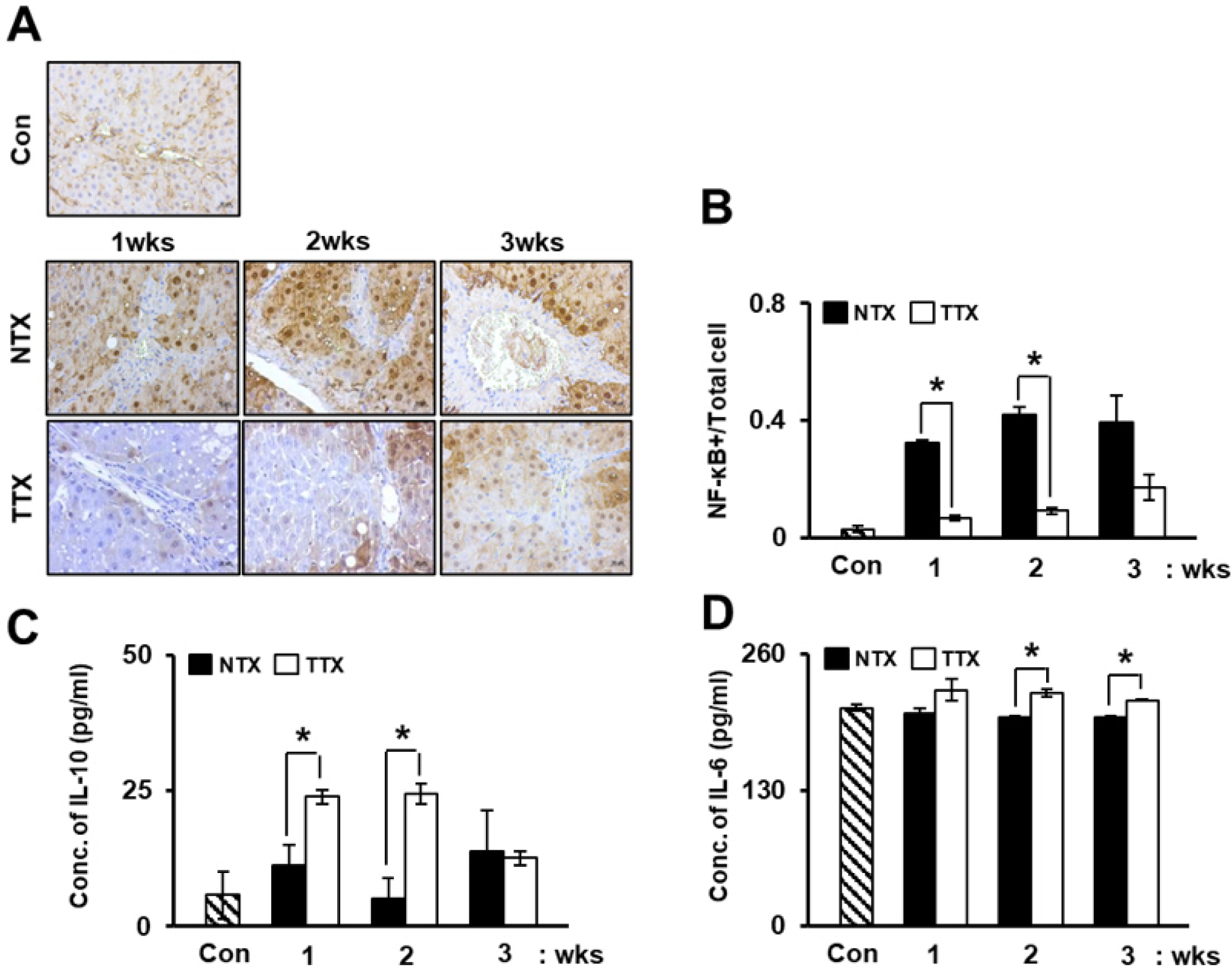
Fig. 1 PD-MSC transplantation attenuates inflammation in CCl4-injured rats. Expression of NF-κB was detected by immunohistochemistry (A). ×400, Scale bars; 20 μm. NF-κB-positive cells were quantified based on the total number of cells (B). Expression of IL-10 (C) and IL-6 (D) was analyzed in rat serum by ELISA. Data are represented as the triplicated mean±SD. p<0.05, *; NTX vs.; NTX and TTX refer to the non-transplanted group and the PD-MSC-transplanted group, respectively.
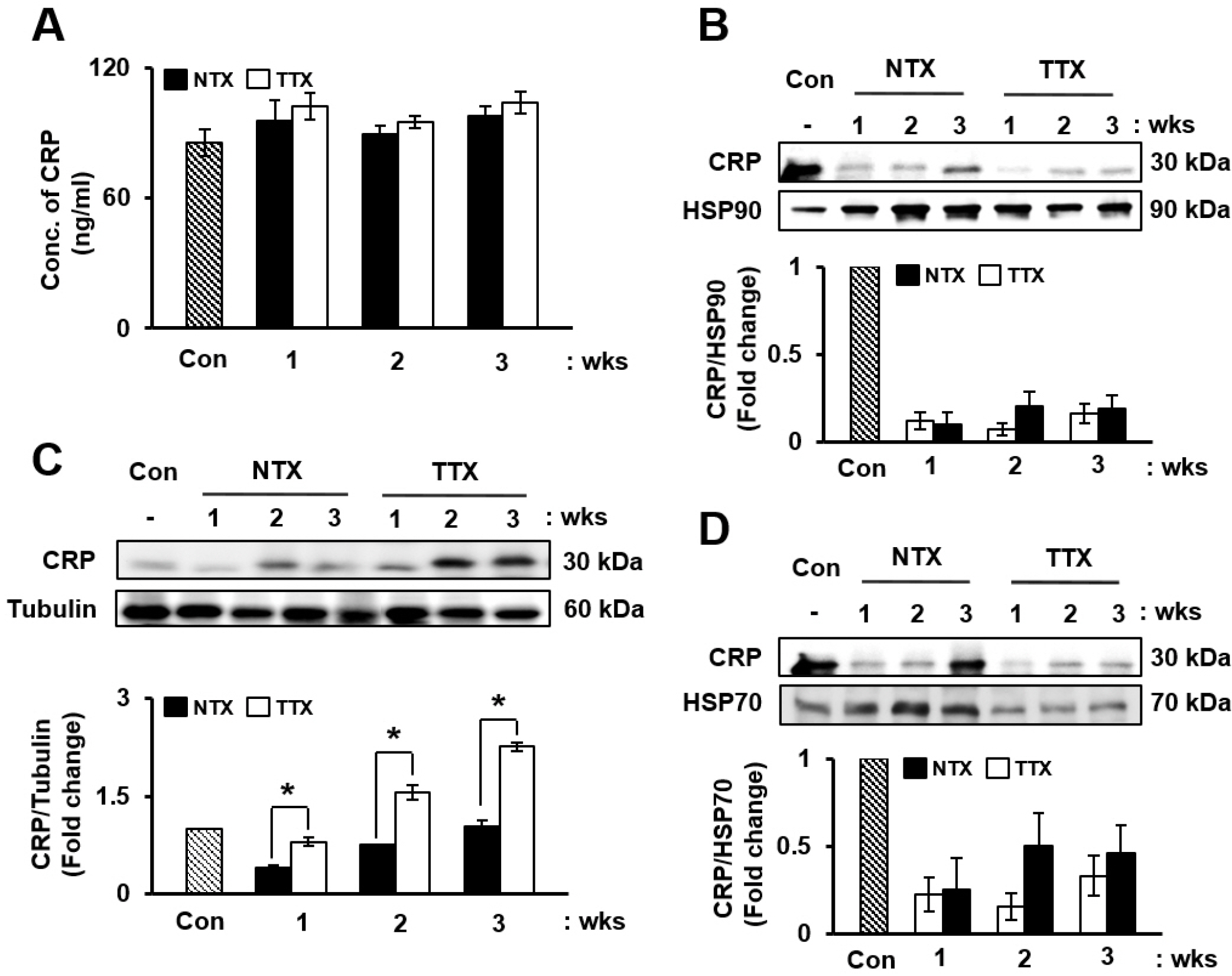
Fig. 2 PD-MSC transplantation improves the expression of CRP. Expre-ssion of CRP was analyzed by ELISA (A) and Western blotting from total proteins (C) and exosomes (B) and (D). Data are represented as the triplicated or duplicated mean±SD. SD; p<0.05, *; NTX vs.; NTX and TTX refer to the non-transplanted group and the PD-MSC-transplanted group, respectively.
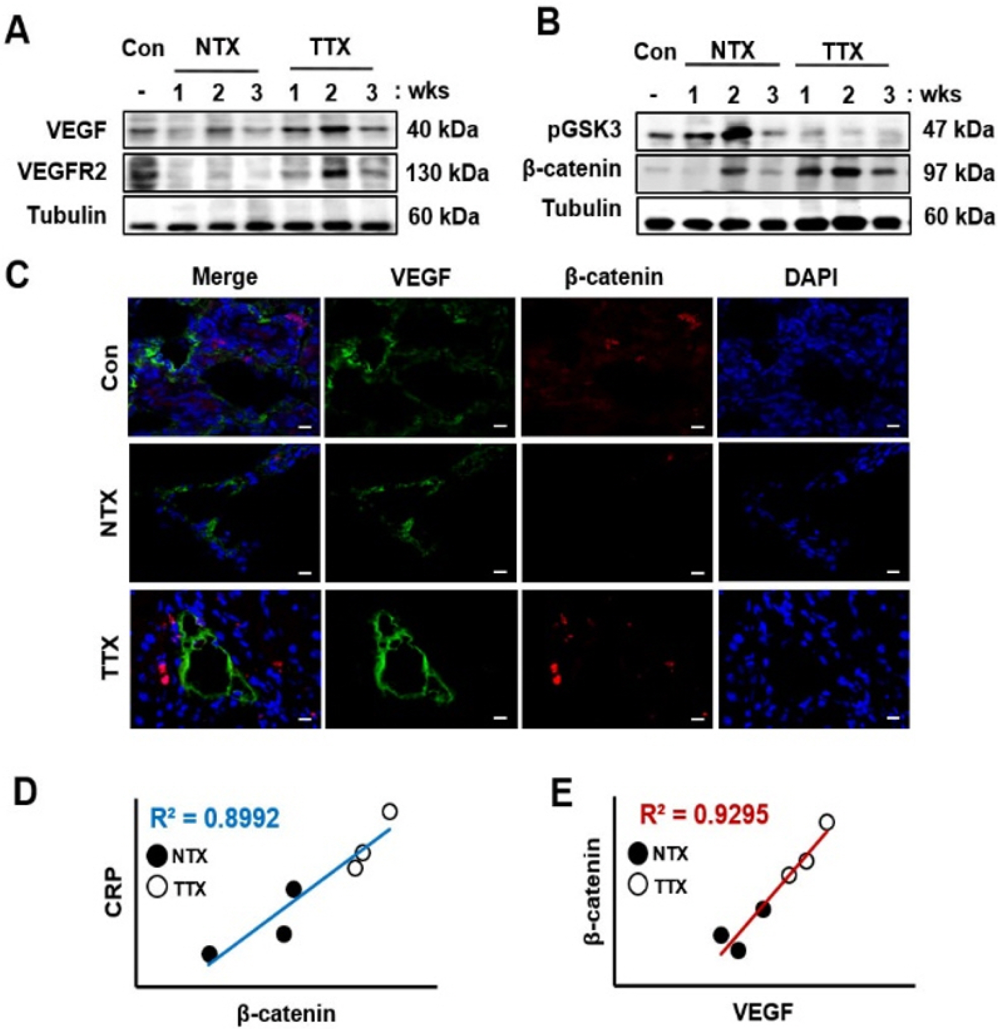
Fig. 3 PD-MSC transplantation induces Wnt signaling and angiogene-sis in CCl4-injured rats. Angiogenic (A) and Wnt signaling (B) factors were detected by Western blotting. Localization and expression of VEGF and β-catenin visualized using immunofluorescence (C). ×630, Scale bars; 20 μm. Positive correlations were found between CRP and β-catenin (D) also, between β-catenin and VEGF (E). NTX and TTX refer to the non-transplanted group and the PD-MSC-transplanted group, respec-tively.
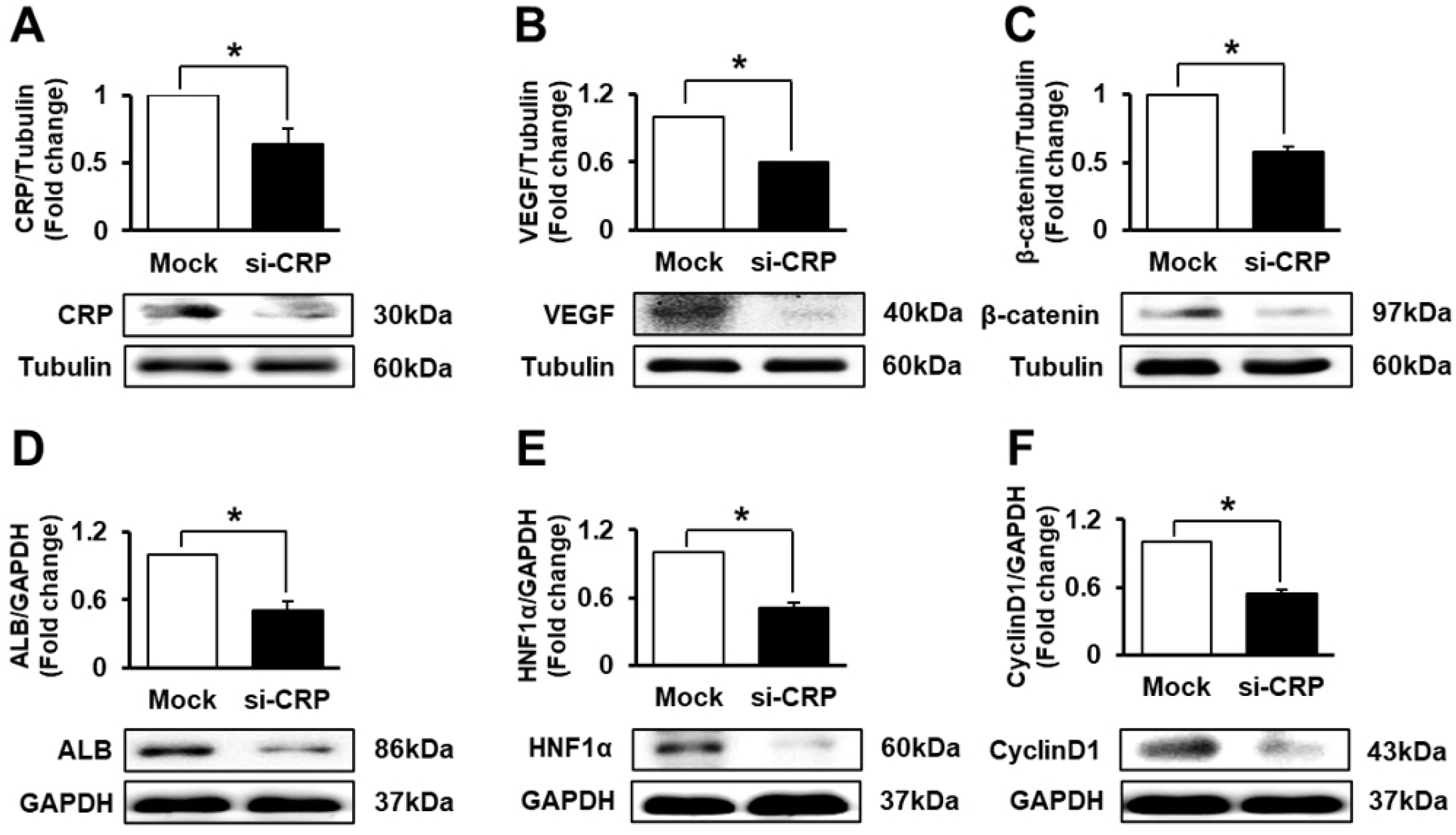
Fig. 4 CRP regulates angiogenesis and Wnt signaling in rat hepatocytes (WB-F344s). Protein levels of CRP (A), VEGF (B), β-catenin (C), ALB (D), HNF1α (E), and CyclinD1 (F) were evaluated in siRNA-CRP-transfected rat hepatocytes by western blot. Data are represented as the triplicated mean±SD of. *, p<0.05. Mock and si-CRP refer to non-transfected and siRNA-CRP-transfected rat hepatocytes, respectively.
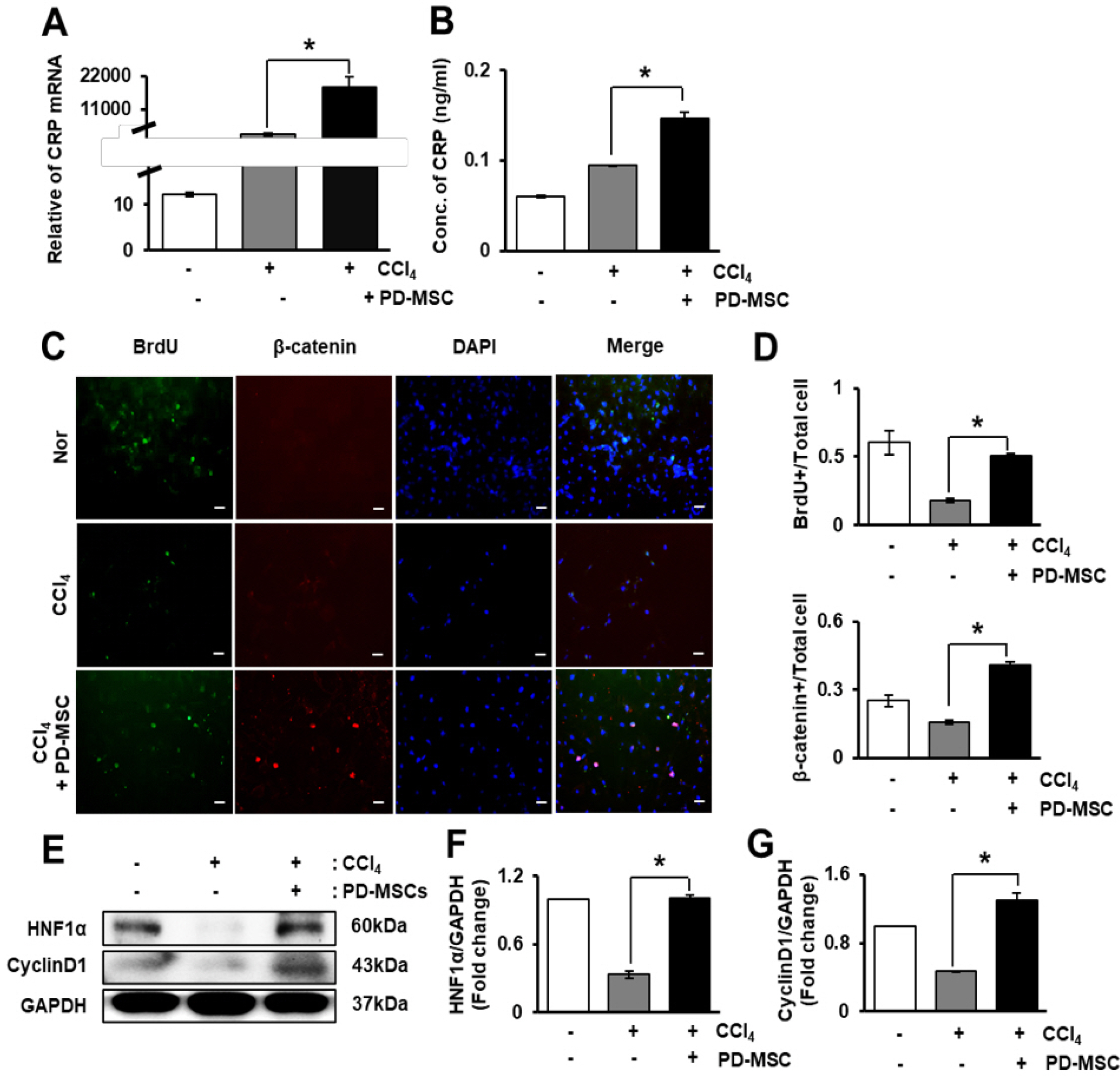
Fig. 5 PD-MSCs promote the expression of CRP and proliferation of hepatocytes through Wnt signaling. Expression of CRP was analyzed by qRT-PCR (A) and ELISA (B) after CCl4 treatment and co-culture with PD-MSCs. Immunofluorescence staining shows the localization and expression of BrdU and β-catenin in rat hepatocytes treated with CCl4 and co-cultured with PD-MSCs (C). ×400, Scale bars; 20 μm. BrdU- or β-ca-tenin-(D) positive rat hepatocytes were quantified after immunofluore-scence staining. Protein levels of HNF1α, and CyclinD1 were detected by western blotting in rat hepatocytes treated with CCl4 and co-cultured with PD-MSCs (E). Intensity of HNF1α (F) and Cyclin D1 (G) protein bands was calculated. Tripli-cated data are represented as the mean±SD. *, p<0.05.
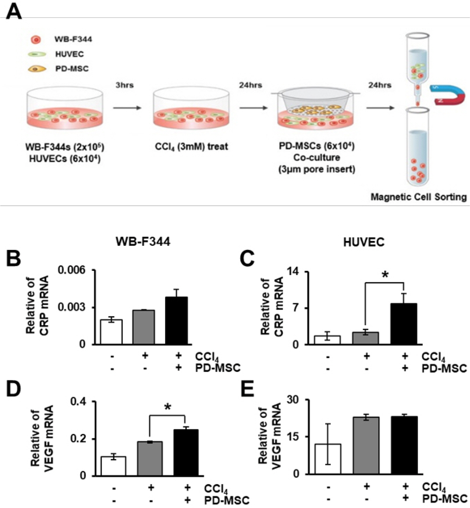
Fig. 6 PD-MSCs induce the expression of CRP and VEGF at the mRNA level. Schematic showing the experimental protocol (A). mRNA level of CRP (B), VEGF (D) was quantified in rat hepatocytes treated with CCl4 and co-cultured with HUVECs and PD-MSCs. mRNA level of CRP (C), VEGF (E) was assayed in HUVECs after CCl4 treatment and co-culture with rat hepatocytes and PD-MSCs. Duplicated data are represented as the mean±SD. *, p<0.05.
Reference
-
References
1. Rui L. 2014; Energy metabolism in the liver. Compr Physiol. 4:177–197. DOI: 10.1002/cphy.c130024. PMID: 24692138. PMCID: PMC4050641.
Article2. Poisson J, Lemoinne S, Boulanger C, Durand F, Moreau R, Valla D, Rautou PE. 2017; Liver sinusoidal endothelial cells: physiology and role in liver diseases. J Hepatol. 66:212–227. DOI: 10.1016/j.jhep.2016.07.009. PMID: 27423426.
Article3. Ding BS, Nolan DJ, Butler JM, James D, Babazadeh AO, Rosenwaks Z, Mittal V, Kobayashi H, Shido K, Lyden D, Sato TN, Rabbany SY, Rafii S. 2010; Inductive angiocrine signals from sinusoidal endothelium are required for liver regene-ration. Nature. 468:310–315. DOI: 10.1038/nature09493. PMID: 21068842. PMCID: PMC3058628.
Article4. Ding BS, Cao Z, Lis R, Nolan DJ, Guo P, Simons M, Penfold ME, Shido K, Rabbany SY, Rafii S. 2014; Divergent angiocrine signals from vascular niche balance liver regene-ration and fibrosis. Nature. 505:97–102. DOI: 10.1038/nature12681. PMID: 24256728. PMCID: PMC4142699.
Article5. Pinzani M, Rosselli M, Zuckermann M. 2011; Liver cirrhosis. Best Pract Res Clin Gastroenterol. 25:281–290. DOI: 10.1016/j.bpg.2011.02.009. PMID: 21497745.
Article6. Salazar J, Martínez MS, Chávez-Castillo M, Núñez V, Añez R, Torres Y, Toledo A, Chacín M, Silva C, Pacheco E, Rojas J, Bermúdez V. 2014; C-reactive protein: an in-depth look into structure, function, and regulation. Int Sch Res Notices. 2014:653045. DOI: 10.1155/2014/653045. PMID: 27433484. PMCID: PMC4897210.
Article7. Eisenhardt SU, Habersberger J, Murphy A, Chen YC, Woollard KJ, Bassler N, Qian H, von Zur Muhlen C, Hagemeyer CE, Ahrens I, Chin-Dusting J, Bobik A, Peter K. 2009; Dissociation of pentameric to monomeric C-reactive protein on activated platelets localizes inflammation to atherosclerotic plaques. Circ Res. 105:128–137. DOI: 10.1161/CIRCRESAHA.108.190611. PMID: 19520972.
Article8. Eisenhardt SU, Thiele JR, Bannasch H, Stark GB, Peter K. 2009; C-reactive protein: how conformational changes influence inflammatory properties. Cell Cycle. 8:3885–3892. DOI: 10.4161/cc.8.23.10068. PMID: 19887916.
Article9. Peña E, de la Torre R, Arderiu G, Slevin M, Badimon L. 2017; mCRP triggers angiogenesis by inducing F3 transcription and TF signalling in microvascular endothelial cells. Thromb Haemost. 117:357–370. DOI: 10.1160/TH16-07-0524. PMID: 27808345.
Article10. Krayem I, Bazzi S, Karam M. 2017; The combination of CRP isoforms with oxLDL decreases TNF-α and IL-6 release by U937-derived macrophages. Biomed Rep. 7:272–276. DOI: 10.3892/br.2017.949. PMID: 28808571. PMCID: PMC5543421.
Article11. Clevers H. 2006; Wnt/beta-catenin signaling in development and disease. Cell. 127:469–480. DOI: 10.1016/j.cell.2006.10.018. PMID: 17081971.12. Thompson MD, Monga SP. 2007; WNT/beta-catenin signaling in liver health and disease. Hepatology. 45:1298–1305. DOI: 10.1002/hep.21651. PMID: 17464972.13. Korn C, Scholz B, Hu J, Srivastava K, Wojtarowicz J, Arnsperger T, Adams RH, Boutros M, Augustin HG, Augustin I. 2014; Endothelial cell-derived non-canonical Wnt ligands control vascular pruning in angiogenesis. Development. 141:1757–1766. DOI: 10.1242/dev.104422. PMID: 24715464.
Article14. Tran KA, Zhang X, Predescu D, Huang X, Machado RF, Göthert JR, Malik AB, Valyi-Nagy T, Zhao YY. 2016; Endothelial β-catenin signaling is required for maintaining adult blood-brain barrier integrity and central nervous system homeostasis. Circulation. 133:177–186. DOI: 10.1161/CIRCULATIONAHA.115.015982. PMID: 26538583. PMCID: PMC4814374.
Article15. Lee HJ, Jung J, Cho KJ, Lee CK, Hwang SG, Kim GJ. 2012; Comparison of in vitro hepatogenic differentiation potential between various placenta-derived stem cells and other adult stem cells as an alternative source of functional hepatocytes. Differentiation. 84:223–231. DOI: 10.1016/j.diff.2012.05.007. PMID: 22885322.
Article16. Zhang D, Jiang M, Miao D. 2011; Transplanted human amniotic membrane-derived mesenchymal stem cells ameliorate carbon tetrachloride-induced liver cirrhosis in mouse. PLoS One. 6:e16789. DOI: 10.1371/journal.pone.0016789. PMID: 21326862. PMCID: PMC3033905.
Article17. Jung J, Moon JW, Choi JH, Lee YW, Park SH, Kim GJ. 2015; Epigenetic alterations of IL-6/STAT3 signaling by placental stem cells promote hepatic regeneration in a rat model with CCl4-induced liver injury. Int J Stem Cells. 8:79–89. DOI: 10.15283/ijsc.2015.8.1.79. PMID: 26019757. PMCID: PMC4445712.
Article18. Jun JH, Choi JH, Bae SH, Oh SH, Kim GJ. 2016; Decreased C-reactive protein induces abnormal vascular structure in a rat model of liver dysfunction induced by bile duct ligation. Clin Mol Hepatol. 22:372–381. DOI: 10.3350/cmh.2016.0032. PMID: 27729629. PMCID: PMC5066379.
Article19. Xu B, Broome U, Uzunel M, Nava S, Ge X, Kumagai-Braesch M, Hultenby K, Christensson B, Ericzon BG, Holgersson J, Sumitran-Holgersson S. 2003; Capillarization of hepatic sinusoid by liver endothelial cell-reactive autoantibodies in patients with cirrhosis and chronic hepatitis. Am J Pathol. 163:1275–1289. DOI: 10.1016/S0002-9440(10)63487-6. PMID: 14507637. PMCID: PMC1868294.
Article20. Iredale JP. 2007; Models of liver fibrosis: exploring the dynamic nature of inflammation and repair in a solid organ. J Clin Invest. 117:539–548. DOI: 10.1172/JCI30542. PMID: 17332881. PMCID: PMC1804370.
Article21. Djonov V, Baum O, Burri PH. 2003; Vascular remodeling by intussusceptive angiogenesis. Cell Tissue Res. 314:107–117. DOI: 10.1007/s00441-003-0784-3. PMID: 14574551.
Article22. Park S, Kim JW, Kim JH, Lim CW, Kim B. 2015; Differential roles of angiogenesis in the induction of fibrogenesis and the resolution of fibrosis in liver. Biol Pharm Bull. 38:980–985. DOI: 10.1248/bpb.b15-00325. PMID: 26133707.
Article23. Yang J, Mowry LE, Nejak-Bowen KN, Okabe H, Diegel CR, Lang RA, Williams BO, Monga SP. 2014; β-catenin signaling in murine liver zonation and regeneration: a Wnt-Wnt situation! Hepatology. 60:964–976. DOI: 10.1002/hep.27082. PMID: 24700412. PMCID: PMC4139486.
Article24. Preziosi M, Okabe H, Poddar M, Singh S, Monga SP. 2018; Endothelial Wnts regulate β-catenin signaling in murine liver zonation and regeneration: a sequel to the Wnt-Wnt situation. Hepatol Commun. 2:845–860. DOI: 10.1002/hep4.1196. PMID: 30027142. PMCID: PMC6049069.
Article25. Leibing T, Géraud C, Augustin I, Boutros M, Augustin HG, Okun JG, Langhans CD, Zierow J, Wohlfeil SA, Olsavszky V, Schledzewski K, Goerdt S, Koch PS. 2018; Angiocrine Wnt signaling controls liver growth and metabolic maturation in mice. Hepatology. 68:707–722. DOI: 10.1002/hep.29613. PMID: 29059455. PMCID: PMC6099291.
Article26. Oishi N, Yamashita T, Kaneko S. 2014; Molecular biology of liver cancer stem cells. Liver Cancer. 3:71–84. DOI: 10.1159/000343863. PMID: 24944998. PMCID: PMC4057789.
Article27. Moutachakkir M, Lamrani Hanchi A, Baraou A, Boukhira A, Chellak S. 2017; Immunoanalytical characteristics of C-reactive protein and high sensitivity C-reactive protein. Ann Biol Clin (Paris). 75:225–229. DOI: 10.1684/abc.2017.1232. PMID: 28377336.
Article28. Su HX, Zhou HH, Wang MY, Cheng J, Zhang SC, Hui F, Chen XZ, Liu SH, Liu QJ, Zhu ZJ, Hu QR, Wu Y, Ji SR. 2014; Mutations of C-reactive protein (CRP) -286 SNP, APC and p53 in colorectal cancer: implication for a CRP-Wnt crosstalk. PLoS One. 9:e102418. DOI: 10.1371/journal.pone.0102418. PMID: 25025473. PMCID: PMC4099363.
Article29. Chen J, Gu Z, Wu M, Yang Y, Zhang J, Ou J, Zuo Z, Wang J, Chen Y. 2016; C-reactive protein can upregulate VEGF expression to promote ADSC-induced angiogenesis by activating HIF-1α via CD64/PI3k/Akt and MAPK/ERK signaling pathways. Stem Cell Res Ther. 7:114. DOI: 10.1186/s13287-016-0377-1. PMID: 27526687. PMCID: PMC4986362.
- Full Text Links
-
- Actions
-
Cited
- CITED
-
- Close
- Share
-
- Similar articles
-
- L-Theanine-Treated Adipose-Derived Mesenchymal Stem Cells Alleviate the Cytotoxicity Induced by N-Nitrosodiethylamine in Liver
- Epigenetic Alterations of IL-6/STAT3 Signaling by Placental Stem Cells Promote Hepatic Regeneration in a Rat Model with CCl4-induced Liver Injury
- Advanced Research on Stem Cell Therapy for Hepatic Diseases: Potential Implications of a Placenta-derived Mesenchymal Stem Cell-based Strategy
- Therapeutic Angiogenesis with Somatic Stem Cell Transplantation
- Mesenchymal Stem Cell Therapy in Pulmonary Disease
