Inhibitory potentials of Cymbopogon citratus oil against aluminium-induced behavioral deficits and neuropathology in rats
- Affiliations
-
- 1Division of Neurobiology, Department of Anatomy, Faculty of Basic Medical Sciences, College of Health Sciences, University of Ilorin, Ilorin, Nigeria
- 2Division of Neurobiology, Department of Anatomy, Faculty of Basic Medical Sciences, Adeleke University, Ede, Nigeria
- 3Center for Studies in Behavioral Neurobiology, Department of Psychology, Concordia University Montreal, Montreal, Quebec, Canada
- KMID: 2507647
- DOI: http://doi.org/10.5115/acb.20.099
Abstract
- Cymbopogon citratus is a tropical phytomedicinal plant that is widely known for its hypoglycemic, hypolipidemic, anxiolytic, sedative, antioxidative and anti-inflammatory properties. In this study, we have examined the neuroprotective effects of the essential oil (ESO) of Cymbopogon citratus, following aluminum chloride (AlCl3)-induced neurotoxicity within the cerebellum of Wistar rats. A total of 40 adult male Wistar rats were assigned into five groups and treated orally as follows: A–phosphate-buffered saline (1 ml daily for 15 days); B–ESO (50 mg/kg daily for 15 days); C–AlCl3 (100 mg/kg daily for 15 days); D–AlCl3 then ESO (100 mg/kg AlCl3 daily for 15 days followed by 50 mg/kg ESO daily for subsequent 15 days); E– ESO then AlCl3 (50 mg/kg ESO daily for 15 days followed by 100 mg/kg AlCl3 daily for following 15 days). To address our questions, we observed the locomotion and exploratory behavior of the rats in the open field apparatus and subsequently evaluated cerebellar oxidative redox parameters, neural bioenergetics, acetylcholinesterase levels, transferrin receptor protein, and total protein profiles by biochemical assays. Furthermore, we investigated cerebellar histomorphology and Nissl profile by H&E and Cresyl violet Nissl staining procedures. ESO treatment markedly attenuated deficits in exploratory activities and rearing behavior following AlCl3 toxicity, indicating its anxiolytic potentials. Additionally, AlCl3 evokedincrease in malondialdehyde and nitric oxide levels, as well as repressed cerebellar catalase, glutathione peroxidase, and superoxide dismutase profiles were normalised to baseline levels by ESO treatment. Treatment with ESO, ergo, exhibits substantial neuroprotective and modulatory potentials in response to AlCl3 toxicity.
Keyword
Figure
-
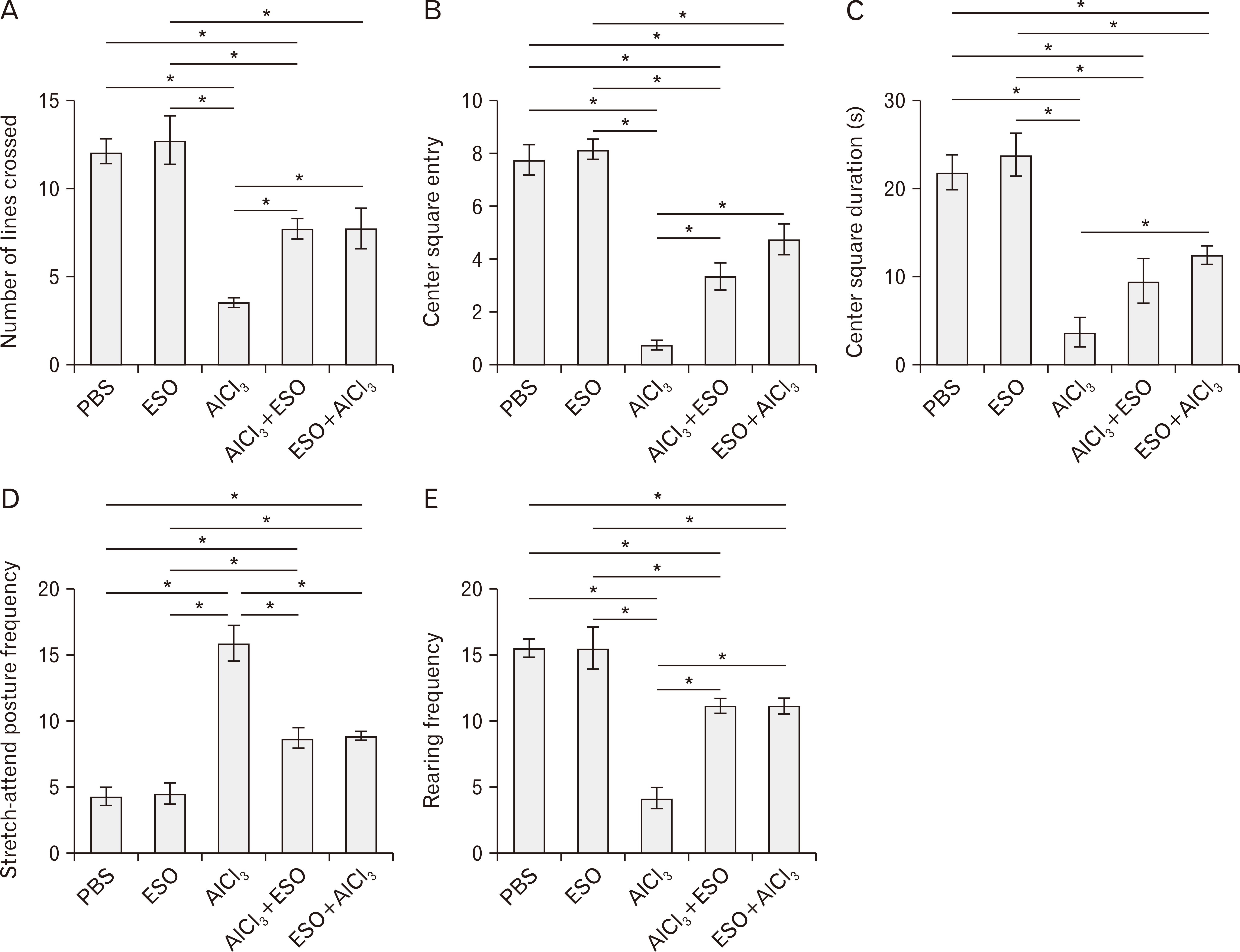
Fig. 1 (A–E) Effects of ESO administration on exploratory drive and locomotor activities in treated animals. (A) Analysis of the number of lines crossed by rats in the open field test showing no significant difference between the numbers of lines crossed by PBS and ESO controls. AlCl3 treated rats crossed the least number of lines relative to other groups. AlCl3+ESO and ESO+AlCl3 groups significantly crossed more lines than the AlCl3 treated rats (both at P<0.05). (B) Analysis of central square entry frequency in the open field test showed that rats treated AlCl3 made the least entry to the central square of the open field compared to all of PBS (P<0.05), ESO (P<0.05), AlCl3+ESO (P<0.05), and ESO+AlCl3 (P<0.05) groups. (C) Analysis of central square duration revealed that the administration of AlCl3 reduced the time rats spent in the central square when compared to the PBS (P<0.05) and ESO (P<0.05) controls. However, ESO significantly improved the central square duration of rats in the ESO+AlCl3 treated rats (P<0.05) compared to the AlCl3 group. (D) Rearing frequency analysis of experimental animals showing significant reductions in both groups treated AlCl3 compared to other groups. ESO intervention improved rearing frequencies in rats in the AlCl3+ESO and ESO+AlCl3 groups (at P<0.05, respectively) compared to the AlCl3 group. (E) Analysis of the stretch-attend posture frequency shows significant elevation in rats treated AlCl3 treated compared to PBS and ESO control (both at P<0.05). However, the AlCl3+ESO and ESO+AlCl3 treated rats showed improved stretch-attend-posture, which is significantly different (both at P<0.05) from the AlCl3 group. The results are presented as mean±standard error of mean. AlCl3, aluminum chloride; ESO, essential oil; PBS, phosphate-buffered saline. *P<0.05.
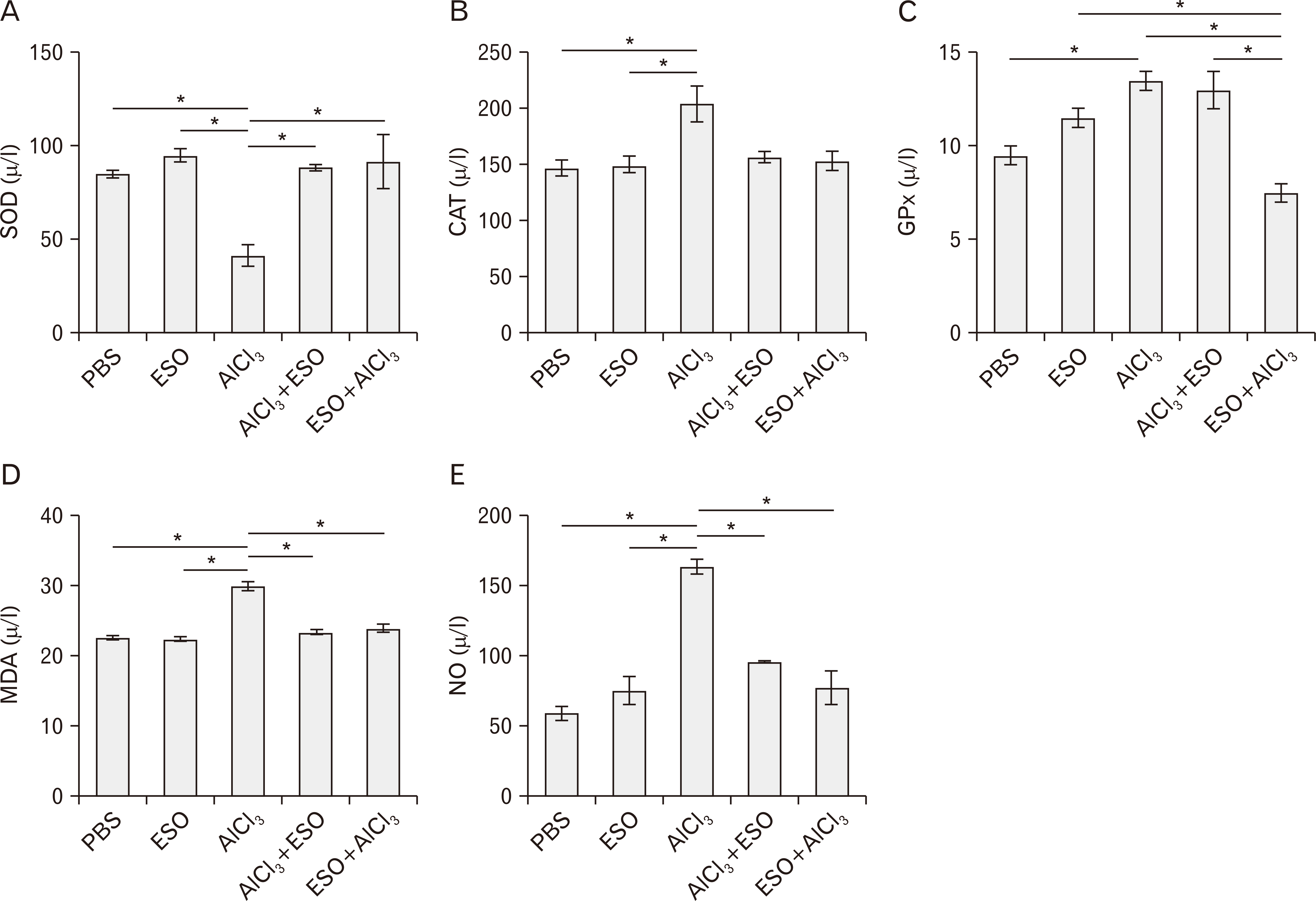
Fig. 2 Cerebellar oxidative redox state characterized by (A) SOD, (B) CAT, (C) GPx, (D) MDA, and (E) NO activities respectively. (A) Cerebellar SOD activity was significantly depleted by AlCl3 treatment relative to both PBS (P<0.05) and ESO groups (P<0.05) but was normalized by ESO treatment in ESO+AlCl3 (P<0.05) and AlCl3 (P<0.05) groups. (B) CAT level was increased by AlCl3 relative to both PBS and ESO controls (P<0.05) but regularized by ESO treatment in both AlCl3+ESO and ESO+AlCl3 groups at P<0.05. (C) AlCl3 treatment induced an increase in GPx level relative to the PBS and ESO groups at P<0.05 for both. ESO treatment restored the level of GPx in AlCl3+ESO (P<0.05) to normal levels but not ESO+AlCl3 group. (D) AlCl3 treatment triggered a significant increase in the level of MDA when compared to the PBS (P<0.05) and ESO (P<0.05) controls. This was normalized in the AlCl3+ESO (P<0.05) and ESO+AlCl3 (P<0.05) groups. (C) AlCl3 treatment elicited a striking increase in NO activities relative to the PBS and ESO groups (both at P<0.05) and was regularized by ESO in both AlCl3+ESO and ESO+AlCl3 groups (at P<0.05 each). AlCl3, aluminum chloride; CAT, catalase; ESO, essential oil; GPx, glutathione peroxidase; MDA, malondialdehyde; NO, nitric oxide; PBS, phosphate-buffered saline; SOD, superoxide dismutase. *P<0.05.

Fig. 3 Cerebellar G6PDH (A), LDH (B), GSA (C), TRP (D), and AChE (E) activities under treatment conditions. (A) AlCl3 treatment caused depletion in G6PDH expression relative to the PBS and ESO groups (P>0.05 in both). Pre- and post-treatment with ESO normalized G6PDH levels in the AlCl3+ESO (P<0.05) and ESO+AlCl3 (P<0.05) groups. (B) Expectantly, AlCl3 treatment induced a significant decrease in the level of LDH compared to the PBS, and ESO controls both at P<0.05. ESO treatment prevented LDH dysregulation in AlCl3+ESO (P<0.05) and ESO+AlCl3 (P<0.05) treated rats. (C) Cerebral GSA activity was significantly depleted in AlCl3 treated rats when compared to PBS (P<0.05), ESO (P<0.05), AlCl3+ESO (P<0.05) and ESO+AlCl3 (P<0.05) treated rats. (D) The expressed level of TRP in AlCl3 treated rats was significantly higher than in PBS, and ESO controls at P<0.05 each, as well as in AlCl3+ESO and ESO+AlCl3 treated rats (both at P<0.05). When compared to all other treated groups, AlCl3 treated rats presented with the highest level of AChE, with significant modulation seen following ESO intervention in the AlCl3+ESO and ESO+AlCl3 both at P<0.05. AChE, acetylcholinesterase; AlCl3, aluminum chloride; ESO, essential oil; GSA, glucokinase specific activity; G6PDH, glucose-6-phosphate dehydrogenase; LDH, lactate dehydrogenase; PBS, phosphate-buffered saline; TRP, transferrin receptor protein.*P<0.05.

Fig. 4 (A–E) Representative photomicrographs showing panoramic views of cerebellar cortex general morphological presentations and Nissl profile in Wistar rats across the various study groups. (A) is H&E (×100, ×400); The ML, Purkinje cells (black arrowhead) layer, GL, and the medullary layer of WM are demonstrated across study groups. PBS and ESO treatments did not alter the morphological presentation of cerebellar layers from this study. Both groups show a fine array of cells that are distinctly arranged from the molecular to the granular layer. Also, cellular density within the groups appears normal across all cortical layers. AlCl3 treatment caused degenerative changes characterized by fragmented GL in cerebellar cortex and poorly stained Purkinje cells. Comparatively, increased cellular density was observed in this group. Groups administered ESO before and after AlCl3-induced toxicity (AlCl3+ESO and ESO+AlCl3) shows mild degenerative changes in line of morphologic appearance of cerebellar sections of AlCl3-treated rats; cerebellar layers in both groups looks better structured and layers are well-delineated. (B) shows Nissl profile as demonstrated by CFV stain (×400). Normal morphological presentations of the cerebellar cortex are observable in PBS and ESO treated groups that are characterized by well-defined staining profiles. Cellular morphology in these groups is characterized by Purkinje cells with conspicuous cell bodies and dendrites that are projecting deep into the MLs (black arrowhead). Also, the granule layer in these groups consists of small granule neurons which are well outlined in contrast to the loosely arranged and cryptic cells in the granule layers of groups AlCl3 treated groups. Degenerating Purkinje cells with chromatolytic cell bodies (red arrowhead) and short dendritic processes can be seen around the indistinctly demarcated cerebellar layers of groups that receive aluminum treatment. The transitional regions between the cerebellar layers of AlCl3 treated with ESO are also generally better delineated. Although a few perineural spaces are present around Purkinje neurons of this groups, the general cytoarchitecture is much similar to that of the PBS and ESO groups. (C–E) show quantitative analysis of the CFV stain. AlCl3, aluminum chloride; CFV, Cresyl Fast Violet; ESO, essential oil; GL, granule cell layer; ML, molecular layer; PBS, phosphate-buffered saline; WM, white matter.*P<0.05.
Reference
-
References
1. Nelson PT, Alafuzoff I, Bigio EH, Bouras C, Braak H, Cairns NJ, Castellani RJ, Crain BJ, Davies P, Del Tredici K, Duyckaerts C, Frosch MP, Haroutunian V, Hof PR, Hulette CM, Hyman BT, Iwatsubo T, Jellinger KA, Jicha GA, Kövari E, Kukull WA, Leverenz JB, Love S, Mackenzie IR, Mann DM, Masliah E, McKee AC, Montine TJ, Morris JC, Schneider JA, Sonnen JA, Thal DR, Trojanowski JQ, Troncoso JC, Wisniewski T, Woltjer RL, Beach TG. 2012; Correlation of Alzheimer disease neuropathologic changes with cognitive status: a review of the literature. J Neuropathol Exp Neurol. 71:362–81. DOI: 10.1097/NEN.0b013e31825018f7. PMID: 22487856. PMCID: PMC3560290.
Article2. Mota SI, Ferreira IL, Rego AC. 2014; Dysfunctional synapse in Alzheimer's disease - a focus on NMDA receptors. Neuropharmacology. 76 Pt A:16–26. DOI: 10.1016/j.neuropharm.2013.08.013. PMID: 23973316. PMCID: PMC7507095.
Article3. Hardy J, Selkoe DJ. 2002; The amyloid hypothesis of Alzheimer's disease: progress and problems on the road to therapeutics. Science. 297:353–6. DOI: 10.1126/science.1072994. PMID: 12130773.
Article4. Wu T, Hallett M. 2013; The cerebellum in Parkinson's disease. Brain. 136(Pt 3):696–709. DOI: 10.1093/brain/aws360. PMID: 23404337.
Article5. Olajide OJ, Ugbosanmi AT, Enaibe BU, Ogunrinola KY, Lewu SF, Asogwa NT, Akapa T, Imam A, Ibrahim A, Gbadamosi IT, Yawson EO. 2017; Cerebellar molecular and cellular characterization in rat models of Alzheimer's disease: neuroprotective mechanisms of Garcinia biflavonoid complex. Ann Neurosci. 24:32–45. DOI: 10.1159/000464421. PMID: 28827919.6. Mathiyazahan DB, Thenmozhi AJ, Manivasagam T. 2015; Protective effect of black tea extract against aluminium chloride-induced Alzheimer's disease in rats: a behavioural, biochemical and molecular approach. J Funct Foods. 16:423–35. DOI: 10.1016/j.jff.2015.05.001.
Article7. Rodella LF, Ricci F, Borsani E, Stacchiotti A, Foglio E, Favero G, Rezzani R, Mariani C, Bianchi R. 2008; Aluminium exposure induces Alzheimer's disease-like histopathological alterations in mouse brain. Histol Histopathol. 23:433–9.8. Kumar A, Prakash A, Dogra S. 2011; Neuroprotective effect of carvedilol against aluminium induced toxicity: possible behavioral and biochemical alterations in rats. Pharmacol Rep. 63:915–23. DOI: 10.1016/S1734-1140(11)70607-7. PMID: 22001979.
Article9. Roskams AJ, Connor JR. 1990; Aluminum access to the brain: a role for transferrin and its receptor. Proc Natl Acad Sci U S A. 87:9024–7. DOI: 10.1073/pnas.87.22.9024. PMID: 2247478. PMCID: PMC55093.
Article10. Crapper DR, Krishnan SS, Quittkat S. 1976; Aluminium, neurofibrillary degeneration and Alzheimer's disease. Brain. 99:67–80. DOI: 10.1093/brain/99.1.67. PMID: 963531.
Article11. McDonald B, Esiri MM, Morris J. 1993; Aluminium and Alzheimer's disease. Age Ageing. 22:392–3. DOI: 10.1093/ageing/22.5.392. PMID: 8237634.
Article12. Adeneye AA, Agbaje EO. 2007; Hypoglycemic and hypolipidemic effects of fresh leaf aqueous extract of Cymbopogon citratus Stapf. in rats. J Ethnopharmacol. 112:440–4. DOI: 10.1016/j.jep.2007.03.034. PMID: 17513076.13. Tapia A, Cheel J, Theoduloz C, Rodríguez J, Schmeda-Hirschmann G, Gerth A, Wilken D, Jordan M, Jiménez-González E, Gomez-Kosky R, Mendoza EQ. 2007; Free radical scavengers from Cymbopogon citratus (DC.) stapf plants cultivated in bioreactors by the temporary immersion (TIS) principle. Z Naturforsch C J Biosci. 62:447–57. DOI: 10.1515/znc-2007-5-620. PMID: 17708453.14. Orrego R, Leiva E, Cheel J. 2009; Inhibitory effect of three C-glycosylflavonoids from Cymbopogon citratus (Lemongrass) on human low density lipoprotein oxidation. Molecules. 14:3906–13. DOI: 10.3390/molecules14103906. PMID: 19924037. PMCID: PMC6254720.15. Campos J, Schmeda-Hirschmann G, Leiva E, Guzmán L, Orrego R, Fernández P, González M, Radojkovic C, Zuñiga FA, Lamperti L, Pastene E, Aguayo C. 2014; Lemon grass (Cymbopogon citratus (D.C) Stapf) polyphenols protect human umbilical vein endothelial cell (HUVECs) from oxidative damage induced by high glucose, hydrogen peroxide and oxidised low-density lipoprotein. Food Chem. 151:175–81. DOI: 10.1016/j.foodchem.2013.11.018. PMID: 24423518.16. Salim E, Kumolosasi E, Jantan I. 2014; Inhibitory effect of selected medicinal plants on the release of pro-inflammatory cytokines in lipopolysaccharide-stimulated human peripheral blood mononuclear cells. J Nat Med. 68:647–53. DOI: 10.1007/s11418-014-0841-0. PMID: 24799081.
Article17. Francisco V, Costa G, Figueirinha A, Marques C, Pereira P, Miguel Neves B, Celeste Lopes M, García-Rodríguez C, Teresa Cruz M, Teresa Batista M. 2013; Anti-inflammatory activity of Cymbopogon citratus leaves infusion via proteasome and nuclear factor-κB pathway inhibition: contribution of chlorogenic acid. J Ethnopharmacol. 148:126–34. DOI: 10.1016/j.jep.2013.03.077. PMID: 23583902.18. Figueirinha A, Paranhos A, Pérez-Alonso JJ, Santos-Buelga C, Batista MT. 2008; Cymbopogon citratus leaves: characterization of flavonoids by HPLC-PDA-ESI/MS/MS and an approach to their potential as a source of bioactive polyphenols. Food Chem. 110:718–28. DOI: 10.1016/j.foodchem.2008.02.045.19. Garcia R, Ferreira JP, Costa G, Santos T, Branco F, Caramona M, de Carvalho M, Dinis AM, Batista MT, Castel-Branco M, Figueiredo IV. 2015; Evaluation of anti-inflammatory and analgesic activities of cymbopogon citratus in vivo-polyphenols contribution. Res J Med Plant. 9:1–13. DOI: 10.3923/rjmp.2015.1.13.20. Ganjewala D. 2009; Cymbopogon essential oils: chemical compositions and bioactivities. Int J Essent Oil Ther. 3:56–65.21. Stanford SC. 2007; The open field test: reinventing the wheel. J Psychopharmacol. 21:134–5. DOI: 10.1177/0269881107073199. PMID: 17329288.
Article22. Nehru B, Bhalla P. 2007; Biochemical alterations in rat brain following aluminum exposure: protection with centrophenoxine. Toxicol Environ Chem. 89:577–85. DOI: 10.1080/02772240701228763.
Article23. Amjad S, Umesalma S. 2015; Protective effect of Centella asiatica against aluminium-induced neurotoxicity in cerebral cortex, striatum, hypothalamus and hippocampus of rat brain histopathological, and biochemical approach. J Mol Biomark Diagn. 6:212. DOI: 10.4172/2155-9929.1000212.
Article24. Lakshmi BV, Sudhakar M, Prakash KS. 2015; Protective effect of selenium against aluminum chloride-induced Alzheimer's disease: behavioral and biochemical alterations in rats. Biol Trace Elem Res. 165:67–74. DOI: 10.1007/s12011-015-0229-3. PMID: 25613582.
Article25. Olajide OJ, Fatoye JO, Idowu OF, Ilekoya D, Gbadamosi IT, Gbadamosi MT, Asogwa NT. 2018; Reversal of behavioral decline and neuropathology by a complex vitamin supplement involves modulation of key neurochemical stressors. Environ Toxicol Pharmacol. 62:120–31. DOI: 10.1016/j.etap.2018.07.005. PMID: 30005307.
Article26. Taïr K, Kharoubi O, Taïr OA, Hellal N, Benyettou I, Aoues A. 2016; Aluminium-induced acute neurotoxicity in rats: treatment with aqueous extract of Arthrophytum (Hammada scoparia). J Acute Disease. 5:470–82. DOI: 10.1016/j.joad.2016.08.028.
Article27. Olajide OJ, Enaibe BU, Bankole OO, Akinola OB, Laoye BJ, Ogundele OM. 2016; Kolaviron was protective against sodium azide (NaN3) induced oxidative stress in the prefrontal cortex. Metab Brain Dis. 31:25–35. DOI: 10.1007/s11011-015-9674-0. PMID: 25916484.
Article28. Kumar V, Gill KD. 2014; Oxidative stress and mitochondrial dysfunction in aluminium neurotoxicity and its amelioration: a review. Neurotoxicology. 41:154–66. DOI: 10.1016/j.neuro.2014.02.004. PMID: 24560992.
Article29. Sayre LM, Perry G, Smith MA. 2008; Oxidative stress and neurotoxicity. Chem Res Toxicol. 21:172–88. DOI: 10.1021/tx700210j. PMID: 18052107.
Article30. Yuan CY, Lee YJ, Hsu GS. 2012; Aluminum overload increases oxidative stress in four functional brain areas of neonatal rats. J Biomed Sci. 19:51. DOI: 10.1186/1423-0127-19-51. PMID: 22613782. PMCID: PMC3404950.
Article31. Guix FX, Uribesalgo I, Coma M, Muñoz FJ. 2005; The physiology and pathophysiology of nitric oxide in the brain. Prog Neurobiol. 76:126–52. DOI: 10.1016/j.pneurobio.2005.06.001. PMID: 16115721.
Article32. Pacher P, Beckman JS, Liaudet L. 2007; Nitric oxide and peroxynitrite in health and disease. Physiol Rev. 87:315–424. DOI: 10.1152/physrev.00029.2006. PMID: 17237348. PMCID: PMC2248324.
Article33. Calabrese V, Mancuso C, Calvani M, Rizzarelli E, Butterfield DA, Stella AM. 2007; Nitric oxide in the central nervous system: neuroprotection versus neurotoxicity. Nat Rev Neurosci. 8:766–75. DOI: 10.1038/nrn2214. PMID: 17882254. PMCID: PMC4903952.
Article34. Chen Z, Zhong C. 2013; Decoding Alzheimer's disease from perturbed cerebral glucose metabolism: implications for diagnostic and therapeutic strategies. Prog Neurobiol. 108:21–43. DOI: 10.1016/j.pneurobio.2013.06.004. PMID: 23850509.
Article35. Zhang X, Yang F, Xu C, Liu W, Wen S, Xu Y. 2008; Cytotoxicity evaluation of three pairs of hexabromocyclododecane (HBCD) enantiomers on Hep G2 cell. Toxicol In Vitro. 22:1520–7. DOI: 10.1016/j.tiv.2008.05.006. PMID: 18644697.
Article36. Ross JM, Öberg J, Brené S, Coppotelli G, Terzioglu M, Pernold K, Goiny M, Sitnikov R, Kehr J, Trifunovic A, Larsson NG, Hoffer BJ, Olson L. 2010; High brain lactate is a hallmark of aging and caused by a shift in the lactate dehydrogenase A/B ratio. Proc Natl Acad Sci U S A. 107:20087–92. DOI: 10.1073/pnas.1008189107. PMID: 21041631. PMCID: PMC2993405.
Article37. Trapp GA. 1983; Plasma aluminum is bound to transferrin. Life Sci. 33:311–6. DOI: 10.1016/S0024-3205(83)80002-2. PMID: 6410138. PMCID: PMC6804145.
Article38. Al-Hashem F. 2009; Camel's milk protects against aluminum chloride-induced normocytic normocromic anemia, lipid peroxidation and oxidative stress in erythrocytes of white albino rats. Am J Biochem Biotechnol. 5:126–36. DOI: 10.3844/ajbbsp.2009.126.136.
Article39. Goldstein ME, Cooper HS, Bruce J, Carden MJ, Lee VM, Schlaepfer WW. 1987; Phosphorylation of neurofilament proteins and chromatolysis following transection of rat sciatic nerve. J Neurosci. 7:1586–94. DOI: 10.1523/JNEUROSCI.07-05-01586.1987. PMID: 3106591. PMCID: PMC6568824.
Article40. Ekong MB, Ekpo MM, Akpanyung EO, Nwaokonko DU. 2017; Neuroprotective effect of Moringa oleifera leaf extract on aluminium-induced temporal cortical degeneration. Metab Brain Dis. 32:1437–47. DOI: 10.1007/s11011-017-0011-7. PMID: 28397152.
Article41. Khalil EA. 2011; Evaluation of the possible protective and therapeutic influence of coriander (Coriandum sativum L.) seed aqueous extract on hippocampal pyramidal cells against Alzheimer's disease induced by aluminum chloride in adult male albino rats. Researcher. 3:22–9.
- Full Text Links
-
- Actions
-
Cited
- CITED
-
- Close
- Share
-
- Similar articles
-
- Colorado Potato Beetle (Leptinotarsa decemlineata Say) Control Potential of Essential Oil Isolated from Iranian Cymbopogon citratus Stapf
- Antimicrobial Activity of Some Essential Oils Against Microorganisms Deteriorating Fruit Juices
- Antimicrobial property of lemongrass (Cymbopogon citratus) oil against pathogenic bacteria isolated from pet turtles
- Essential Oil Prepared from Cymbopogon citrates Exerted an Antimicrobial Activity Against Plant Pathogenic and Medical Microorganisms
- Effect of administration of etretinate and fish oil on plasma cholesterol levels in rats
