Korean J Gastroenterol.
2020 Jul;76(1):46-48. 10.4166/kjg.2020.76.1.46.
Pancreatic Hemangioma Suspected of Neuroendocrine Tumor
- Affiliations
-
- 1Departments of Internal Medicine, Dongguk University Ilsan Hospital, Dongguk University College of Medicine, Goyang, Korea
- 2Departments of Pathology, Dongguk University Ilsan Hospital, Dongguk University College of Medicine, Goyang, Korea
- KMID: 2504594
- DOI: http://doi.org/10.4166/kjg.2020.76.1.46
Abstract
- Adult pancreatic hemangioma is an extremely rare disease, with only 22 cases reported since 1939. Pancreatic hemangioma has no specific symptoms, diagnostic imaging, or laboratory findings, making it difficult to be clinically suspected and diagnosed. The majority are confirmed after surgery. In this report, a 61-year-old woman presented with melena and showed multiple small hyper- vascular lesions in the pancreas. A pancreatic neuroendocrine tumor was suspected, and the patient underwent a distal pancreatectomy. The pathology examination and immunohistochemical study revealed a pancreatic hemangioma.
Keyword
Figure
-
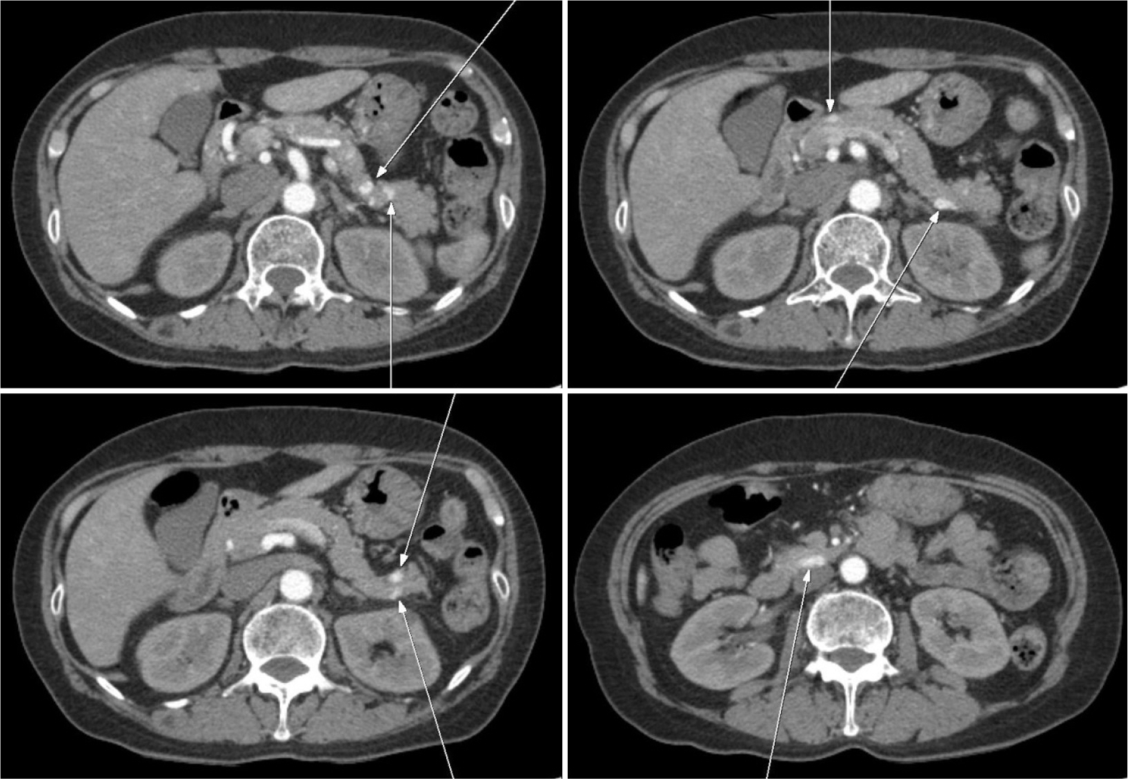
Fig. 1 Contrast-enhanced computed tomography scan during the arterial phase shows multiple hypervascular lesions in the pancreas (arrows), and the largest lesion was 14 mm in the uncinate process.

Fig. 2 Magnetic resonance imaging after gadolinium enhancement during the arterial phase shows multiple hyperintense lesions in the pancreas (arrows).
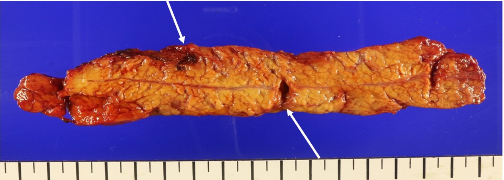
Fig. 3 Gross specimen shows two well-demarcated multilocular cystic lesions (arrows).

Fig. 4 Microscopic findings. (A) Hematoxylin and eosin stain of the tumor showing large dilated vessels lined by flattened endothelium (×100) and (B) immunohistochemical stain for CD34 is positive (×40).
Reference
-
1. Raymundo SRO, Hussain KMK, Hussein KG, Kuga ML. 2018; Rare case of adult pancreatic haemangioma and literature review. BMJ Case Rep. 2018:bcr2018226456. DOI: 10.1136/bcr-2018-226456. PMID: 30297496.
Article2. Kim SH, Kim JY, Choi JY, Choi YD, Kim KS. 2016; Incidental detection of pancreatic hemangioma mimicking a metastatic tumor of renal cell carcinoma. Korean J Hepatobiliary Pancreat Surg. 20:93–96. DOI: 10.14701/kjhbps.2016.20.2.93. PMID: 27212999. PMCID: PMC4874050.
Article3. Lu T, Yang C. 2015; Rare case of adult pancreatic hemangioma and review of the literature. World J Gastroenterol. 21:9228–9232. DOI: 10.3748/wjg.v21.i30.9228. PMID: 26290651. PMCID: PMC4533056.
Article4. Mondal U, Henkes N, Henkes D, Rosenkranz L. 2015; Cavernous hemangioma of adult pancreas: a case report and literature review. World J Gastroenterol. 21:9793–9802. DOI: 10.3748/wjg.v21.i33.9793. PMID: 26361427. PMCID: PMC4562964.
Article5. Lu ZH, Wu M. 2013; Unusual features in an adult pancreatic hemangioma:CT and MRI demonstration. Korean J Radiol. 14:781–785. DOI: 10.3348/kjr.2013.14.5.781. PMID: 24043972. PMCID: PMC3772258.6. Jarboui S, Salem A, Gherib BS, et al. 2010; Hemangioma of the pancreas in a 60-year-old woman: a report of a new case. Gastroenterol Clin Biol. 34:569–571. DOI: 10.1016/j.gcb.2010.06.001. PMID: 20609542.
Article7. Mundinger GS, Gust S, Micchelli ST, Fishman EK, Hruban RH, Wolfgang CL. 2009; Adult pancreatic hemangioma: case report and literature review. Gastroenterol Res Pract. 2009:839730. DOI: 10.1155/2009/839730. PMID: 19421421. PMCID: PMC2676326.
Article8. Naito Y, Nishida N, Nakamura Y, et al. 2014; Adult pancreatic hemangioma:a case report. Oncol Lett. 8:642–644. DOI: 10.3892/ol.2014.2206. PMID: 25013478. PMCID: PMC4081133.9. Naili RE, Nicolas M. 2015; Hemangioma of the pancreas: a rare tumor in adults. Am J Clin Pathol. 144(Suppl 2):A350–A350. DOI: 10.1093/ajcp/144.suppl2.350.
Article
- Full Text Links
-
- Actions
-
Cited
- CITED
-
- Close
- Share
-
- Similar articles
-
- Surgical Results of Pancreatic Neuroendocrine Tumors
- A Case of Pancreatic Neuroendocrine Tumor Mimicked Intraductal Papillary Mucinous Neoplasm
- Non-Functioning, Malignant Pancreatic Neuroendocrine Tumor in a 16-Year-old Boy: A Case Report
- Pancreatic Collision Tumor of Desmoid-Type Fibromatosis and Mucinous Cystic Neoplasm: A Case Report
- A Clinical Experience with Pancreatic Cavernous Hemangioma
