Non-Functioning, Malignant Pancreatic Neuroendocrine Tumor in a 16-Year-old Boy: A Case Report
- Affiliations
-
- 1Department of Radiology, Wonkwang University School of Medicine and Hospital, Korea. yjyh@wonkwang.ac.kr
- 2Department of Radiology, Inje University Sanggyepaik Hospital, Korea.
- KMID: 2206913
- DOI: http://doi.org/10.13104/jksmrm.2010.14.2.145
Abstract
- We report the case of a 16-year-old boy with a solid pancreatic mass which proved to be a nonfunctioning, malignant pancreatic neuroendocrine tumor (PNET). In pediatric patients, malignant pancreatic tumors are rare, especially malignant PNET. When dynamic contrast enhanced MRI showed a well enhancing solid pancreatic tumor on arterial and delayed phases and combined with malignant features, such as vascular invasion, invasion of adjascent organs, and lymphadenopathy, we should include malignant pancreatic neuroendocrine tumor in the differential diagnosis of childhood pancreatic tumors.
MeSH Terms
Figure
-
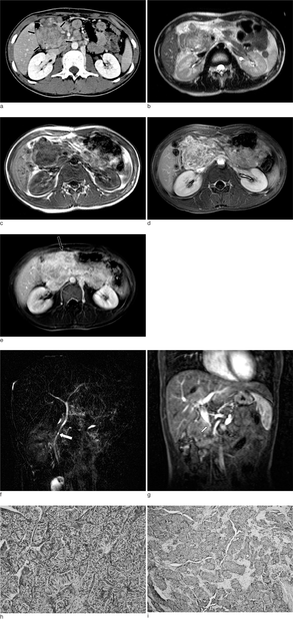
Fig. 1 A 16-year-old boy with epigastric pain for one year. (a) Ccontrast-enhanced CT of the abdomen showed a multilobulated contoured solid mass with inhomogeneous enhancement in the region of the pancreatic head (arrows). (b) The axial T2-weighted MR image obtained at the same level as in A, depicts a multilobulated mass with heterogeneous high signal intensity. (c) The mass showed intermediate to low signal intensity on the axial T1-weighted MR image. (d) The mass revealed a reticular pattern of inhomogeneous enhancement on the late portal phase of dynamic gadolinium-enhanced T1-weighted MR image. (e) On the delayed phase of dynamic gadolinium-enhanced T1-weighted MR image, persistent and intense enhancement of the mass and multiple conglomerated metastatic lymphadenopathy were seen (arrow). (f) MRCP showed that despite the large size of the mass, the common bile duct (arrow) was not dilated but had been displaced by the mass. (g) The coronal T1-weighted MR image showed encasement of the main portal vein (arrow) by the mass, which suggested the malignant features of the mass. (h) Photomicrograph (H & E,×400 ) of the specimen showed that the tumor cells had a trabacular pattern over the fibrotic and hyaline stroma. (i) The immunohistochemistry staining for synaptophysin (×100) was positive which confirmed the diagnosis of malignant endocrine neoplasm of the pancreas.
Reference
-
1. Shorter NA, Glick RD, Klimstra DS, Brennan MF, LaQuaglia MP. Malignant pancreatic tumors in childhood and adolescence: the Memorial Sloan-Kettering experience, 1967 to present. J Pediatr Surg. 2002. 37:887–892.2. Roebuck DJ, Yuen MK, Wong YC, Shing MK, Lee CW, Li CK. Imaging features of pancreatoblastoma. Pediatr Radiol. 2001. 31:501–506.3. Chung EM, Travis MD, Conran RM. Pancreatic tumors in children: radiologic-pathologic correlation. Radiographics. 2006. 26:1211–1238.4. Grosfeld JL, Vane DW, Rescorla FJ, McGuire W, West KW. Pancreatic tumors in childhood: analysis of 13 cases. J Pediatr Surg. 1990. 25:1057–1062.5. Verhoef S, van Diemen-Steenvoorde R, Akkersdijk WL, Bax NM, Ariyurek Y, Hermans CJ, et al. Malignant pancreatic tumour within the spectrum of tuberous sclerosis complex in childhood. Eur J Pediatr. 1999. 158:284–287.6. Francalanci P, Diomedi-Camassei F, Purificato C, Santorelli FM, Giannotti A, Dominici C, et al. Malignant pancreatic endocrine tumor in a child with tuberous sclerosis. Am J Surg Pathol. 2003. 27:1386–1389.7. Procacci C, Carbognin G, Accordini S, Biasiutti C, Bicego E, Romano L, et al. Nonfunctioning endocrine tumors of the pancreas: possibilities of spiral CT characterization. Eur Radiol. 2001. 11:1175–1183.8. Stafford-Johnson DB, Francis IR, Eckhauser FE, Knol JA, Chang AE. Dual-phase helical CT of nonfunctioning islet cell tumors. J Comput Assist Tomogr. 1998. 22:335–339.9. Koito K, Namieno T, Nagakawa T, Morita K. Delayed enhancement of islet cell carcinoma on dynamic computed tomography: a sign of its malignancy. Abdom Imaging. 1997. 22:304–306.10. Ichikawa T, Peterson MS, Federle MP, Baron RL, Haradome H, Kawamori Y, et al. Islet cell tumor of the pancreas: biphasic CT versus MR imaging in tumor detection. Radiology. 2000. 216:163–171.
- Full Text Links
-
- Actions
-
Cited
- CITED
-
- Close
- Share
-
- Similar articles
-
- Pancreatic Collision Tumor of Desmoid-Type Fibromatosis and Mucinous Cystic Neoplasm: A Case Report
- Nonfunctioning Neuroendocrine Tumor of the Pancreas in a 15-year-old Girl: a Case Report
- Laparoscopic Enucleation of a Nonfunctioning Neuroendocrine Tumor of the Pancreas
- Malignant Melanoma of the Pancreas and Liver Mimicking a Neuroendocrine Tumor
- Surgical Experiences of Neuroendocrine Neoplasms of the Pancreas : Comparative Study of Functioning vs. Non-functioning Neoplasms
