Apparent diffusion coefficient as a valuable quantitative parameter for predicting clinical outcomes in patients with newly diagnosed primary CNS lymphoma
- Affiliations
-
- 1Department of Hematology/Oncology, Kyungpook National University Hospital, School of Medicine, Kyungpook National University, Daegu, Korea
- 2Department of Radiology, Kyungpook National University Hospital, School of Medicine, Kyungpook National University, Daegu, Korea
- KMID: 2503484
- DOI: http://doi.org/10.5045/br.2020.2020032
Abstract
- Background
This study attempted to identify novel prognostic factors in patients with newly diagnosed primary central nervous system lymphoma (PCNSL) using magnetic resonance imaging (MRI).
Methods
We retrospectively evaluated 67 patients diagnosed with central nervous system (CNS) tumors. The enrollment criteria were as follows: i) pathologic diagnosis of CNS lymphoma, ii) no evidence of systemic involvement, iii) no evidence of human immunodeficiency virus-1 infection or other immunodeficiencies, and iv) MRI scans available at diagnosis. Fifty-two patients met these criteria and were enrolled.
Results
The 3-year overall survival (OS) and failure-free survival rates were 69.7% and 45.6%, respectively, with a median follow-up duration of 36.2 months. OS of patients with low apparent diffusion coefficient (ADC) was lower than those with higher ADC. Multivariate analysis revealed that old age (>60 yr) [hazard ratio (HR), 20.372; P=0.001], Eastern Cooperative Oncology Group performance status (ECOG PS) ≥2 (HR, 10.429; P < 0.001), higher lactate dehydrogenase (LDH) levels (HR, 7.408; P =0.001), and low ADC (HR, 0.273; P=0.009) were associated with lower OS. We modified the conventional prognostic scoring system using low ADC, old age (>60 yr), ECOG PS ≥2, and higher LDH. The risk of death was categorized as high (score 3-4), intermediate-2 (score 2), intermediate- 1 (score 1), and low (score 0), with three-year OS rates of 33.5%, 55.4%, 88.9%, and 100%, respectively.
Conclusion
ADC demonstrated significant prognostic value for long-term survival in patients with newly diagnosed PCNSL. Low ADC was an independent unfavorable prognostic factor, suggesting that ADC obtained from MRI can improve the current prognostic scoring system.
Figure
-
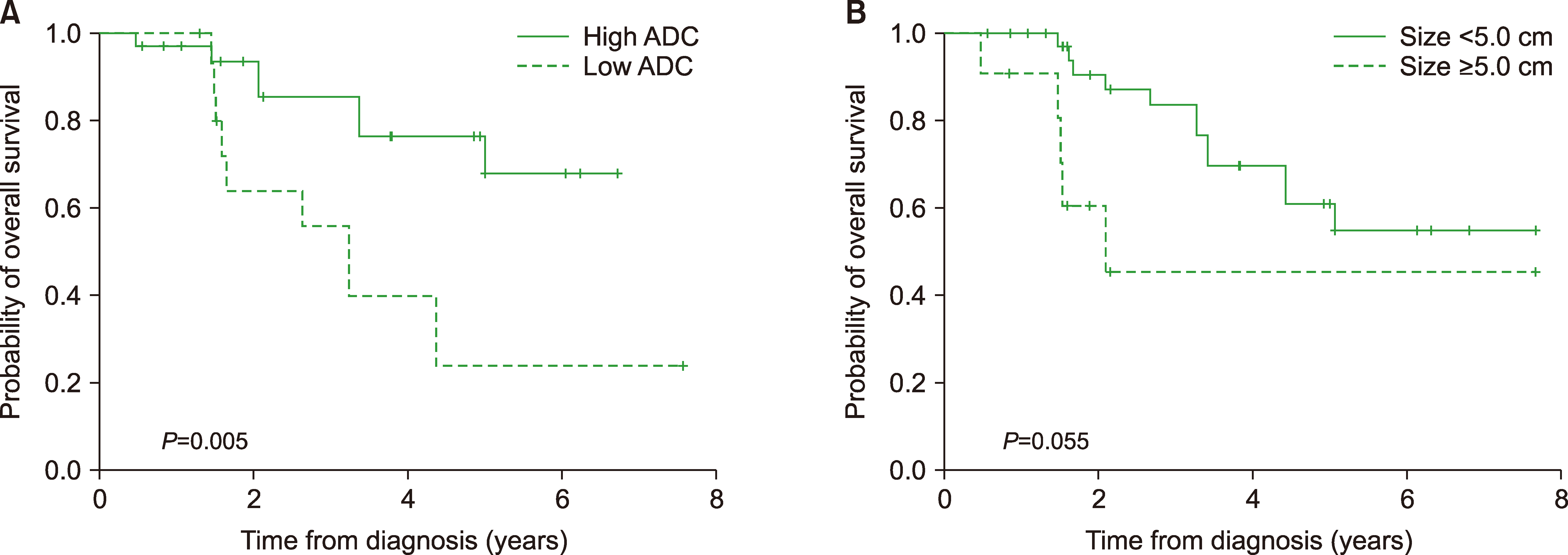
Fig. 1 Kaplan-Meier curves for overall survival (OS). Patients with low ADC had lower OS (P=0.005) (A), while patients with tumors measuring less than 5 cm exhibited a tendency toward better OS (P=0.055) (B). Abbreviation: ADC, apparent diffusion coefficient.

Fig. 2 Kaplan-Meier curves for failure-free survival (FFS). Patients with hyperintense signal on T2-weighted imaging (P=0.001) (A) and homogenous enhancement (P=0.011) (B) had better FFS, while patients with low ADC (P=0.018) (C) and necrosis had poor FFS (P<0.001) (D). Abbreviation: ADC, apparent diffusion coefficient.

Fig. 3 Modified prognostic scoring system. The prognostic scoring system comprising old age, ECOG PS, and LDH (A) and the modified scoring system comprising old age, ECOG PS, LDH, and ADC (B). Abbreviations: ADC, apparent diffusion coefficient; ECOG PS, Eastern Cooperative Oncology Group performance status; LDH, lactate dehydrogenase.
Reference
-
1. Hoffman S, Propp JM, McCarthy BJ. 2006; Temporal trends in incidence of primary brain tumors in the United States, 1985-1999. Neuro Oncol. 8:27–37. DOI: 10.1215/S1522851705000323. PMID: 16443945. PMCID: PMC1871920.2. Haldorsen IS, Krossnes BK, Aarseth JH, et al. 2007; Increasing incidence and continued dismal outcome of primary central nervous system lymphoma in Norway 1989-2003 : time trends in a 15-year national survey. Cancer. 110:1803–14. DOI: 10.1002/cncr.22989. PMID: 17721992.3. van der Sanden GA, Schouten LJ, van Dijck JA, et al. 2002; Primary central nervous system lymphomas: incidence and survival in the Southern and Eastern Netherlands. Cancer. 94:1548–56. DOI: 10.1002/cncr.10357. PMID: 11920513.4. Morris PG, Abrey LE. 2009; Therapeutic challenges in primary CNS lymphoma. Lancet Neurol. 8:581–92. DOI: 10.1016/S1474-4422(09)70091-2. PMID: 19446277.
Article5. Ferreri AJ, DeAngelis L, Illerhaus G, et al. 2011; Whole-brain radiotherapy in primary CNS lymphoma. Lancet Oncol. 12:118–9. author reply 119–20. DOI: 10.1016/S1470-2045(11)70018-3. PMID: 21277546.
Article6. Grommes C, DeAngelis LM. 2017; Primary CNS lymphoma. J Clin Oncol. 35:2410–8. DOI: 10.1200/JCO.2017.72.7602. PMID: 28640701. PMCID: PMC5516483.
Article7. Niparuck P, Boonsakan P, Sutthippingkiat T, et al. 2019; Treatment outcome and prognostic factors in PCNSL. Diagn Pathol. 14:56. DOI: 10.1186/s13000-019-0833-1. PMID: 31189479. PMCID: PMC6563360.
Article8. DeAngelis LM, Seiferheld W, Schold SC, Fisher B, Schultz CJ. Radiation Therapy Oncology Group Study 93-10. 2002; Combination chemotherapy and radiotherapy for primary central nervous system lymphoma:. J Clin Oncol. 20:4643–8. DOI: 10.1200/JCO.2002.11.013. PMID: 12488408.9. Ferreri AJ, Dell'Oro S, Foppoli M, et al. 2006; MATILDE regimen followed by radiotherapy is an active strategy against primary CNS lymphomas. Neurology. 66:1435–8. DOI: 10.1212/01.wnl.0000210464.94122.e1. PMID: 16682682.
Article10. Ferreri AJ, Blay JY, Reni M, et al. 2003; Prognostic scoring system for primary CNS lymphomas: the International Extranodal Lymphoma Study Group experience. J Clin Oncol. 21:266–72. DOI: 10.1200/JCO.2003.09.139. PMID: 12525518.
Article11. Han CH, Batchelor TT. 2017; Diagnosis and management of primary central nervous system lymphoma. Cancer. 123:4314–24. DOI: 10.1002/cncr.30965. PMID: 28950405.
Article12. Coulon A, Lafitte F, Hoang-Xuan K, et al. 2002; Radiographic findings in 37 cases of primary CNS lymphoma in immunocompetent patients. Eur Radiol. 12:329–40. DOI: 10.1007/s003300101037. PMID: 11870430.
Article13. Haldorsen IS, Espeland A, Larsson EM. 2011; Central nervous system lymphoma: characteristic findings on traditional and advanced imaging. AJNR Am J Neuroradiol. 32:984–92. DOI: 10.3174/ajnr.A2171. PMID: 20616176.
Article14. Nabavizadeh SA, Vossough A, Hajmomenian M, Assadsangabi R, Mohan S. 2016; Neuroimaging in central nervous system lymphoma. Hematol Oncol Clin North Am. 30:799–821. DOI: 10.1016/j.hoc.2016.03.005. PMID: 27443998.
Article15. Ahn SJ, Shin HJ, Chang JH, Lee SK. 2014; Differentiation between primary cerebral lymphoma and glioblastoma using the apparent diffusion coefficient: comparison of three different ROI methods. PLoS One. 9:e112948. DOI: 10.1371/journal.pone.0112948. PMID: 25393543. PMCID: PMC4231099.
Article16. Barajas RF Jr, Rubenstein JL, Chang JS, Hwang J, Cha S. 2010; Diffusion-weighted MR imaging derived apparent diffusion coefficient is predictive of clinical outcome in primary central nervous system lymphoma. AJNR Am J Neuroradiol. 31:60–6. DOI: 10.3174/ajnr.A1750. PMID: 19729544. PMCID: PMC3376760.
Article17. Guo AC, Cummings TJ, Dash RC, Provenzale JM. 2002; Lymphomas and high-grade astrocytomas: comparison of water diffusibility and histologic characteristics. Radiology. 224:177–83. DOI: 10.1148/radiol.2241010637. PMID: 12091680.
Article18. O'Neill BP, Decker PA, Tieu C, Cerhan JR. 2013; The changing incidence of primary central nervous system lymphoma is driven primarily by the changing incidence in young and middle-aged men and differs from time trends in systemic diffuse large B-cell non-Hodgkin's lymphoma. Am J Hematol. 88:997–1000. DOI: 10.1002/ajh.23551. PMID: 23873804. PMCID: PMC4020348.19. Bataille B, Delwail V, Menet E, et al. 2000; Primary intracerebral malignant lymphoma: report of 248 cases. J Neurosurg. 92:261–6. DOI: 10.3171/jns.2000.92.2.0261. PMID: 10659013.
Article20. Jiang T, Xu JH, Zou Y, et al. 2017; Diffusion-weighted imaging (DWI) of hepatocellular carcinomas: a retrospective analysis of the correlation between qualitative and quantitative DWI and tumour grade. Clin Radiol. 72:465–72. DOI: 10.1016/j.crad.2016.12.017. PMID: 28109531.
Article21. Padhani AR, Liu G, Koh DM, et al. 2010; Diffusion-weighted magnetic resonance imaging as a cancer biomarker: consensus and recommendations. Neoplasia. 11:102–25. DOI: 10.1593/neo.81328. PMID: 19186405. PMCID: PMC2631136.
Article22. Chikarmane SA, Gombos EC, Jagadeesan J, Raut C, Jagannathan JP. 2015; MRI findings of radiation-associated angiosarcoma of the breast (RAS). J Magn Reson Imaging. 42:763–70. DOI: 10.1002/jmri.24822. PMID: 25504856. PMCID: PMC4539138.
Article23. Yamada S, Morine Y, Imura S, et al. 2020; Prognostic prediction of apparent diffusion coefficient obtained by diffusion-weighted MRI in mass-forming intrahepatic cholangiocarcinoma. J Hepatobiliary Pancreat Sci. [Epub ahead of print]. DOI: 10.1002/jhbp.732. PMID: 32162483.
Article24. Wang Y, Chen ZE, Nikolaidis P, et al. 2011; Diffusion-weighted magnetic resonance imaging of pancreatic adenocarcinomas: association with histopathology and tumor grade. J Magn Reson Imaging. 33:136–42. DOI: 10.1002/jmri.22414. PMID: 21182131.
Article25. Rubenstein JL, Shen A, Batchelor TT, Kadoch C, Treseler P, Shuman MA. 2009; Differential gene expression in central nervous system lymphoma. Blood. 113:266–7. author reply 267–8. DOI: 10.1182/blood-2008-04-152835. PMID: 19122120. PMCID: PMC2614638.
Article26. Rubenstein JL, Fridlyand J, Shen A, et al. 2006; Gene expression and angiotropism in primary CNS lymphoma. Blood. 107:3716–23. DOI: 10.1182/blood-2005-03-0897. PMID: 16418334. PMCID: PMC1895776.
Article27. Zacharia TT, Law M, Naidich TP, Leeds NE. 2008; Central nervous system lymphoma characterization by diffusion-weighted imaging and MR spectroscopy. J Neuroimaging. 18:411–7. DOI: 10.1111/j.1552-6569.2007.00231.x. PMID: 18494774.
Article28. Schroeder PC, Post MJ, Oschatz E, Stadler A, Bruce-Gregorios J, Thurnher MM. 2006; Analysis of the utility of diffusion-weighted MRI and apparent diffusion coefficient values in distinguishing central nervous system toxoplasmosis from lymphoma. Neuroradiology. 48:715–20. DOI: 10.1007/s00234-006-0123-y. PMID: 16947010.
Article29. Poortmans PM, Kluin-Nelemans HC, Haaxma-Reiche H, et al. 2003; High-dose methotrexate-based chemotherapy followed by consolidating radiotherapy in non-AIDS-related primary central nervous system lymphoma: European Organization for Research and Treatment of Cancer Lymphoma Group Phase II Trial 20962. J Clin Oncol. 21:4483–8. DOI: 10.1200/JCO.2003.03.108. PMID: 14597741.
Article
- Full Text Links
-
- Actions
-
Cited
- CITED
-
- Close
- Share
-
- Similar articles
-
- Reversal of a Large Ischemic Lesion with Low Apparent Diffusion Coefficient Value by Rapid Spontaneous Recanalization
- A Case of Panhypopituitarism and Central Diabetes Insipidus Caused by Primary Central Nervous System Lymphoma
- CNS Involvement in the Non-odgkin's Lymphoma
- Effects of MR Parameter Changes on the Quantification of Diffusion Anisotropy and Apparent Diffusion Coefficient in Diffusion Tensor Imaging: Evaluation Using a Diffusional Anisotropic Phantom
- Clinical applications and characteristics of apparent diffusion coefficient maps for the brain of two dogs
