Clinical applications and characteristics of apparent diffusion coefficient maps for the brain of two dogs
- Affiliations
-
- 1College of Veterinary Medicine and the Research Institute for Veterinary Science at Seoul National University, Seoul 151-742, Korea. heeyoon@snu.ac.kr
- KMID: 2155632
- DOI: http://doi.org/10.4142/jvs.2014.15.3.455
Abstract
- Diffusion-weighted imaging (DWI) and apparent diffusion coefficient (ADC) mapping are functional magnetic resonance imaging techniques for detecting water diffusion. DWI and the ADC map were performed for intracranial lesions in two dogs. In necrotizing leukoencephalitis, cavitated lesions contained a hypointense center with a hyperintense periphery on DWI, and hyperintense signals on the ADC maps. In metastatic sarcoma, masses including a necrotic region were hypointense with DWI, and hyperintense on the ADC map with hyperintense perilesional edema on DWI and ADC map. Since DWI and ADC data reflect the altered water diffusion, they can provide additional information at the molecular level.
Keyword
MeSH Terms
Figure
-

Fig. 1 Transverse magnetic resonance (MR) images of Case 1. There was a marked cavitary lesion in the left occipital lobe. This cavitated lesion appeared hypointense on the T1-weighted (T1W) image (A) without contrast enhancement (B). The T2-weighted (T2W) image (C) had marked hyperintensity in this region. On the fluid-attenuated inversion recovery image (D), the lesion contained a hypointense center with a hyperintense periphery. On the diffusion images, the cavitated lesion contained a hypointense center with a hyperintense periphery on the diffusion-weighted imaging (DWI) (E) and a hyperintense signal on the apparent diffusion coefficient (ADC) maps (F).
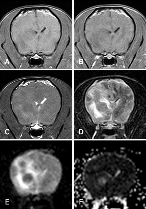
Fig. 2 Transverse MR images of Case 2. There were multiple round masses in the forebrain with a mass effect. The masses contained an isointense signal on the T1W image (A) without contrast enhancement (B) along with isointense to hypointense signals on the T2W images (C). These lesions had a low signal on DWI analysis (E) and a high signal on the ADC map (F). Perilesional edema generated a hyperintense signal with DWI (E) and on the ADC map (F).
Cited by 1 articles
-
Magnetic resonance imaging characteristics of ischemic brain infarction over time in a canine stroke model
Sooyoung Choi, Daji Noh, Youngwhan Kim, Inseong Jeong, Hojung Choi, Youngwon Lee, Kija Lee
J Vet Sci. 2018;19(1):137-143. doi: 10.4142/jvs.2018.19.1.137.
Reference
-
1. Chang SC, Lai PH, Chen WL, Weng HH, Ho JT, Wang JS, Chang CY, Pan HB, Yang CF. Diffusion-weighted MRI features of brain abscess and cystic or necrotic brain tumors: comparison with conventional MRI. Clin Imaging. 2002; 26:227–236.
Article2. Desprechins B, Stadnik T, Koerts G, Shabana W, Breucq C, Osteaux M. Use of diffusion-weighted MR imaging in differential diagnosis between intracerebral necrotic tumors and cerebral abscesses. AJNR Am J Neuroradiol. 1999; 20:1252–1257.3. Garosi L, McConnell JF, Platt SR, Barone G, Baron JC, de Lahunta A, Schatzberg SJ. Clinical and topographic magnetic resonance characteristics of suspected brain infarction in 40 dogs. J Vet Intern Med. 2006; 20:311–321.
Article4. Holtås S, Geijer B, Strömblad LG, Maly-Sundgren P, Burtscher IM. A ring-enhancing metastasis with central high signal on diffusion-weighted imaging and low apparent diffusion coefficients. Neuroradiology. 2000; 42:824–827.
Article5. Karaarslan E, Arslan A. Diffusion weighted MR imaging in non-infarct lesions of the brain. Eur J Radiol. 2008; 65:402–416.
Article6. Koh DM, Collins DJ. Diffusion-weighted MRI in the body: applications and challenges in oncology. AJR Am J Roentgenol. 2007; 188:1622–1635.
Article7. Krabbe K, Gideon P, Wagn P, Hansen U, Thomsen C, Madsen F. MR diffusion imaging of human intracranial tumours. Neuroradiology. 1997; 39:483–489.
Article8. McConnell JF, Garosi L, Platt SR. Magnetic resonance imaging findings of presumed cerebellar cerebrovascular accident in twelve dogs. Vet Radiol Ultrasound. 2005; 46:1–10.
Article9. Nadal Desbarats L, Herlidou S, de Marco G, Gondry-Jouet C, Le Gars D, Deramond H, Idy-Peretti I. Differential MRI diagnosis between brain abscesses and necrotic or cystic brain tumors using the apparent diffusion coefficient and normalized diffusion-weighted images. Magn Reson Imaging. 2003; 21:645–650.
Article10. Park SH, Chang KH, Song IC, Kim YJ, Kim SH, Han MH. Diffusion-weighted MRI in cystic or necrotic intracranial lesions. Neuroradiology. 2000; 42:716–721.
Article11. Schaefer PW, Grant PE, Gonzalez RG. Diffusion-weighted MR imaging of the brain. Radiology. 2000; 217:331–345.
Article12. Sener RN. Herpes simplex encephalitis: diffusion MR imaging findings. Comput Med Imaging Graph. 2001; 25:391–397.
Article13. Sutherland-Smith J, King R, Faissler D, Ruthazer R, Sato A. Magnetic resonance imaging apparent diffusion coefficients for histologically confirmed intracranial lesions in dogs. Vet Radiol Ultrasound. 2011; 52:142–148.
Article14. Tsuchiya K, Katase S, Yoshino A, Hachiya J. Diffusionweighted MR imaging of encephalitis. AJR Am J Roentgenol. 1999; 173:1097–1099.
Article15. Wisner ER, Dickinson PJ, Higgins RJ. Magnetic resonance imaging features of canine intracranial neoplasia. Vet Radiol Ultrasound. 2011; 52:1 Suppl 1. S52–S61.
Article
- Full Text Links
-
- Actions
-
Cited
- CITED
-
- Close
- Share
-
- Similar articles
-
- Diffusion-weighted Imaging and Apparent Diffusion Coefficient Maps for the Evaluation of Pyogenic Ventriculitis
- Reversal of a Large Ischemic Lesion with Low Apparent Diffusion Coefficient Value by Rapid Spontaneous Recanalization
- Effect of Gd-DTPA on Diffusion in Canine Brain with Hyperacute Stroke
- Diffusion Tensor Imaging in Schizophrenia with Statistical Probabilistic Anatomical Mapping: A study of the Apparent Diffusion Coefficient Map
- Evaluating Spinal Canal Lesions Using Apparent Diffusion Coefficient Maps with Diffusion-Weighted Imaging
