Alveolar bone thickness and fenestration of incisors in untreated Korean patients with skeletal class III malocclusion: A retrospective 3-dimensional cone-beam computed tomography study
- Affiliations
-
- 1Department of Oral and Maxillofacial Radiology, Graduate School, Kyung Hee University, Seoul, Korea.
- 2Department of Orthodontics, Graduate School, Kyung Hee University, Seoul, Korea. bravortho@khu.ac.kr
- KMID: 2471840
- DOI: http://doi.org/10.5624/isd.2020.50.1.9
Abstract
- PURPOSE
The purpose of this study was to evaluate vertical bone loss and alveolar bone thickness in the maxillary and mandibular incisors of patients with skeletal class III malocclusion. This study also aimed to evaluate the periodontal condition of class III malocclusion patients who had not undergone orthodontic treatment.
MATERIALS AND METHODS
The sample included cone-beam computed tomography scans of 24 Korean subjects (3 male and 21 female). Alveolar bone thickness (ABT), alveolar bone area (ABA), alveolar bone loss (ABL), and fenestration of the maxillary and mandibular incisors were measured using 3-dimensional imaging software.
RESULTS
All incisors displayed an ABT of less than 1.0 mm from the labial surface to root level 7 (70% of the root length). A statistically significant difference was observed between the mandibular labial and lingual ABAs and between the maxillary labial and mandibular labial ABAs. The lingual ABA of the mandibular lateral incisors was larger than that of the mandibular central incisors. ABL was severe on the labial surface. A statistically significant difference was observed between the maxillary and mandibular labial ABL values(21.8% and 34.4%, respectively). Mandibular lingual ABL (27.6%) was significantly more severe than maxillary lingual ABL (18.3%) (P<0.05). Eighty-two fenestrations were found on the labial surfaces of the incisors, while only 2 fenestrations were observed on the lingual surfaces. Fenestrations were most commonly observed at root level 6.
CONCLUSION
Careful evaluation is needed before orthodontic treatment to avoid iatrogenic damage of periodontal support when treating patients with class III malocclusion.
MeSH Terms
Figure
-
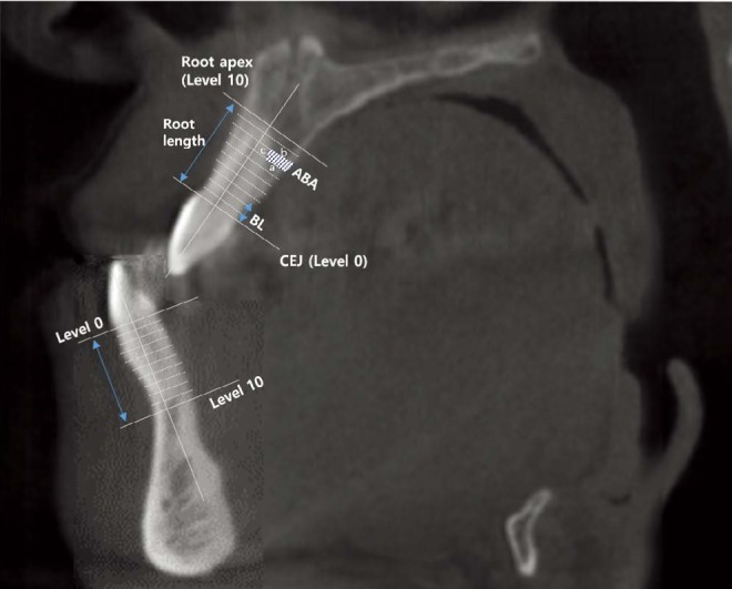
Fig. 1 A schematic diagram of the maxillary and mandibular reference points, lines, and measurement variables used in the present study. ABA: alveolar bone area (mm2)=[12(a+b)×c], level 0: line perpendicular to the root axis in the cementoenamel junction (CEJ) area, level 10: line perpendicular to the root axis in the root apex area, BL: bone loss.
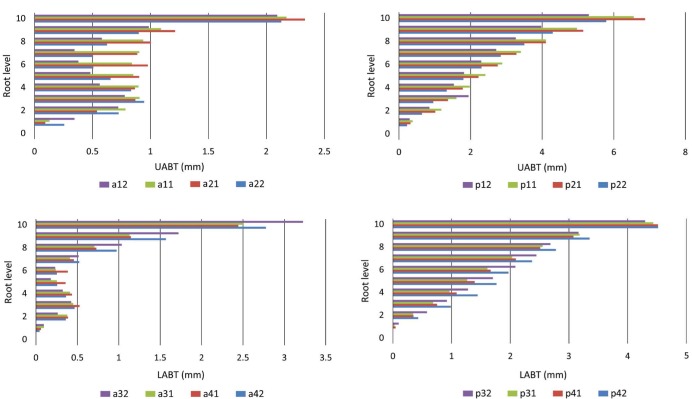
Fig. 2 Alveolar bone thickness(ABT) at each root level(0–10) for each of the incisors(#12–22, #32–42). a: labial side, p: lingual side.
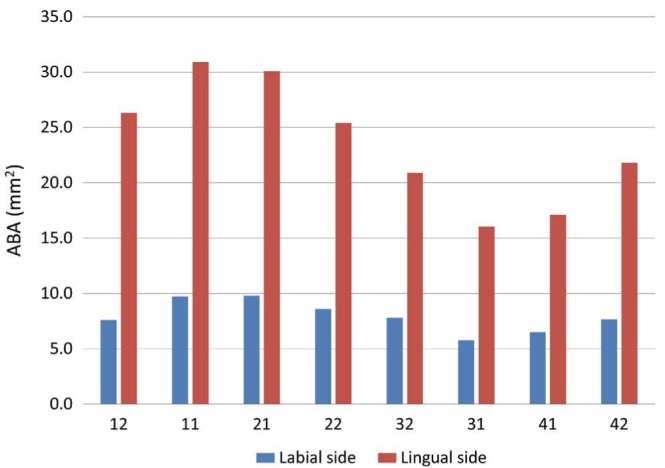
Fig. 3 Alveolar bone area (ABA, mm2) of incisor cross-sections. A statistically significant difference was observed between the upper and lower incisors.
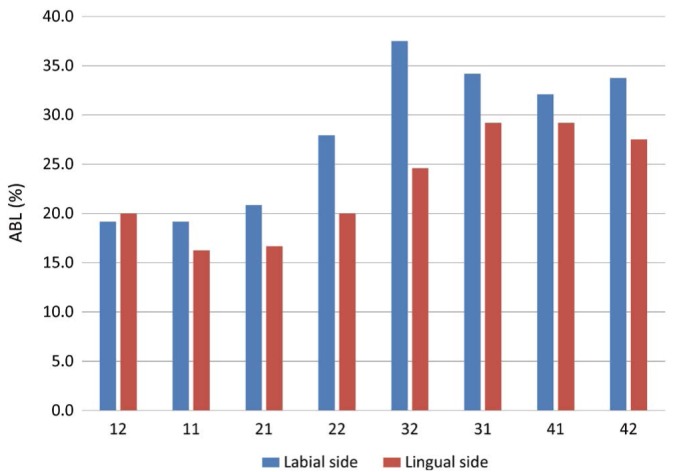
Fig. 4 Alveolar bone loss(ABL) of the labial and lingual sides(%).
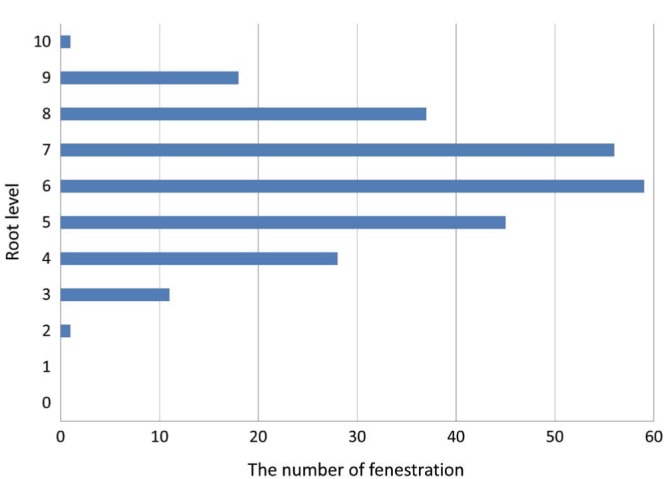
Fig. 5 Prevalence of fenestrations according to root level.
Reference
-
1. Wennström JL. Mucogingival considerations in orthodontic treatment. Semin Orthod. 1996; 2:46–54. PMID: 9161283.
Article2. Handelman CS. The anterior alveolus: its importance in limiting orthodontic treatment and its influence on the occurrence of iatrogenic sequelae. Angle Orthod. 1996; 66:95–110. PMID: 8712499.3. Wehrbein H, Bauer W, Diedrich P. Mandibular incisors, alveolar bone, and symphysis after orthodontic treatment. A retrospective study. Am J Orthod Dentofacial Orthop. 1996; 110:239–246. PMID: 8814023.4. Vardimon AD, Oren E, Ben-Bassat Y. Cortical bone remodeling/tooth movement ratio during maxillary incisor retraction with tip versus torque movements. Am J Orthod Dentofacial Orthop. 1998; 114:520–529. PMID: 9810048.
Article5. Kim Y, Park JU, Kook YA. Alveolar bone loss around incisors in surgical skeletal Class III patients. Angle Orthod. 2009; 79:676–682. PMID: 19537864.
Article6. Nahm KY, Kang JH, Moon SC, Choi YS, Kook YA, Kim SH, et al. Alveolar bone loss around incisors in Class I bidentoalveolar protrusion patients: a retrospective three-dimensional cone beam CT study. Dentomaxillofac Radiol. 2012; 41:481–488. PMID: 22184474.
Article7. Nelson PA, Artun J. Alveolar bone loss of maxillary anterior teeth in adult orthodontic patients. Am J Orthod Dentofacial Orthop. 1997; 111:328–334. PMID: 9082856.
Article8. Janson G, Bombonatti R, Brandão AG, Henriques JF, de Freitas MR. Comparative radiographic evaluation of the alveolar bone crest after orthodontic treatment. Am J Orthod Dentofacial Orthop. 2003; 124:157–164. PMID: 12923511.
Article9. Baumrind S, Frantz RC. The reliability of head film measurements. 2. Conventional angular and linear measures. Am J Orthod. 1971; 60:505–517. PMID: 5286677.10. Nakajima K, Yamaguchi T, Maki K. Surgical orthodontic treatment for a patient with advanced periodontal disease: evaluation with electromyography and 3-dimensional cone-beam computed tomography. Am J Orthod Dentofacial Orthop. 2009; 136:450–459. PMID: 19732680.
Article11. Lee SL, Kim HJ, Son MK, Chung CH. Anthropometric analysis of maxillary anterior buccal bone of Korean adults using conebeam CT. J Adv Prosthodont. 2010; 2:92–96. PMID: 21165276.
Article12. Yagci A, Veli I, Uysal T, Ucar FI, Ozer T, Enhos S. Dehiscence and fenestration in skeletal Class I, II, and III malocclusions assessed with cone-beam computed tomography. Angle Orthod. 2012; 82:67–74. PMID: 21696298.
Article13. Evangelista K, Vasconcelos Kde F, Bumann A, Hirsch E, Nitka M, Silva MA. Dehiscence and fenestration in patients with Class I and Class II Division 1 malocclusion assessed with cone-beam computed tomography. Am J Orthod Dentofacial Orthop. 2010; 138:133.e1–133.e7. PMID: 20691344.
Article14. Choe HY, Park W, Jeon JK, Kim YH, Shon BW. Differences in mandibular anterior alveolar bone thickness according to age in a normal skeletal group. Korean J Orthod. 2007; 37:220–230.15. Kim YS, Cha JY, Yu HS, Hwang CJ. Comparison of mandibular anterior alveolar bone thickness in different facial skeletal types. Korean J Orthod. 2010; 40:314–324.
Article16. Chakeres DW. Clinical significance of partial volume averaging of the temporal bone. AJNR Am J Neuroradiol. 1984; 5:297–302. PMID: 6426283.
- Full Text Links
-
- Actions
-
Cited
- CITED
-
- Close
- Share
-
- Similar articles
-
- Assessment of lower incisor alveolar bone width using cone-beam computed tomography images in skeletal Class III adults of different vertical patterns
- Alveolar bone thickness around maxillary central incisors of different inclination assessed with cone-beam computed tomography
- Analysis of the root position of the maxillary incisors in the alveolar bone using cone-beam computed tomography
- Comparison of interradicular distances and cortical bone thickness in Thai patients with Class I and Class II skeletal patterns using cone-beam computed tomography
- Quantitative evaluation of palatal bone thickness in patients with normal and open vertical skeletal configurations using cone-beam computed tomography
