False-Negative Results of Real-Time Reverse-Transcriptase Polymerase Chain Reaction for Severe Acute Respiratory Syndrome Coronavirus 2: Role of Deep-Learning-Based CT Diagnosis and Insights from Two Cases
- Affiliations
-
- 1Department of Radiology, Beijing Haidian Section of Peking University Third Hospital (Beijing Haidian Hospital), Beijing, China. 724501143@qq.com
- 2Institute of Advanced Research, Infervision, Beijing, China.
- 3Department of Infection, Beijing Haidian Section of Peking University Third Hospital (Beijing Haidian Hospital), Beijing, China.
- KMID: 2471817
- DOI: http://doi.org/10.3348/kjr.2020.0146
Abstract
- The epidemic of 2019 novel coronavirus, later named as severe acute respiratory syndrome coronavirus 2 (SARS-CoV-2), is still gradually spreading worldwide. The nucleic acid test or genetic sequencing serves as the gold standard method for confirmation of infection, yet several recent studies have reported false-negative results of real-time reverse-transcriptase polymerase chain reaction (rRT-PCR). Here, we report two representative false-negative cases and discuss the supplementary role of clinical data with rRT-PCR, including laboratory examination results and computed tomography features. Coinfection with SARS-COV-2 and other viruses has been discussed as well.
Keyword
MeSH Terms
Figure
-
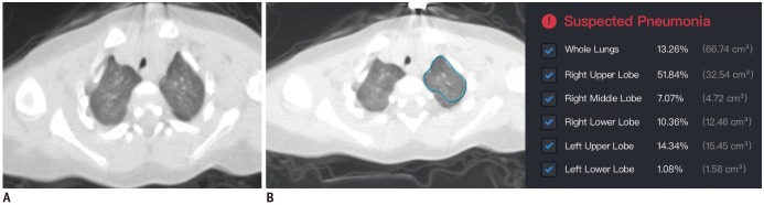
Fig. 1 Chest CT scans for 10-month-old patient in Case 1.A. Thin-slice CT scan that shows glimpse of lesions (breathing-induced motion artifacts are heavy for patient). CT shows diffuse ill-defined ground-glass opacities in both upper lung lobes. B. Representative of DL-based segmentation of lesions in left lung that shows overview of automatically calculated abnormality proportions. Artificial intelligence alarms suspected pneumonia based on relatively large proportion of abnormalities in lung. Detailed abnormality proportions in whole lungs, right upper lobe, right middle lobe, right lower lobe, left upper lobe, and left lower lobe were calculated and listed. CT = computed tomography, DL = deep learning
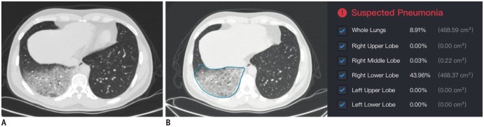
Fig. 2 Chest CT scans for patient in Case 2.A. Thin-slice CT scan that shows glimpse of lesions. CT shows diffuse ground-glass opacities in dependent area of right lower lobe. B. Representative of DL-based segmentation of lesions in lower lobe of right lung that shows overview of automatically calculated ratios. Artificial intelligence alarms suspected pneumonia based on relatively large proportion of abnormalities in lung. Detailed abnormality proportions in whole lungs, right upper lobe, right middle lobe, right lower lobe, left upper lobe, and left lower lobe were calculated and listed.
Reference
-
1. Gorbalenya AE, Baker SC, Baric RS, de Groot RJ, Drosten C, Gulyaeva AA, et al. Severe acute respiratory syndrome-related coronavirus: the species and its viruses - a statement of the Coronavirus Study Group. bioRxiv;2020. Accessed February 11,2020. Available at: . DOI: 10.1101/2020.02.07.937862.2. Zhu N, Zhang D, Wang W, Li X, Yang B, Song J, et al. China Novel Coronavirus Investigating and Research Team. A novel Coronavirus from patients with pneumonia in China, 2019. N Engl J Med. 2020; 382:727–773. PMID: 31978945.
Article3. Xie X, Zhong Z, Zhao W, Zheng C, Wang F, Liu J. Chest CT for typical 2019-nCoV pneumonia: relationship to negative RT-PCR testing. Radiology. 2020; 2. 12. DOI: 10.1148/radiol.2020200343. [Epub].4. Chung M, Bernheim A, Mei X, Zhang N, Huang M, Zeng X, et al. CT imaging features of 2019 novel Coronavirus (2019-nCoV). Radiology. 2020; 2. 04. DOI: 10.1148/radiol.2020200230. [Epub].5. National Health Commission of the People's Republic of China. Diagnosis and treatment protocols of pneumonia caused by a novel coronavirus (trial version 5). Beijing: National Health Commission of the People's Republic of China;2020.
- Full Text Links
-
- Actions
-
Cited
- CITED
-
- Close
- Share
-
- Similar articles
-
- Laboratory Diagnosis of COVID-19 in Korea
- Detection of Severe Acute Respiratory Syndrome Coronavirus 2 in the Pleural Fluid
- Variability in the Cycle Threshold Values of SARS-CoV-2 Real-Time Reverse Transcription Polymerase Chain Reaction Test: A Review of the Nationwide Proficiency Testing Survey in Korea
- Universal Screening of Severe Acute Respiratory Syndrome Coronavirus 2 with Polymerase Chain Reaction Testing after Rally of Trainee Doctors
- A Case of Severe Enterovirus Pneumonia in an Immunocompetent Adult
