Histological analysis of explanted implant-bone interface: a case report
- Affiliations
-
- 1Department of Periodontology, College of Dentistry, Dankook University, Cheonan, Republic of Korea. jcp@dent.dku.edu
- 2Department of Prosthodontics, College of Dentistry, Dankook University, Cheonan, Republic of Korea.
- KMID: 2470629
- DOI: http://doi.org/10.14368/jdras.2019.35.4.235
Abstract
- Osseointegration has been reported to be a dynamic process in which the alveolar bone comes in direct contact with the implant. Various methods were tried to evaluate degree of osseointegration and the measurement of bone-implant contact (BIC) have been commonly used among them. To properly assess the BIC, only histologic analysis is available. However, few studies evaluated BIC of successfully osseointegrated implants in humans. Thus, this is a unique opportunity when implants should be explanted due to inappropriate positioning of implant, presence of pain or sensory disturbance, or broken screw or fixture. This report presents a case of the implant underwent 3-year functional load and a histologic analysis after the fixture fracture. The histomorphometric analysis revealed 53.1% of BIC measured along the whole implant and 70.9% measured only in subcrestal area, respectively. In the present study, although the implant was fractured, a high degree of BIC was observed.
Keyword
Figure
-
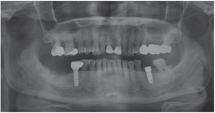
Fig. 1 Panoramic radiograph after implant placement (#36).
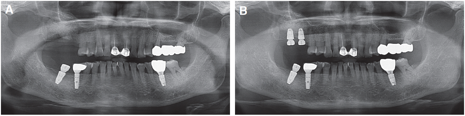
Fig. 2 Panoramic radiograph. (A) 1 year recall after implant placement, (B) 1.5 years recall.
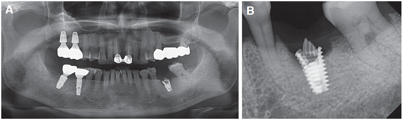
Fig. 3 Radiographs when returned to the clinic with the complaint of loose implant 3.5 years after implant placement. (A) Paroramic view, (B) Periapical view.

Fig. 4 Clinical photographs illustrating surgical procedure. (A) After flap reflection. Granulation tissue around fractured implant fixture can be observed, (B) After explantation using trephine bur, (C) Removed fragment of implant fixture, (D) After alveolar ridge preservation with deproteinized bovine bone mineral with 10% porcine collagen (Bio-Oss Collagen, Geislitch, Wolhusen, Switzerland) and resorbable collagen membrane (Collagen graft, GenOss, Seoul, Korea).
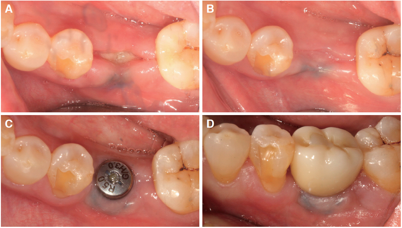
Fig. 5 Clinical photographs illustrating surgical procedure. (A) 2 weeks after alveolar ridge preservation, (B) 5 months after alveolar ridge preservation, (C) After 1-stage implant surgery, (D) After final crown placement.
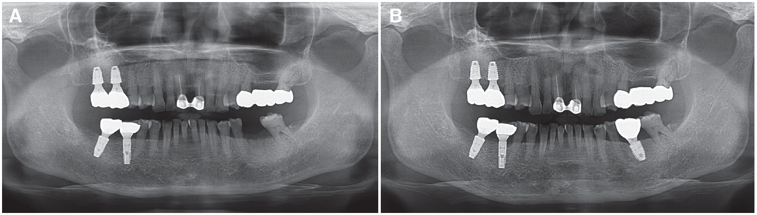
Fig. 6 Paroramic radiographs. (A) Immediately after explantation and alveolar ridge preservation, (B) After final crown placement.
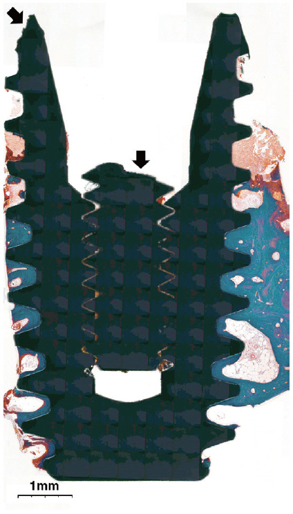
Fig. 7 A general overview of the sectioned specimen (×15). The arrows indicate implant fracture line and abutment screw fracture line, respectively.
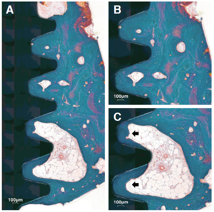
Fig. 8 Medium magnification views of bone tissue in the sectioned specimen. (A) The implant threads are well surrounded by compact mature bone (×40). (B) Close contact is continued between the implant surface and the compact bone (×50). (C) Implant threads are surrounded by band-shaped bone with marrow space (Black arrows, ×50).
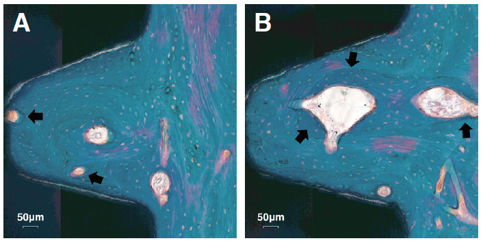
Fig. 9 High magnification views of the sectioned specimen (×100). (A) Osteons in contact with the implant surface are observed (Black arrows). (B) Newlyformed bone is demarcated from previously-formed bone by darker staining color (Black arrows).
Reference
-
References
1. Iezzi G, Piattelli A, Mangano C, Degidi M, Testori T, Vantaggiato G, Fiera E, Frosecchi M, Floris P, Perroni R, Ravera L, Moreno GG, De Martinis E, Perrotti V. 2016; Periimplant bone response in human-retrieved, clinically stable, successful, and functioning dental implants after a long-term loading period: a report of 17 cases from 4 to 20 years. Implant Dent. 25:380–6. DOI: 10.1097/ID.0000000000000415. PMID: 27002775.2. Cordioli G, Majzoub Z, Piattelli A, Scarano A. 2000; Removal torque and histomorphometric investigation of 4 different titanium surfaces: an experimental study in the rabbit tibia. Int J Oral Maxillofac Implants. 15:668–74. PMID: 11055134.3. Sakakura CE, Margonar R, Holzhausen M, Nociti FH Jr, Alba RC Jr, Marcantonio E Jr. 2003; Influence of cyclosporin A therapy on bone healing around titanium implants: a histometric and biomechanic study in rabbits. J Periodontol. 74:976–81. DOI: 10.1902/jop.2003.74.7.976. PMID: 12931759.4. Degidi M, Petrone G, Iezzi G, Piattelli A. 2003; Bone contact around acid-etched implants: a histological and histomorphometrical evaluation of two human-retrieved implants. J Oral Implantol. 29:13–8. DOI: 10.1563/1548-1336(2003)029<0013:BCAAIA>2.3.CO;2. PMID: 12614080.5. Brunel G, Armand S, Miller N, Rue J. 2000; Histologic analysis of a fractured implant: a case report. Int J Periodontics Restorative Dent. 20:520–6.6. Degidi M, Petrone G, Iezzi G, Piattelli A. 2002; Histologic evaluation of a human immediately loaded titanium implant with a porous anodized surface. Clin Implant Dent Relat Res. 4:110–4. DOI: 10.1111/j.1708-8208.2002.tb00160.x. PMID: 12121611.7. Sennerby L, Ericson LE, Thomsen P, Lekholm U, Astrand P. 1991; Structure of the bone-titanium interface in retrieved clinical oral implants. Clin Oral Implants Res. 2:103–11. DOI: 10.1034/j.1600-0501.1991.020302.x. PMID: 1843463.8. Johansson CB, Hansson HA, Albrektsson T. 1990; Qualitative interfacial study between bone and tantalum, niobium or commercially pure titanium. Biomaterials. 11:277–80. DOI: 10.1016/0142-9612(90)90010-N. PMID: 2383624.9. Donath K, Breuner G. 1982; A method for the study of undecalcified bones and teeth with attached soft tissues. The Sage-Schliff (sawing and grinding) technique. 11:318–26. DOI: 10.1111/j.1600-0714.1982.tb00172.x. PMID: 6809919.10. Shah FA, Nilson B, Branemark R, Thomsen P, Palmquist A. 2014; The bone-implant interface - nanoscale analysis of clinically retrieved dental implants. Nanomedicine. 10:1729–37. DOI: 10.1016/j.nano.2014.05.015. PMID: 24941460.11. Mangano FG, Pires JT, Shibli JA, Mijiritsky E, Iezzi G, Piattelli A, Mangano C. 2017; Early bone response to dual acid-etched and machined dental implants placed in the posterior maxilla: a histologic and histomorphometric human study. Implant Dent. 26:24–9. DOI: 10.1097/ID.0000000000000511. PMID: 27861190.12. Schwartz Z, Lohmann C, Oefinger J, Bonewald LF, Dean DD, Boyan BD. 1999; Implant surface characteristics modulate differentiation behavior of cells in the osteoblastic lineage. Adv Dent Res. 13:38–48. DOI: 10.1177/08959374990130011301. PMID: 11276745.13. Buser D, Schenk RK, Steinemann S, Fiorellini JP, Fox CH, Stich H. 1991; Influence of surface characteristics on bone integration of titanium implants. A histomorphometric study in miniature pigs. 25:889–902. DOI: 10.1002/jbm.820250708. PMID: 1918105.14. Hayakawa T, Kiba H, Yasuda S, Yamamoto H, Nemoto K. 2002; A histologic and histomorphometric evaluation of two types of retrieved human titanium implants. Int J Periodontics Restorative Dent. 22:164–71.15. Schwarz MS. 2000; Mechanical complications of dental implants. Clin Oral Implants Res. 11:156–8. DOI: 10.1034/j.1600-0501.2000.011S1156.x. PMID: 11168264.16. Rangert B, Krogh PH, Langer B, Van Roekel N. 1995; Bending overload and implant fracture: a retrospective clinical analysis. Int J Oral Maxillofac Implants. 10:326–34. PMID: 7615329.17. Balshi TJ, Wolfinger GJ. 1997; Two-implant-supported single molar replacement: interdental space requirements and comparison to alternative options. Int J Periodontics Restorative Dent. 17:426–35. PMID: 9497731.18. Steflik DE, Parr GR, Singh BB, Lake FT, Sisk AL, Howell FV, Shelton TW. 1994; Light microscopic and scanning electron microscopic analyses of dental implants retrieved from humans. J Oral Implantol. 20:8–24. DOI: 10.1097/00008505-199412000-00017. PMID: 7932859.19. Piattelli A, Scarano A, Piattelli M, Vaia E, Matarasso S. 1998; Hollow implants retrieved for fracture: a light and scanning electron microscope analysis of 4 cases. J Periodontol. 69:185–9. DOI: 10.1902/jop.1998.69.2.185. PMID: 9526918.20. Eckert SE, Meraw SJ, Cal E, Ow RK. 2000; Analysis of incidence and associated factors with fractured implants: a retrospective study. Int J Oral Maxillofac Implants. 15:662–7. PMID: 11055133.
- Full Text Links
-
- Actions
-
Cited
- CITED
-
- Close
- Share
-
- Similar articles
-
- Marginal bone level changes in association with different vertical implant positions: a 3-year retrospective study
- Finite element study on the effect of abutment length and material on implant bone interface against dynamic loading
- Torque and mechanical failure of orthodontic micro-implant influenced by implant design parameters
- Stress-strain distribution at bone-implant interface of two splinted overdenture systems using 3D finite element analysis
- Finite element analysis of the effect of cantilever and implant orientation on stress distribution in a mandibular implant-supported bar overdenture
