Ann Lab Med.
2020 Jul;40(4):312-316. 10.3343/alm.2020.40.4.312.
Development of an Automated Image Analyzer for Microvessel Density Measurement in Bone Marrow Biopsies
- Affiliations
-
- 1Department of Laboratory Medicine, Kangdong Sacred Heart Hospital, Seoul, Korea.
- 2Optical Research Team, Magok R&D Campus, Z-tec Co., Ltd., Seoul, Korea.
- 3Department of Laboratory Medicine, Center for Diagnostic Oncology, Hospital and Research Institute, National Cancer Center, Goyang, Korea. ksy@ncc.re.kr
- 4Department of Laboratory Medicine, Eone Laboratories, Incheon, Korea.
- 5Biostatistics Collaboration Team, Research Institute, National Cancer Center, Goyang, Korea.
- 6Department of Hematology-Oncology, Center for Hematologic Malignancy, National Cancer Center, Goyang, Korea.
- 7Department of Medical Engineering, Gachon University, Incheon, Korea. kimkg@gachon.ac.kr
- KMID: 2470327
- DOI: http://doi.org/10.3343/alm.2020.40.4.312
Abstract
- Angiogenesis is important for the proliferation and survival of multiple myeloma (MM) cells. Bone marrow (BM) microvessel density (MVD) is a useful marker of angiogenesis and an increase in MVD can be used as a marker of poor prognosis in MM patients. We developed an automated image analyzer to assess MVD from images of BM biopsies stained with anti-CD34 antibodies using two color models. MVD was calculated by merging images from the red and hue channels after eliminating non-microvessels. The analyzer results were compared with those obtained by two experienced hematopathologists in a blinded manner using the 84 BM samples of MM patients. Manual assessment of the MVD by two hematopathologists yielded mean±SD values of 19.4±11.8 and 20.0±11.8. The analyzer generated a mean±SD of 19.5±11.2. The intraclass correlation coefficient (ICC) and Bland-Altman plot of the MVD results demonstrated very good agreement between the automated image analyzer and both hematopathologists (ICC=0.893 [0.840-0.929] and ICC=0.906 [0.859-0.938]). This automated analyzer can provide time- and labor-saving benefits with more objective results in hematology laboratories.
MeSH Terms
Figure
-
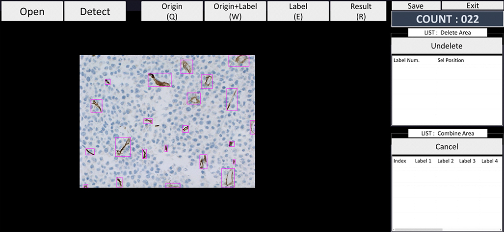
Fig. 1 MVD being calculated by the automated image analyzer using an image (×400) of a hot spot from BM section stained with anti-CD34 antibodies. Microvessels are marked by a pink box and the MVD count is displayed at the right upper corner. Abbreviations: MVD, microvessel density; BM, bone marrow.
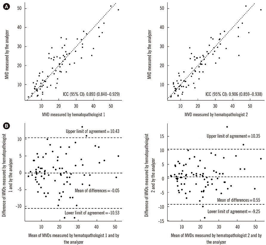
Fig. 2 Comparison of MVD between manual counting and the automated image analyzer for 84 BM biopsy samples from multiple myeloma patients. (A) intraclass correlation coefficient. (B) Bland-Altman plot. Abbreviations: MVD, microvessel density; BM, bone marrow; CI, confidence interval.
Reference
-
1. Bergers G, Benjamin LE. Tumorigenesis and the angiogenic switch. Nat Rev Cancer. 2003; 3:401–410.
Article2. Uzzan B, Nicolas P, Cucherat M, Perret GY. Microvessel density as a prognostic factor in women with breast cancer: a systematic review of the literature and meta-analysis. Cancer Res. 2004; 64:2941–2955.3. Galindo-Gallego M, Fernández-Aceñero MJ, Sanz-Ortega J, Aljama A, López-Elzaurdia C. Prognostic significance of microvascular counts in rectal carcinoma. Pathol Res Pract. 2000; 196:607–612.
Article4. Graham CH, Rivers J, Kerbel RS, Stankiewicz KS, White WL. Extent of vascularization as a prognostic indicator in thin (< 0.76 mm) malignant melanomas. Am J Pathol. 1994; 145:510–514.5. Hussong JW, Rodgers GM, Shami PJ. Evidence of increased angiogenesis in patients with acute myeloid leukemia. Blood. 2000; 95:309–313.
Article6. Aguayo A, Kantarjian H, Manshouri T, Gidel C, Estey E, Thomas D, et al. Angiogenesis in acute and chronic leukemias and myelodysplastic syndromes. Blood. 2000; 96:2240–2245.
Article7. Kini AR, Kay NE, Peterson LC. Increased bone marrow angiogenesis in B cell chronic lymphocytic leukemia. Leukemia. 2000; 14:1414–1418.
Article8. Vacca A, Ribatti D, Roncali L, Ranieri G, Serio G, Silvestris F, et al. Bone marrow angiogenesis and progression in multiple myeloma. Br J Haematol. 1994; 87:503–508.
Article9. Sezer O, Niemöller K, Eucker J, Jakob C, Kaufmann O, Zavrski I, et al. Bone marrow microvessel density is a prognostic factor for survival in patients with multiple myeloma. Ann Hematol. 2000; 79:574–577.
Article10. Rajkumar SV, Leong T, Roche PC, Fonseca R, Dispenzieri A, Lacy MQ, et al. Prognostic value of bone marrow angiogenesis in multiple myeloma. Clin Cancer Res. 2000; 6:3111–3116.11. Weidner N. Current pathologic methods for measuring intratumoral microvessel density within breast carcinoma and other solid tumors. Breast Cancer Res Treat. 1995; 36:169–180.
Article12. Bhatti SS, Kumar L, Dinda AK, Dawar R. Prognostic value of bone marrow angiogenesis in multiple myeloma: use of light microscopy as well as computerized image analyzer in the assessment of microvessel density and total vascular area in multiple myeloma and its correlation with various clinical, histological, and laboratory parameters. Am J Hematol. 2006; 81:649–656.
Article13. Yang JZ, Wu XD, Meng JB, Zhang JQ, Sun LX. Association of increased microvessel density with skeletal extramedullary disease relapse in multiple myeloma patients who have skeletal extramedullary disease at diagnosis. Pathol Res Pract. 2018; 214:1694–1699.
Article14. Rao L, De Veirman K, Giannico D, Saltarella I, Desantis V, Frassanito MA, et al. Targeting angiogenesis in multiple myeloma by the VEGF and HGF blocking DARPin protein MP0250: a preclinical study. Oncotarget. 2018; 9:13366–13381.
Article15. Rozic G, Paukov L, Cohen Z, Shapira I, Duek A, Bejamini O, et al. STK405759 as a combination therapy with bortezomib or dexamethasone, in in vitro and in vivo multiple myeloma models. Oncotarget. 2018; 9:31367–31379.16. Ntellas P, Perivoliotis K, Dadouli K, Koukoulis GK, Ioannou M. Microvessel density as a surrogate prognostic marker in patients with multiple myeloma: A meta-analysis. Acta Haematol. 2017; 138:77–84.
Article17. Shi P, Zhong J, Hong J, Huang R, Wang K, Chen Y. Automated Ki-67 quantification of immunohistochemical staining image of human nasopharyngeal carcinoma xenografts. Sci Rep. 2016; 6:32127.
Article18. Irshad H, Oh EY, Schmolze D, Quintana LM, Collins L, Tamimi RM, et al. Crowdsourcing scoring of immunohistochemistry images: evaluating performance of the crowd and an automated computational method. Sci Rep. 2017; 7:43286.
Article19. Guirado R, Carceller H, Castillo-Gómez E, Castrén E, Nacher J. Automated analysis of images for molecular quantification in immunohistochemistry. Heliyon. 2018; 4:e00669.
Article
- Full Text Links
-
- Actions
-
Cited
- CITED
-
- Close
- Share
-
- Similar articles
-
- Image Analysis Quantifying Microvessel Density in Renal Cell Carcinoma
- Clinical significance of microvessel density in multiple myeloma patients
- Analysis of the Bone Marrow Aspirates with Automated Hematology Analyzer for Assessment of the Bone Marrow Cellularity and Effective Hematopoiesis
- The determination of reference material for bone density by using bone phantom
- A Study of Bone Marrow Density in Korean Children of Normal Growth and Development
