Full mouth rehabilitation on the patient with deep bite and posterior bite collapse using re-establishment of occlusal vertical dimension
- Affiliations
-
- 1Department of Prosthodontics, School of Dentistry, Chonnam National University, Gwangju, Republic of Korea. yhsdent@chonnam.ac.kr
- KMID: 2469921
- DOI: http://doi.org/10.4047/jkap.2020.58.1.50
Abstract
- The loss of posterior support and the abnormal jaw relation can cause pathologic findings. If deep bite patients with multiple missing teeth, can not have the stable posterior contact, the mandible moves posteriorly, and consequently the overjet and overbite get worse. And when the mandibular irregular occlusal plane is corrected, it is easier to have the bilateral balanced occlusion with the maxilla. So the treatment goal is to give proper posetrior support and establish appropriate anterior guidance, and ultimately provide improved mastication and esthetics recovery. In this case, a 68 year old man, having deep bite without posterior support was evaluated by the vertical dimesion decision flow-chart. An available prosthetic height, anterior occlusal relation such as overjet, overbite and the esthetic part such as facial height and the cephalometric analysis are the factors to be considered.
MeSH Terms
Figure
-
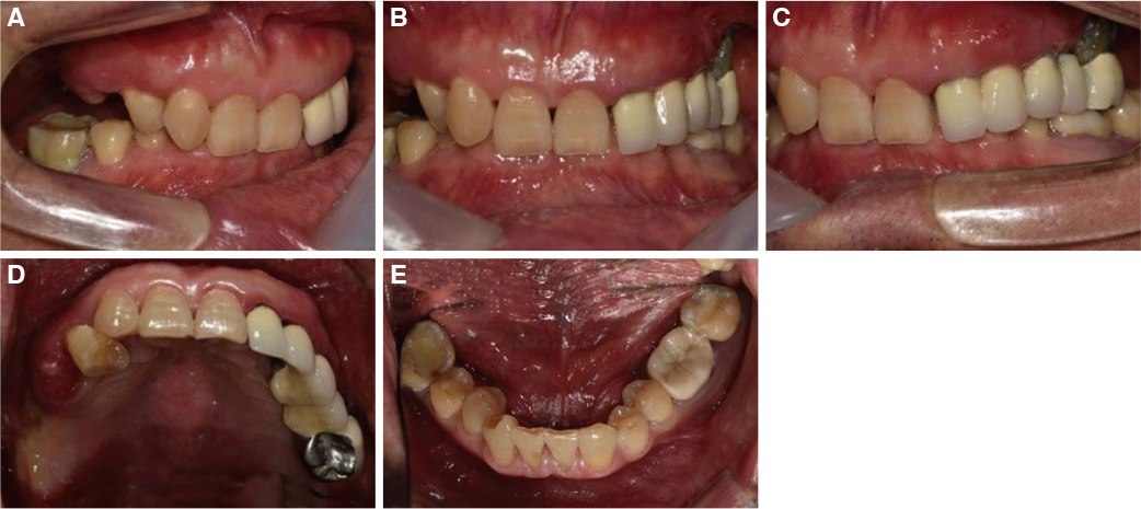
Fig. 1 Initial intraoral photographs. (A) Right lateral view, (B) Frontal view, (C) Left lateral view, (D) Maxillary occlusal view, (E) Mandibular occlusal view.
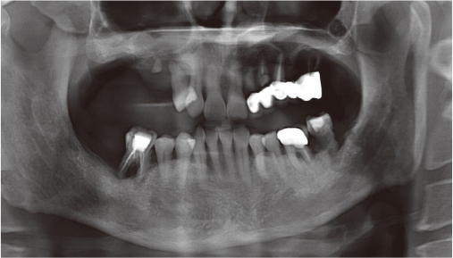
Fig. 2 Initial panoramic radiograph.
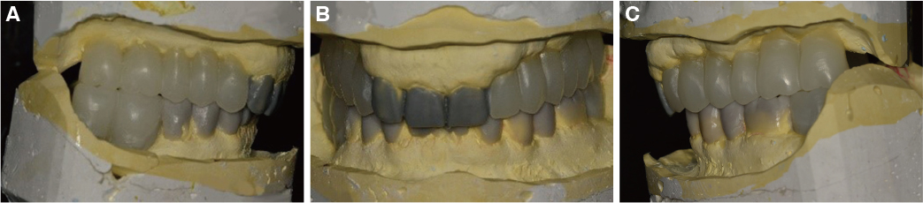
Fig. 3 Diagnostic wax up model with new occlusal vertical dimension. (A) Right lateral view, (B) Frontal view, (C) Left lateral view.
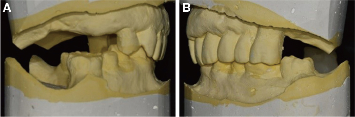
Fig. 4 Initial cast analysis (A) Right lateral view, (B) Left lateral view.
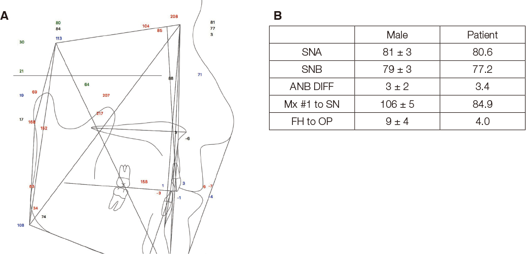
Fig. 5 Cephalometric analysis (A) Lateral Cephalography (B) Comparison of the cephalometric analysis on the patient and the male.
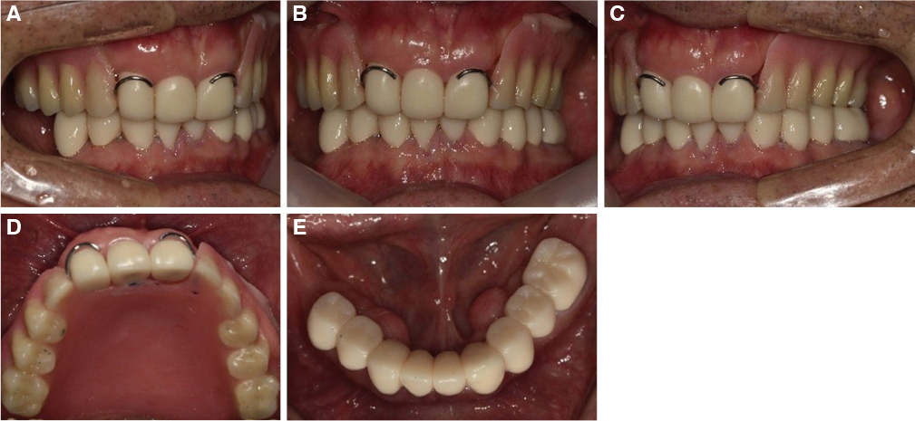
Fig. 6 Provisional restoration. (A) Right lateral view, (B) Frontal view, (C) Left lateral view, (D) Maxillary occlusal view, (E) Mandibular occlusal view.
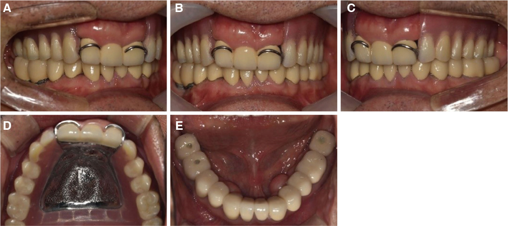
Fig. 7 Definitive restoration. (A) Right lateral view, (B) Frontal view, (C) Left lateral view, (D) Maxillary occlusal view, (E) Mandibular occlusal view.
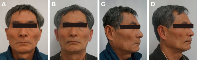
Fig. 8 Patient's profile. (A) Frontal profile of first visit, (B) Frontal profile of after definitive denture delivery, (C) Lateral profile of first visit, (D) Lateral profile of after definitive denture delivery.
Reference
-
1. Turner KA, Missirlian DM. Restoration of the extremely worn dentition. J Prosthet Dent. 1984; 52:467–474.
Article2. Willis FM. Features of the face involved in full denture prosthesis. Dent Cosmos. 1935; 77:851–854.3. Hull CA, Junghans JA. A cephalometric approach to establishing the facial vertical dimension. J Prosthet Dent. 1968; 20:37–42.
Article4. Ibbetson RJ, Setchell DJ. Treatment of the worn dentition: 2. Dent Update. 1989; 16:300–302. 305–307.5. Rebibo M, Darmouni L, Jouvin J, Orthelieb JD. Vertical dimension of occlusion: The keys to decision. Inter J Stomat Occ Med. 2009; 2:147–159.
Article6. Johnson A, Wildgoose DG, Wood DJ. The determination of freeway space using two different methods. J Oral Rehabil. 2002; 29:1010–1013.
Article7. Park JH, Jeong CM, Jeon YC, Lim JS. A study on the occlusal plane and the vertical dimension in Korean adults with natural dentition. J Korean Acad Prosthodont. 2005; 43:41–51.8. Turner KA, Missirlian DM. Restoration of the extremely worn dentition. J Prosthet Dent. 1984; 52:467–474.
Article9. Okeson JP. Management of temporomandibular disorders and occlusion. 7th ed. St. Louis: CV Mosby;2013. p. 459–468.10. Dawson PE. Functional occlusion from TMJ to smile design. 1st ed. New York: Elsevier Inc.;2007. p. 114–119.11. Rivera-Morales WC, Mohl ND. Relationship of occlusal vertical dimension to the health of the masticatory system. J Prosthet Dent. 1991; 65:547–553.
Article12. Orthlieb JD, Laurent M, Laplanche O. Cephalometric estimation of vertical dimension of occlusion. J Oral Rehabil. 2000; 27:802–807.
Article13. Edwards CL, Richards MW, Billy EJ, Neilans LC. Using computerized cephalometrics to analyze the vertical dimension of occlusion. Int J Prosthodont. 1993; 6:371–376.14. Obana J. Prosthodontic treatment for maxillary and mandibular teeth cross each other. 1st ed. Tokyo: Ishiyaku Publishers;1994. p. 2–4.15. Sierpinska T, Kuc J, Golebiewska M. Morphological and functional parameters in patients with tooth wear before and after treatment. Open Dent J. 2013; 7:55–61.
Article
- Full Text Links
-
- Actions
-
Cited
- CITED
-
- Close
- Share
-
- Similar articles
-
- Full mouth rehabilitation of patient with decreased occlusal vertical dimension due to severely worn dentition and posterior bite collapse
- Full mouth rehabilitation on the patient with class II jaw relation and posterior bite collapse using reestablishment of occlusal vertical dimension: a case report
- Full mouth rehabilitation of deep bite patient with segmental osteotomy and orthodontic treatment
- Full-mouth rehabilitation of partial edentulism in a deep bite patient
- Full mouth rehabilitation in patient with deep bite, inter-dental arch discrepancy and loss of vertical dimension: a case report
