Full-mouth rehabilitation of partial edentulism in a deep bite patient
- Affiliations
-
- 1Department of Prosthodontics and Research Institute of Oral Science, College of Dentistry, Gangneung-Wonju National University, Gangneung, Republic of Korea. doctorcj@gwnu.ac.kr
- KMID: 2377188
- DOI: http://doi.org/10.4047/jkap.2017.55.2.187
Abstract
- Deep overbite patients who do not have proper occlusal relationship may cause problems such as teeth wear and antagonist extrusion. These lead to the collapse of occlusal plane and esthetic problem. Increasing vertical dimension is frequently essential to resolve those problems. This case report demonstrates a full-mouth rehabilitation for a patient with severe deep bite that contacts surface to surface by increasing vertical dimension. Treatment procedures included diagnosis, treatment planning, implant surgery, and prosthodontic rehabilitation. Satisfactory results were obtained in functional and esthetic aspects.
Figure
-
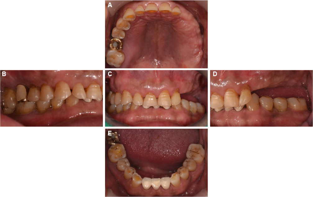
Fig. 1 Intraoral photographs. (A) Anterior dentition attrition, (B) #14,15 non-carious cervical lesion, (C) Anterior deep bite, (D) #23,24,25,26,27 missing, (E) #32=42 PFM bridge fracture.
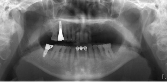
Fig. 2 Preoperative panoramic radiograph.
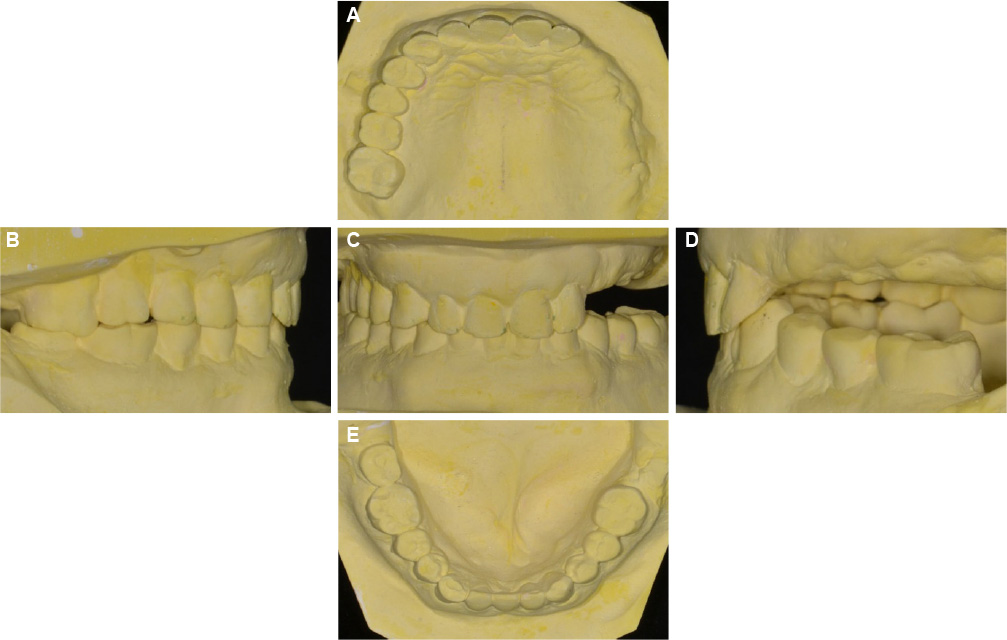
Fig. 3 Diagnostic cast analysis. (A) Anterior dentition attrition (B) Reverse curve of Spee (C, D) Anterior deep bite (E) Mandibular occlusal view.

Fig. 4 Diagnostic wax-up. (A) Maxillary occlusal view, (B) Left lateral view at centric occlusion, (C) Frontal view at centric occlusion, (D) Right lateral view at centric occlusion, (E) Mandibular occlusal view.

Fig. 5 Die preparation. (A) Occlusal view (maxilla), (B) Occlusal view (mandible).

Fig. 6 Interocclusal relationship registrations using provisional restorations. (A) registration of provisional restoration, (B, C, D) registration of maxillary abutment, (E, F) registration of mandibular abutment.

Fig. 7 Implant customized abutment. (A) Implant axis (yellow line), (B) Customized abutment.
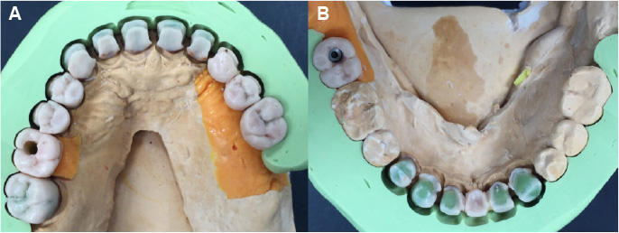
Fig. 8 Full contour wax-up cut back. (A) Occlusal view (maxilla), (B) Occlusal view (mandible).
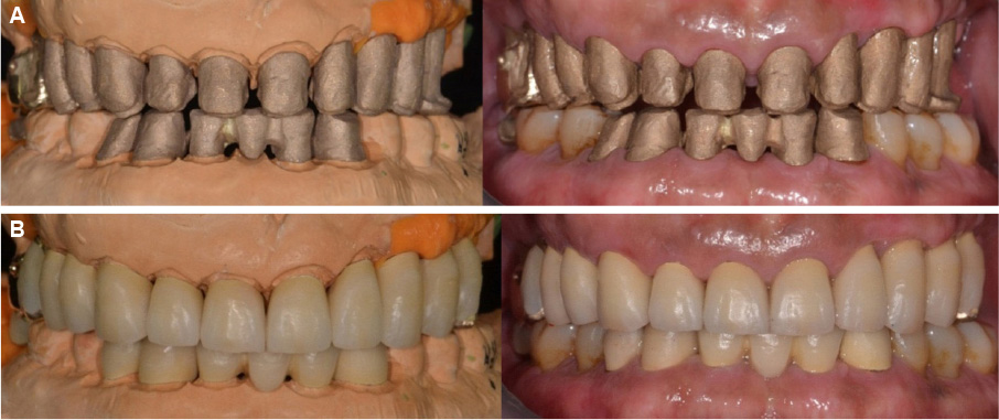
Fig. 9 Gold coping try-in and porcelain build-up. (A) Gold coping, (B) Porcelain build-up.
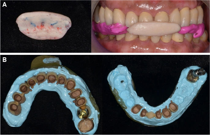
Fig. 10 Remount procedure. (A) Anterior programming device, (B) Pick-up impression.
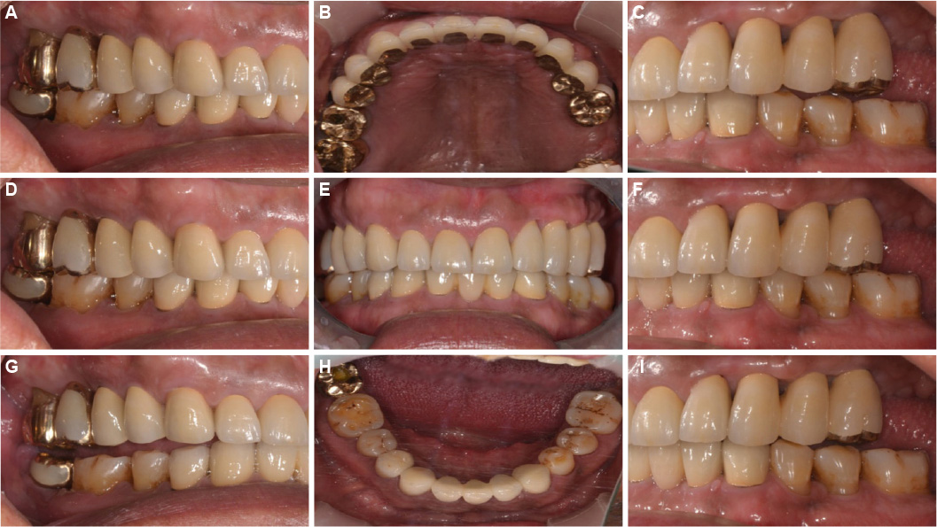
Fig. 11 Definitive prosthesis. (A) Working side during right lateral excursion, (B) Maxillary occlusal view, (C) Non-working side during right lateral excursion, (D) Left lateral view at centric occlusion, (E) Frontal view at centric occlusion, (F) Right lateral view at centric occlusion, (G) Non-working side during left lateral excursion, (H) Mandibular occlusal view, (I) Working side during left lateral excursion.
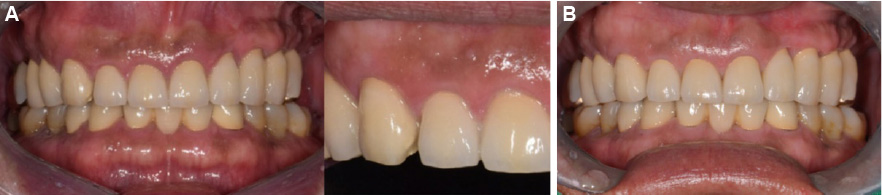
Fig. 12 Intraoral photographs. (A) #13 porcelain fracture, (B) #13 monolithic zirconia crown restoration.
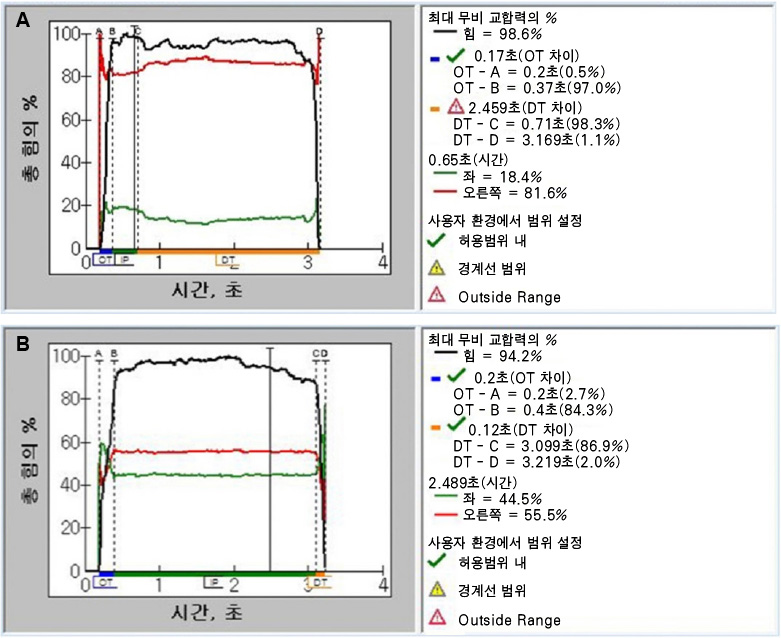
Fig. 13 T-scan data. (A) Before occlusal adjustment, (B) After occlusal adjustment.
Reference
-
1. The glossary of prosthodontic terms. J Prosthet Dent. 2005; 94:10–92.2. Dawson PE. Functional occlusion: from TMJ to smile design. St. Louis, Mo.: Mosby;2007. p. xiii. p. 630.3. McDonagh S, Chadwick J. The combined orthodontic and surgical treatment of traumatic Class II division 2 in the adult. Dent Update. 2004; 31:83–88. 90–91.
Article4. Beddis HP, Durey K, Alhilou A, Chan MF. The restorative management of the deep overbite. Br Dent J. 2014; 217:509–515.
Article5. Ambard A, Mueninghoff L. Planning restorative treatment for patients with severe Class II malocclusions. J Prosthet Dent. 2002; 88:200–207.
Article6. Akerly WB. Prosthodontic treatment of traumatic overlap of the anterior teeth. J Prosthet Dent. 1977; 38:26–34.
Article7. Willis FM. Features of the face involved in full denture prosthesis. Dent Cosmos. 1935; 77:851–854.8. McGee AM, Skinner M. Facial asymmetry and the attribution of personality traits. Br J Soc Psychol. 1987; 26:181–184.
Article9. Curtis TA, Langer Y, Curtis DA, Carpenter R. Occlusal considerations for partially or completely edentulous skeletal class II patients. Part I: Background information. J Prosthet Dent. 1988; 60:202–211.
Article10. Burnett CA, Clifford TJ. The mandibular speech envelope in subjects with and without incisal tooth wear. Int J Prosthodont. 1999; 12:514–518.11. Ash MM, Ramfjord SP. Occlusion. 4th ed. Philadelphia: W.B. Saunders;1995. p. viii. p. 472.12. Capp NJ, Warren K. Restorative treatment for patients with excessive vertical overlap. Int J Prosthodont. 1991; 4:353–360.13. Gopi Chander N, Venkat R. An appraisal on increasing the occlusal vertical dimension in full occlusal rehabilitation and its outcome. J Indian Prosthodont Soc. 2011; 11:77–81.
Article14. Ratnasari A, Hasegawa K, Oki K, Kawakami S, Yanagi Y, Asaumi JI, et al. Manifestation of preferred chewing side for hard food on TMJ disc displacement side. J Oral Rehabil. 2011; 38:12–17.
Article15. Diernberger S, Bernhardt O, Schwahn C, Kordass B. Self-reported chewing side preference and its associations with occlusal, temporomandibular and prosthodontic factors: results from the population-based Study of Health in Pomerania (SHIP-0). J Oral Rehabil. 2008; 35:613–620.
Article16. Wilding RJ, Adams LP, Lewin A. Absence of association between a preferred chewing side and its area of functional occlusal contact in the human dentition. Arch Oral Biol. 1992; 37:423–428.
Article17. Bates JF, Stafford GD, Harrison A. Masticatory function--a review of the literature. 1. The form of the masticatory cycle. J Oral Rehabil. 1975; 2:281–301.18. Yamasaki Y, Kuwatsuru R, Tsukiyama Y, Oki K, Koyano K. Objective assessment of mastication predominance in healthy dentate subjects and patients with unilateral posterior missing teeth. J Oral Rehabil. 2016; 43:575–582.
Article19. Williamson EH, Lundquist DO. Anterior guidance: its effect on electromyographic activity of the temporal and masseter muscles. J Prosthet Dent. 1983; 49:816–823.
Article20. Sulaiman TA, Abdulmajeed AA, Donovan TE, Cooper LF, Walter R. Fracture rate of monolithic zirconia restorations up to 5 years: A dental laboratory survey. J Prosthet Dent. 2016; 116:436–439.
Article
- Full Text Links
-
- Actions
-
Cited
- CITED
-
- Close
- Share
-
- Similar articles
-
- A case of full mouth rehabilitation in patient with loss of vertical dimension and deep bite due to tooth wear
- Full mouth rehabilitation of deep bite patient with segmental osteotomy and orthodontic treatment
- The evaluation of maximum bite force in the occlusal rehabilitation of patient with Angle Class III malocclusion: a case report
- Full mouth rehabilitation on the patient with deep bite and posterior bite collapse using re-establishment of occlusal vertical dimension
- Full mouth rehabilitation in patient with loss of vertical dimension and deep bite due to tooth wear
