A case of full mouth rehabilitation in patient with loss of vertical dimension and deep bite due to tooth wear
- Affiliations
-
- 1Department of Prosthodontics, Veterans Health Service Medical Center, Seoul, Republic of Korea. 2nysnow@gmail.com
- KMID: 2402985
- DOI: http://doi.org/10.4047/jkap.2018.56.1.31
Abstract
- The collapse of the posterior occlusion destroys the normal occlusal plane and causes excessive wear reducing the vertical dimension. Reduced vertical dimension of occlusion causes not only aesthetic and functional problems but also overloading on the temporomandibular joints and abnormalities of muscle nerve system. In order to improve the collapsed occlusal relationship, it is necessary to consider the change of the vertical dimension. It is necessary to make a precise diagnosis and analysis before the treatment and to evaluate the adaption of patient to the new vertical dimension of occlusion. A patient with excessive overbite often has occlusal problems of tooth wear and tooth eruption. Considering these considerations, overall prosthodontic restoration is required to solve the problem. A patient of 68 year old man in this case who suffered major tooth wear and maxillary posterior teeth loss was treated with elevation of vertical dimension of occlusion by maxillary removable dental prosthesis and mandibular fixed prosthesis.
Keyword
MeSH Terms
Figure
-
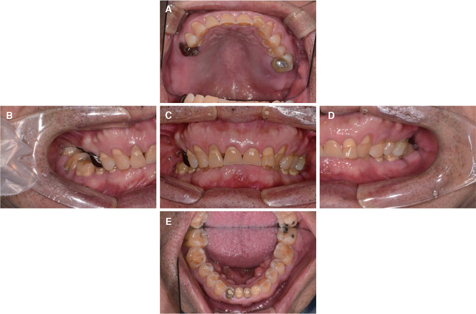
Fig. 1 Intraoral photograph before treatment. (A) Upper, (B) Right, (C) Frontal, (D) Left, (E) Lower.
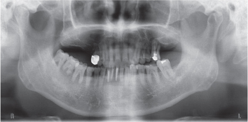
Fig. 2 Panoramic radiograph before treatment.

Fig. 3 TMJ series before treatment. (A) Rt. close, (B) Rt. opening, (C) Lt. opening, (D) Lt. close.
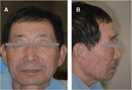
Fig. 4 Facial photographs before treatment. (A) Frontal view, (B) Lateral view.
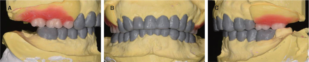
Fig. 5 Diagnostic wax-up model. (A) Right, (B) Frontal, (C) Left.

Fig. 6 Vertical dimension of occlusion evaluation.

Fig. 7 Provisional restoration. (A) Occlusal view (maxillary), (B) Frontal view, (C) Occlusal view (mandible).

Fig. 8 Interocclusal relationship registrations using provisional restorations. (A) Registration of posterior restoration, (B) Registration of anterior restoration.
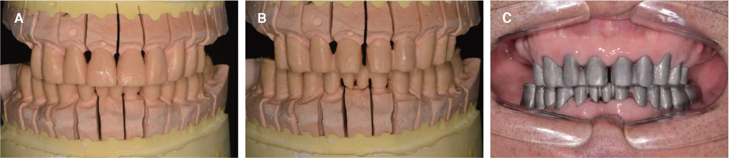
Fig. 9 Full contour wax-up & cut back & coping try in. (A) Full contour wax-up (B) Cut back (C) Coping try in.
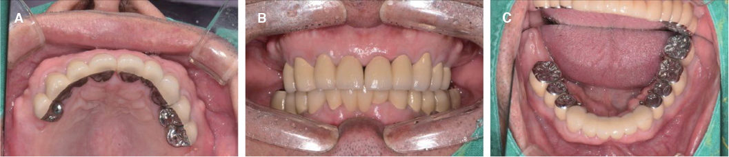
Fig. 10 Porcelain fused to metal crown try in. (A) Occlusal view (maxilla), (B) Frontal view (C) Occlusal view (mandible).
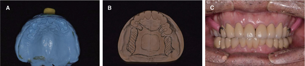
Fig. 11 Maxillary removable partial denture fabrication. (A) Impression taking, (B) Working cast of maxilla, (C) Wax denture.
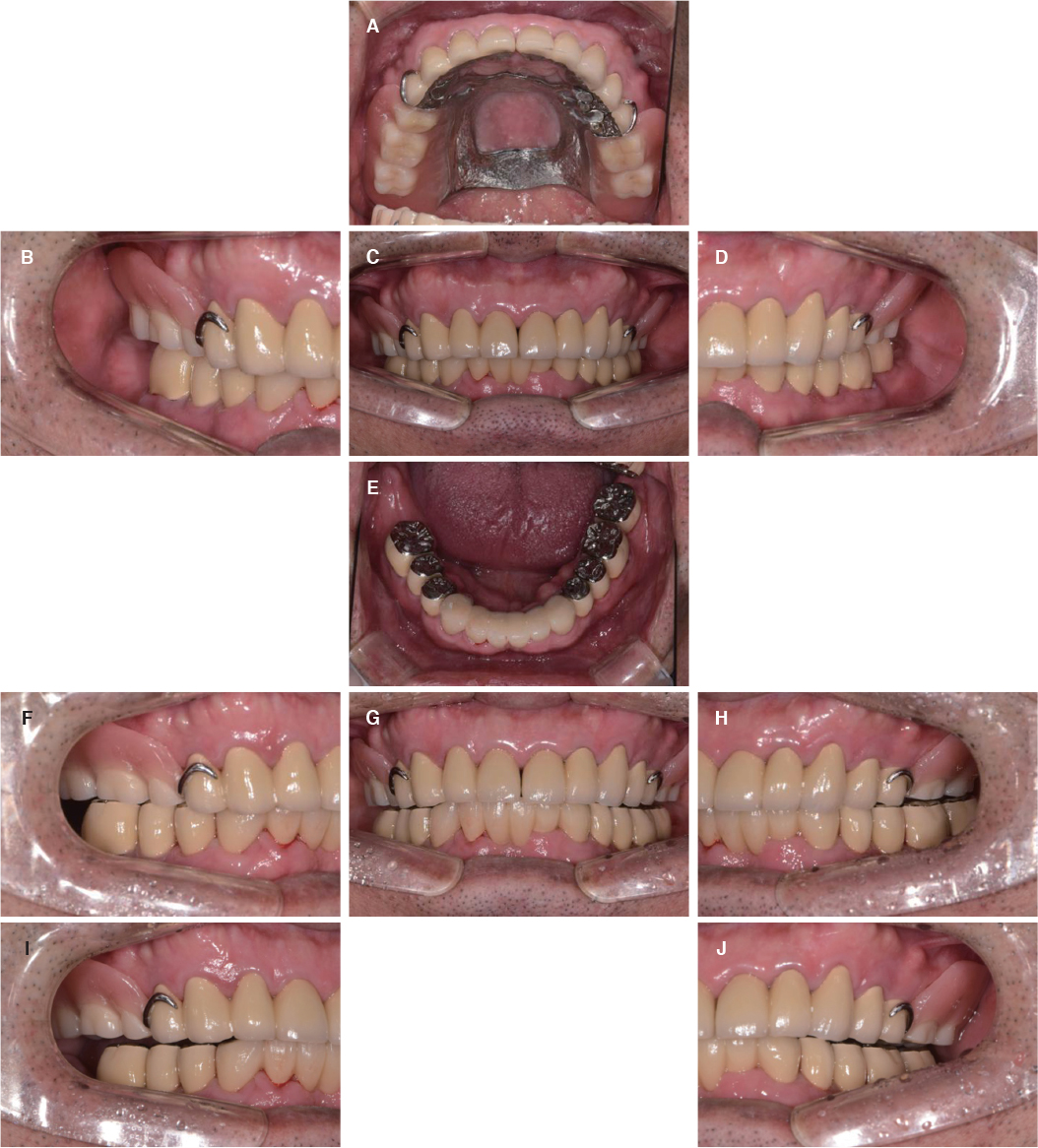
Fig. 12 Definitive prosthesis. (A) Upper, (B) Right, (C) Frontal, (D) Left, (E) Lower, (F) Right working, (G) Protrusive, (H) Left working, (I) Right balancing, (J) Left balancing.
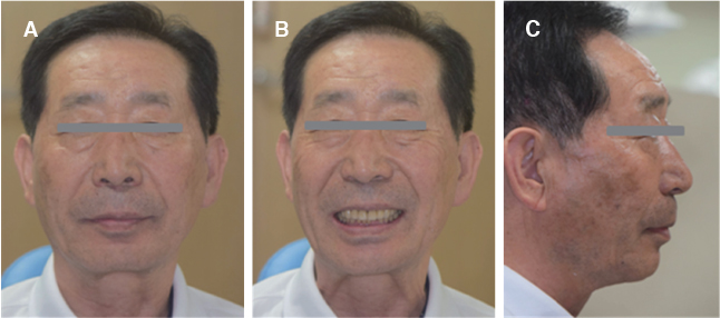
Fig. 13 Facial photographs after treatment. (A) Frontal view, (B) Frontal view smile, (C) Lateral view.

Fig. 14 Panoramic radiograph after treatment.

Fig. 15 TMJ series after treatment. (A) Rt. close, (B) Rt. opening, (C) Lt. opening, (D) Lt. close.
Reference
-
1. Dawson PE. Functional occlusion: From TMJ to smile design. St. Louis: Mosby Elsevier;2007. p. 430–452.2. Murphy T. Compensatory mechanisms in facial height adjustment to functional tooth attrition. Aust Dent J. 1959; 4:312–323.
Article3. Briggs P, Bishop K. Fixed prostheses in the treatment of tooth wear. Eur J Prosthodont Restor Dent. 1997; 5:175–180.4. Hemmings KW, Darbar UR, Vaughan S. Tooth wear treated with direct composite restorations at an increased vertical dimension: results at 30 months. J Prosthet Dent. 2000; 83:287–293.
Article5. Sato S, Hotta TH, Pedrazzi V. Removable occlusal overlay splint in the management of tooth wear: a clinical report. J Prosthet Dent. 2000; 83:392–395.
Article6. Dahl BL, Krogstad O. The effect of a partial bite-raising splint on the inclination of upper and lower front teeth. Acta Odontol Scand. 1983; 41:311–314.
Article7. Ramfjord SP, Blankenship JR. Increased occlusal vertical dimension in adult monkeys. J Prosthet Dent. 1981; 45:74–83.
Article8. Turner KA, Missirlian DM. Restoration of the extremely worn dentition. J Prosthet Dent. 1984; 52:467–474.
Article9. Rivera-Morales WC, Mohl ND. Relationship of occlusal vertical dimension to the health of the masticatory system. J Prosthet Dent. 1991; 65:547–553.
Article10. Park JH, Jeong CM, Jeon YC, Lim JS. A study on the occlusal plane and the vertical dimension in Korean adults with natural dentition. J Korean Acad Prosthodont. 2005; 43:41–51.11. Silverman MM. The speaking method in measuring vertical dimension. J Prosthet Dent. 1953; 3:193–199.
Article12. Rivera-Morales WC, Mohl ND. Restoration of the vertical dimension of occlusion in the severely worn dentition. Dent Clin North Am. 1992; 36:651–664.13. Willis FM. Features of the face involved in full denture prosthesis. Dental Cosmos. 1935; 77:851–854.14. Abduo J, Lyons K. Clinical considerations for increasing occlusal vertical dimension: a review. Aust Dent J. 2012; 57:2–10.
Article15. Abduo J. Safety of increasing vertical dimension of occlusion: a systematic review. Quintessence Int. 2012; 43:369–380.16. Carlsson GE, Ingervall B, Kocak G. Effect of increasing vertical dimension on the masticatory system in subjects with natural teeth. J Prosthet Dent. 1979; 41:284–289.
Article17. Naylor CK. Fabrication of a custom anterior guide table. J Prosthet Dent. 1979; 42:466–469.
Article18. Akerly WB. Prosthodontic treatment of traumatic overlap of the anterior teeth. J Prosthet Dent. 1977; 38:26–34.
Article19. Beddis HP, Durey K, Alhilou A, Chan MF. The restorative management of the deep overbite. Br Dent J. 2014; 217:509–515.
Article20. Torbjörner A, Fransson B. Biomechanical aspects of prosthetic treatment of structurally compromised teeth. Int J Prosthodont. 2004; 17:135–141.
Article21. Curtis TA, Langer Y, Curtis DA, Carpenter R. Occlusal considerations for partially or completely edentulous skeletal class II patients. Part I: Background information. J Prosthet Dent. 1988; 60:202–211.
Article22. Ash MM. Occlusion. 4th ed. Saunders (W.B.) Co. Ltd.;1995. p. 72. p. 3. p. 6.23. Hellsing G. Functional adaptation to changes in vertical dimension. J Prosthet Dent. 1984; 52:867–870.
Article24. Jensen WO. Occlusion for the Class II jaw relations patient. J Prosthet Dent. 1990; 64:432–434.
Article25. Curtis TA, Langer Y, Curtis DA, Carpenter R. Occlusal considerations for partially or completely edentulous skeletal Class II patients. Part II: Treatment concepts. J Prosthet Dent. 1988; 60:334–342.
Article
- Full Text Links
-
- Actions
-
Cited
- CITED
-
- Close
- Share
-
- Similar articles
-
- Full mouth rehabilitation of patient with decreased occlusal vertical dimension due to severely worn dentition and posterior bite collapse
- Full mouth rehabilitation in patient with loss of vertical dimension and deep bite due to tooth wear
- Full mouth rehabilitation of the patient with severe tooth loss and tooth wear with vertical dimension gaining: A case report
- Full-mouth rehabilitation with increasing minimum vertical dimension in the patient with severely worn dentition and deep bite
- A case of full mouth rehabilitation with orthodontic treatment in patient with extensive tooth erosion and wear using monolithic zirconia prostheses
