Full mouth rehabilitation in patient with deep bite, inter-dental arch discrepancy and loss of vertical dimension: a case report
- Affiliations
-
- 1Department of Prosthodontics & Research Institute of Oral Science, College of Dentistry, Gangneung-Wonju National University, Gangneung, Republic of Korea
- KMID: 2525990
- DOI: http://doi.org/10.14368/jdras.2021.37.3.157
Abstract
- Loss of posterior bite support might cause unstable occlusal relationship and when the mandible slides forward in the centric or habitual occlusion, excessive load is applied to the anterior region followed by causing the occlusal plane to collapse or leading to a decrease in occlusal vertical dimension. In addition, disorder of temporomandibular joint function may occur. The inter-dental arch discrepancy causes a mismatch in the vertical and horizontal overlap of the anterior and posterior regions. The deep bite in the anterior region and the scissor bite in the posterior region cause unstable occlusal contact and insufficient occlusal contact area. This report was to rehabilitate a patient with above-mentioned complex problems. Physiologic adaptation to increased vertical dimension and new occlusal plane were evaluated using provisional prostheses, and definitive prostheses was fabricated using cross-mounting technique. Stable occlusion, harmonious teeth overlap and adequate occlusal plane were established, so functionally and aesthetically satisfactory results are obtained.
Keyword
Figure
-
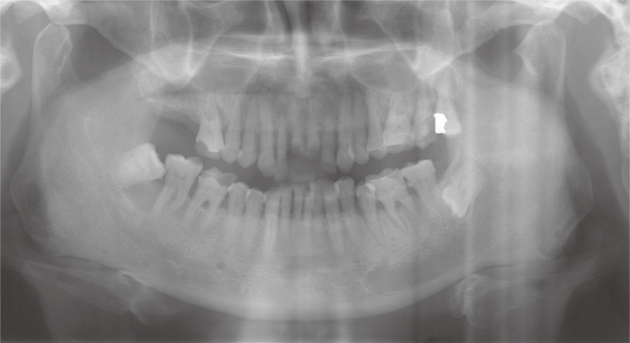
Fig. 1 Initial panoramic radiograph.

Fig. 2 Intraoral photograph. (A - C) At first visit, (D - F) At re-visit after extraction of teeth.

Fig. 3 Diagnostic cast analysis on semi-adjustable articulator at re-visit after extraction of teeth. (A) Right unstable occlusal contact, (B) Maxillary occlusal view, (C) Left scissor bite, (D) Right lateral view, (E) Frontal view, (F) Left lateral view, (G) Irregular occlusal plane, (H) Mandibular occlusal view.

Fig. 4 Vertical dimension evaluation. (A) Facial appearance evaluation, (B) interocclusal rest distance evaluation (maximum intercuspation), (C) interocclusal rest distance evaluation (physiologic rest position).
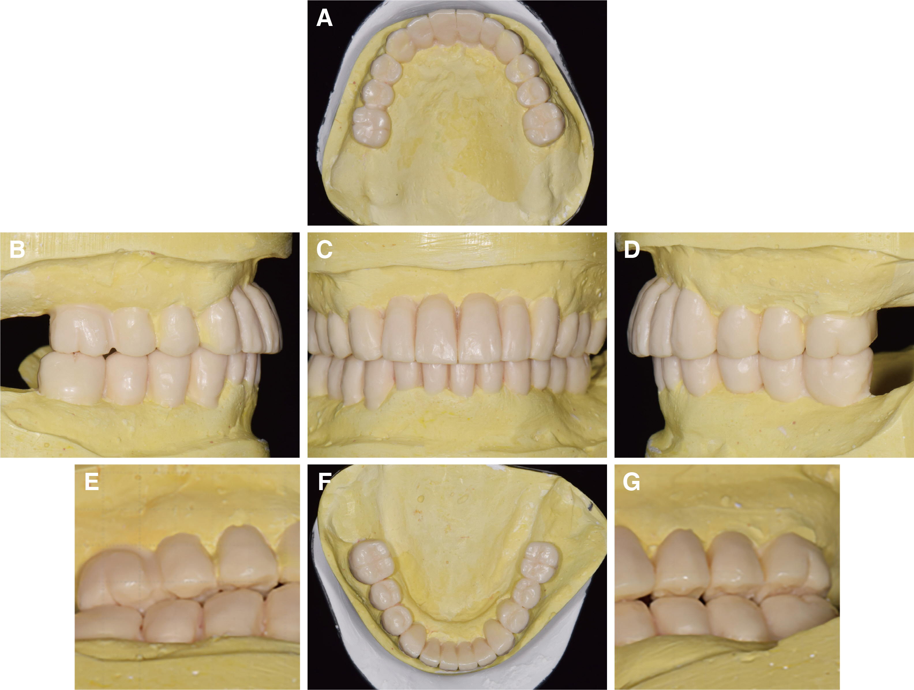
Fig. 5 Diagnostic wax-up. (A) Maxillary occlusal view, (B) Right lateral view, (C) Frontal view, (D) Left lateral view, (E) Right posterior teeth overlap, (F) Mandibular occlusal view, (G) Left posterior teeth overlap.

Fig. 6 Teeth preparation procedures. (A) Occlusal view (maxilla), (B) Frontal view with centric occlusion, (C) Occlusal view (mandible), (D) Frontal view of matrix.

Fig. 7 Mandibular movement registration. (A) Chewing pattern, (B) Centric relation.
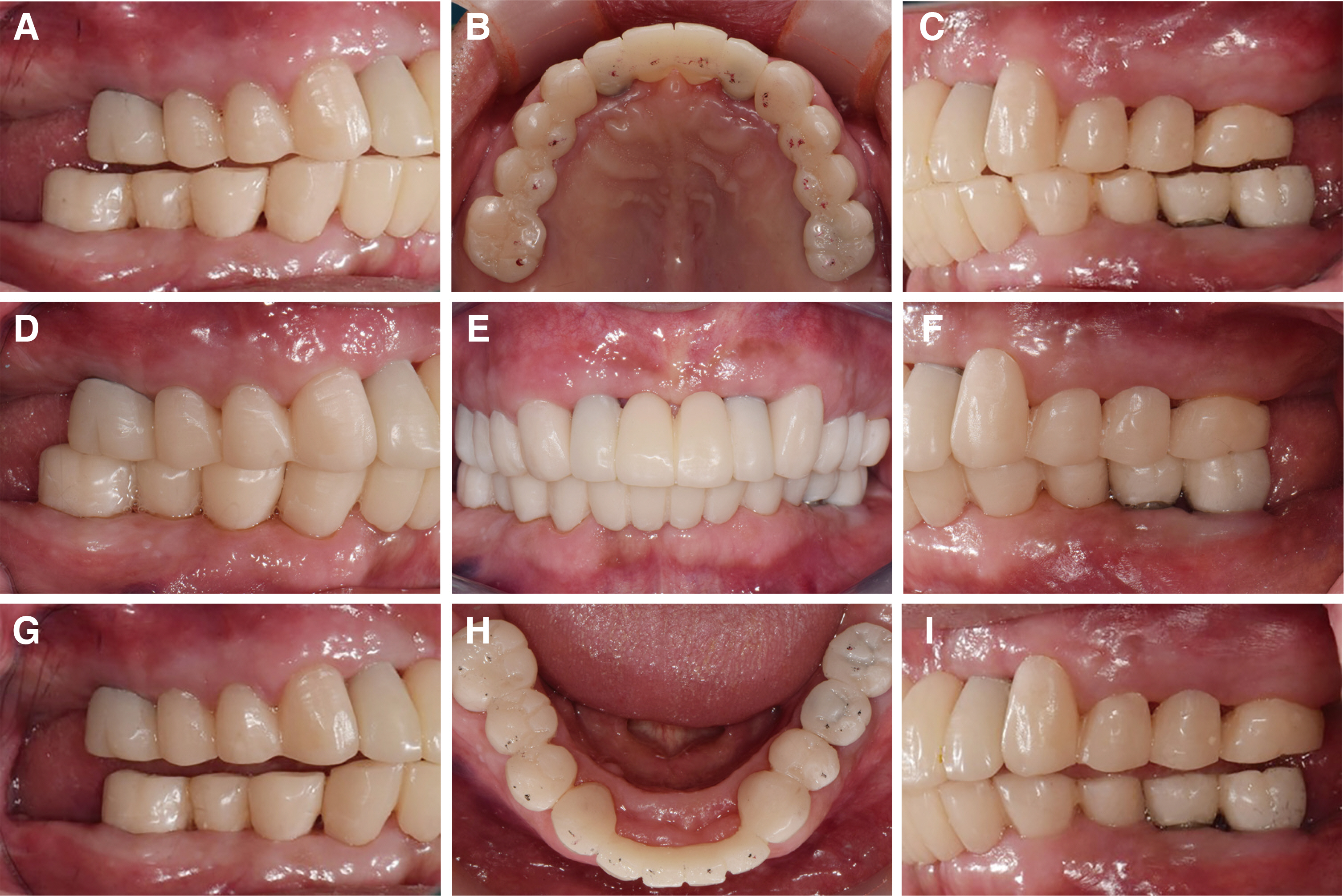
Fig. 8 Intraoral photograph after the placement of 2nd provisional restoration. (A) Working side during right lateral excursion, (B) Maxillary occlusal view, (C) Non-working side during right lateral excursion, (D) Left lateral view at centric occlusion, (E) Frontal view at centric occlusion, (F) Right lateral view at centric occlusion, (G) Non-working side during left lateral excursion, (H) Mandibular occlusal view, (I) Working side during left lateral excursion.
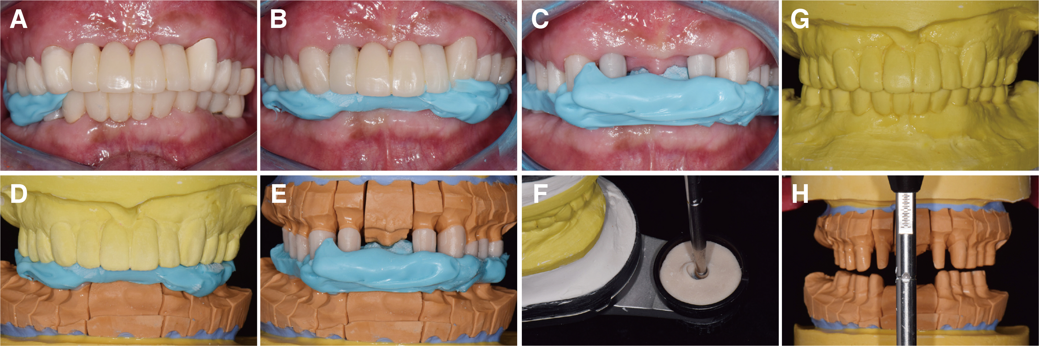
Fig. 9 Cross-mounting procedure. (A - C) Interocclusal relationship registrations using 2nd provisional restorations and milled bonnet crown, (D - F) Cross-mounting of master cast with 2nd provisional restoration cast, (G) Anterior guide table, (H) Mounted mast cast.
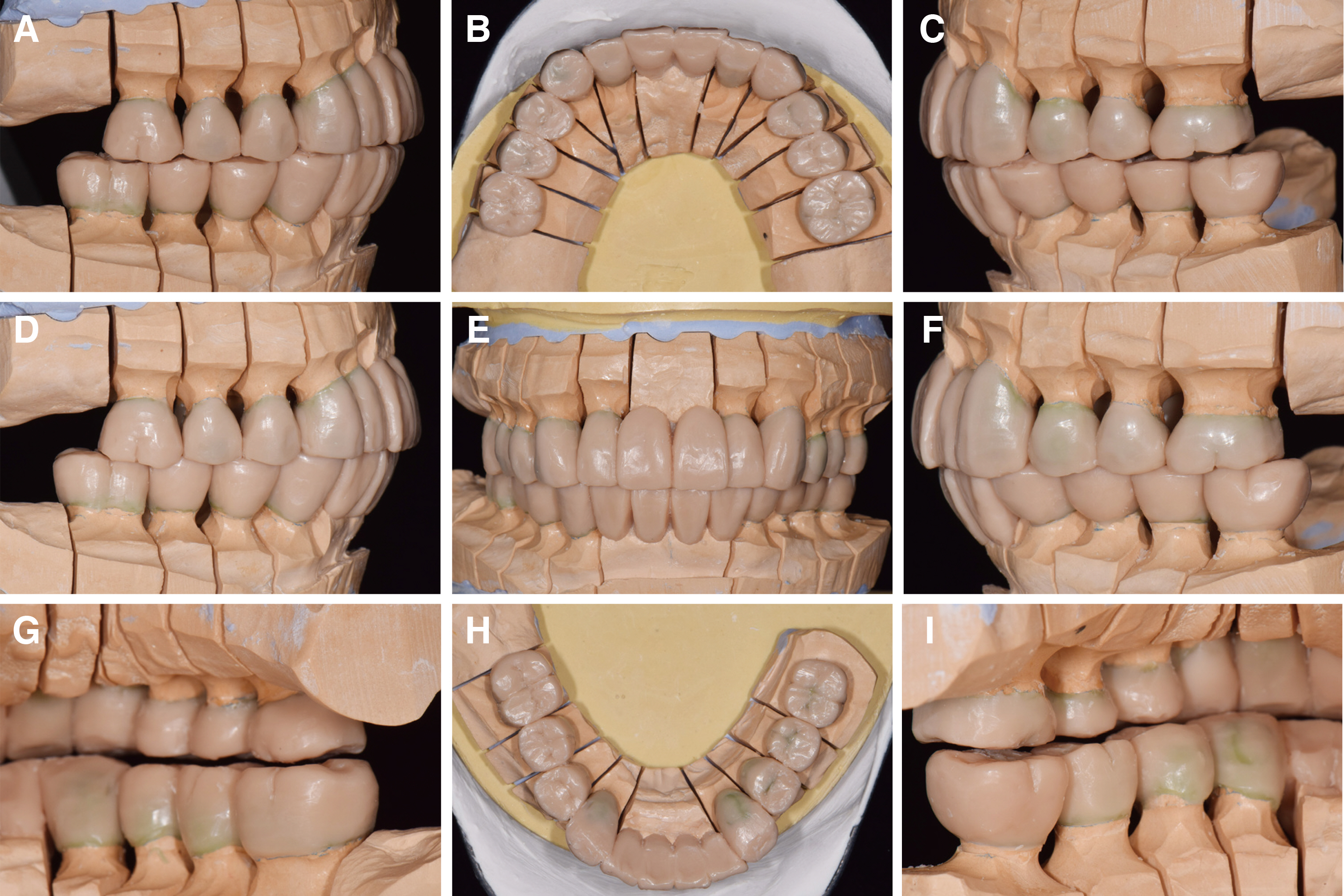
Fig. 10 Wax-up pattern for definite prosthesis. (A) Working side during right lateral excursion, (B) Maxillary occlusal view, (C) Non-working side during right lateral excursion, (D) Left lateral view at centric occlusion, (E) Frontal view at centric occlusion, (F) Right lateral view at centric occlusion, (G) Non-working side during left lateral excursion, (H) Mandibular occlusal view, (I) Working side during left lateral excursion.
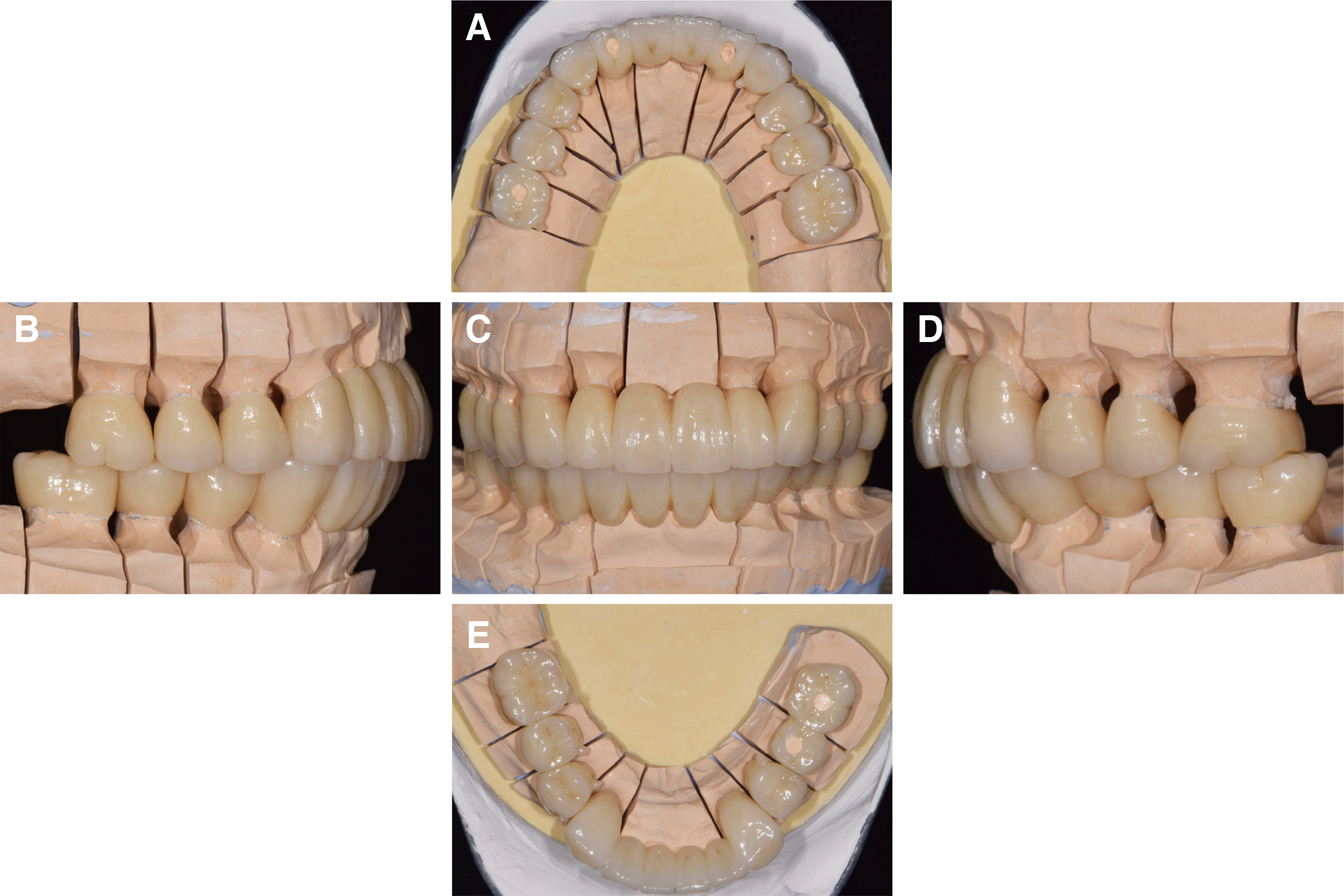
Fig. 11 Definitive prosthesis on master cast. (A) Maxillary occlusal view, (B) Right lateral view, (C) Frontal view, (D) Left lateral view, (E) Mandibular occlusal view.

Fig. 12 Intraoral photograph after the placement of definitive prosthesis. (A) Working side during right lateral excursion, (B) Maxillary occlusal view, (C) Non-working side during right lateral excursion, (D) Left lateral view at centric occlusion, (E) Frontal view at centric occlusion, (F) Right lateral view at centric occlusion, (G) Non-working side during left lateral excursion, (H) Mandibular occlusal view, (I) Working side during left lateral excursion.
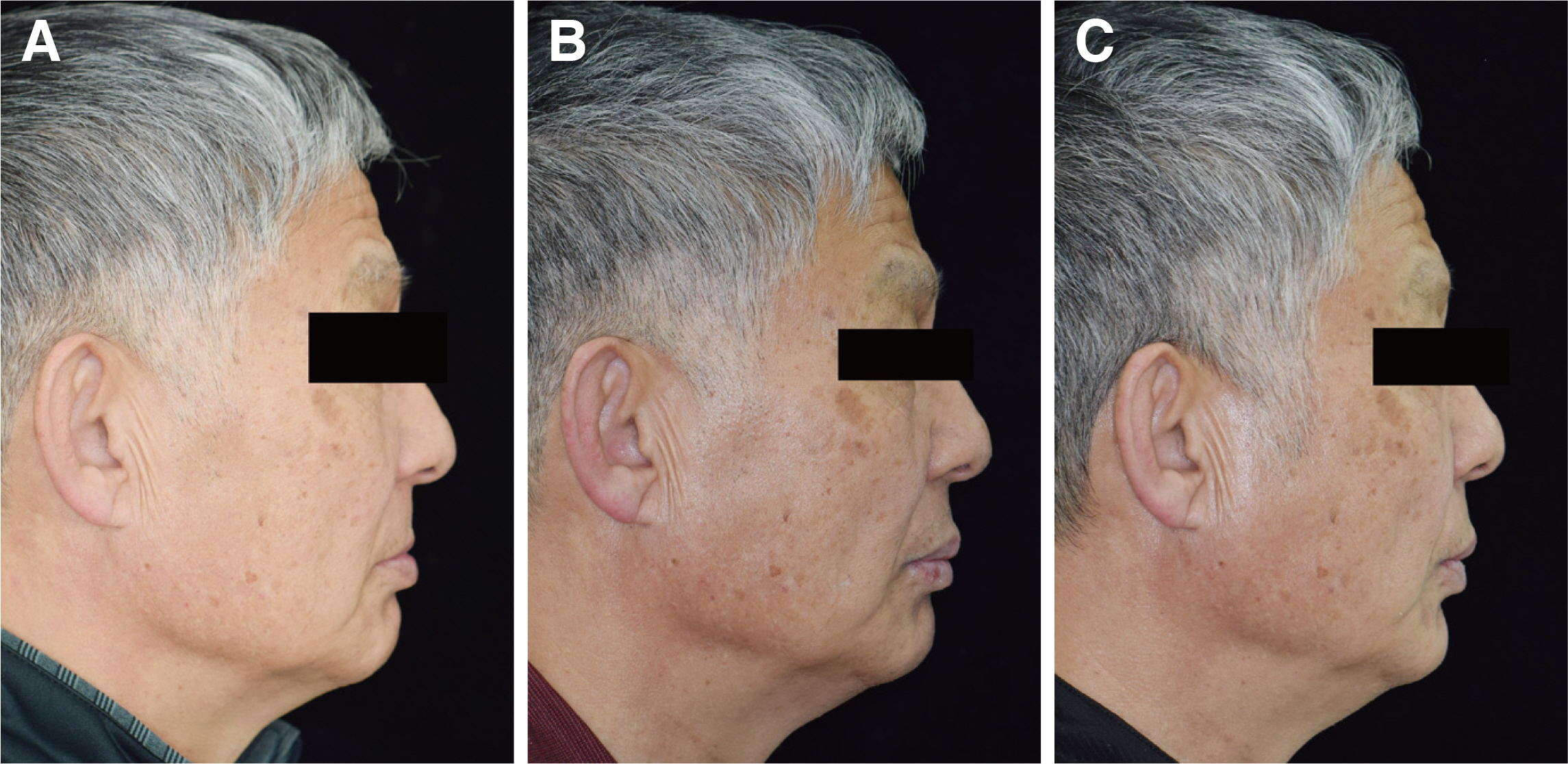
Fig. 13 Facial evaluation at lateral view. (A) Before treatment, (B) Provisional prosthesis, (C) Definitive prosthesis.
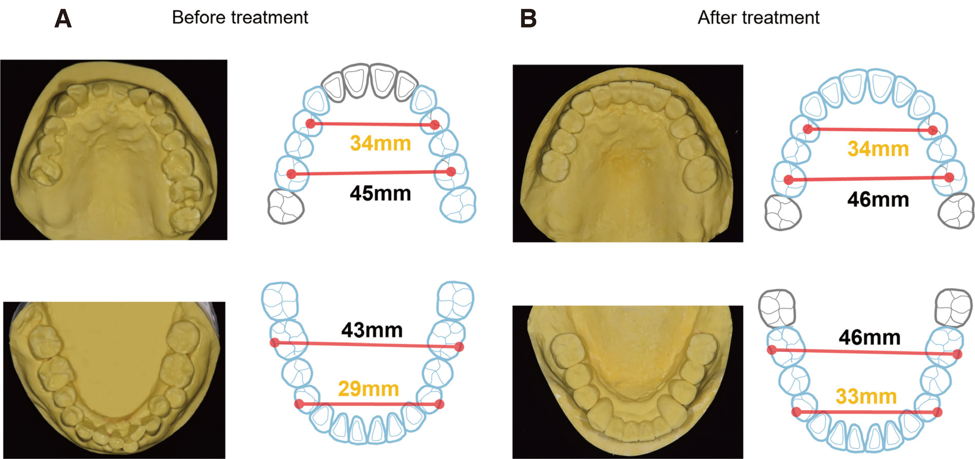
Fig. 14 Inter-dental arch discrepancy. (A) Before treatment, (B) After treatment.

Fig. 15 Chewing pattern analysis. (A) Initial stage of right side, (B) Initial stage of left side, (C) Definitive restoration stage of right side, (D) Definitive restoration stage of left side.

Fig. 16 T-scan (T-scan III, Tekscan Inc., Boston, USA) examination at Maximal Intercuspal Position (MICP). (A) Immediately after definitive restoration, (B) 3 months follow-up after definitive restoration.
Reference
-
References
1. Amsterdam M. 1974; Periodontal prosthesis. Twenty-five years in retrospect. Alpha Omegan. 67:8–52. PMID: 4532467.2. Ramfjord SP, Ash MM. 1966. Occlusion. WB Saunders Co;Philadelphia:3. Shifman A, Laufer BZ, Chweidan H. 1998; Posterior bite collapse-revisited. J Oral Rehabil. 25:376–85. DOI: 10.1046/j.1365-2842.1998.00243.x. PMID: 9639163.4. Okeson JP. 2008. Management of temporomandibular disorders and occlusion. 7th ed. Mosby Elsevier;St. Louis:5. The glossary of prosthodontic terms. 2017; J Prosthet Dent. 117:e1–105. DOI: 10.1016/j.prosdent.2016.12.001. PMID: 28418832.6. Sicher H, DuBrul EL. 1970. Oral Anatomy. 5th. Ed. The C. V. Mosby Company;St. Louis:7. Akerly WB. 1977; Prosthodontic treatment of traumatic overlap of the anterior teeth. J Prosthet Dent. 38:26–34. DOI: 10.1016/0022-3913(77)90263-3. PMID: 267769.8. Dawson PE. 2006. Functional occlusion: From TMJ to smile design. Mosby;St. Louis:9. The council of professors of dental universities. 2006. Textbook of orthodontics. 3rd ed. DaehanNarae Publishing;Seoul:10. Gupta ND, Mahesgwari S, Chaudhari P, Goyal L. 2012; A critical review of the management of deep overbite complicated by periodontal diseases. Eur J Gen Dent. 1:2–5. DOI: 10.4103/2278-9626.101343.11. Tomonari H, Kubota T, Yagi T, Kuninori T, Kitashima F, Uehara S, Miyawaki S. 2014; Posterior scissors-bite: masticatory jaw movement and muscle activity. J Oral Rehabil. 41:257–65. DOI: 10.1111/joor.12148. PMID: 24612226.12. Eichner K. 1990; Renewed examination of the group classification of partially edentulous arches by Eichner and application advices for studies on morbidity statistics. Stomatol DDR. 40:321–5. DOI: 10.1007/978-3-663-13215-8_4. PMID: 2270610.13. Willis FM. 1935; Features of the face involved in full denture prosthesis. Dent Cosmos. 77:851–4.14. Turner KA, Missirlian DM. 1984; Restoration of the extremely worn dentition. J Prosthet Dent. 52:467–74. DOI: 10.1016/0022-3913(84)90326-3. PMID: 6389829.15. Proeschel PA. 2006; Chewing Patterns in Subjects with Normal Occlusion and With Malocclusions. Semin Orthod. 12:138–49. DOI: 10.1053/j.sodo.2006.01.007.16. Won S, An K, Park CJ, Cho LR, Huh YH. 2015; Rehabilitation of unstable occlusion caused by inter-dental arch discrepancy. J Korean Acad Prosthodont. 53:377–91. DOI: 10.4047/jkap.2015.53.4.377.17. Chae HS, Jeon BS, Lee JJ, Ahn SG, Seo JM. 2019; Full mouth rehabilitation in patient with loss of vertical dimension and deep bite due to tooth wear. J Korean Acad Prosthodont. 57:405–15. DOI: 10.4047/jkap.2019.57.4.405.18. Ong MM, Wang HL. 2002; Periodontic and orthodontic treatment in adults. Am J Orthod Dentofacial Orthop. 122:420–8. DOI: 10.1067/mod.2002.126597. PMID: 12411890.19. Ambard A, Mueninghoff L. 2002; Planning restorative treatment for patients with severe Class II malocclusions. J Prosthet Dent. 88:200–7. DOI: 10.1067/mpr.2002.127713. PMID: 12397248.20. Gopi Chander N, Venkat R. 2011; An appraisal on increasing the occlusal vertical dimension in full occlusal rehabilitation and its outcome. J Indian Prosthodont Soc. 11:77–81. DOI: 10.1007/s13191-011-0066-9. PMID: 22654346. PMCID: PMC3120960.
- Full Text Links
-
- Actions
-
Cited
- CITED
-
- Close
- Share
-
- Similar articles
-
- Full mouth rehabilitation of patient with decreased occlusal vertical dimension due to severely worn dentition and posterior bite collapse
- Full mouth rehabilitation of deep bite patient with segmental osteotomy and orthodontic treatment
- A case of full mouth rehabilitation in patient with loss of vertical dimension and deep bite due to tooth wear
- Full-mouth rehabilitation of partial edentulism in a deep bite patient
- Prosthetic Rehabilitation in Patients with Severe Hearing Disability and Loss of Occlusal Vertical Dimension: A Case Report
