Korean J Orthod.
2020 Jan;50(1):3-12. 10.4041/kjod.2020.50.1.3.
Comparison of three midsagittal planes for three-dimensional cone beam computed tomography head reorientation
- Affiliations
-
- 1Department of Orthodontics, Korea University Anam Hospital, Seoul, Korea.
- 2Department of Orthodontics and The Institute of Craniofacial Deformity, College of Dentistry, Yonsei University, Seoul, Korea. hwang@yuhs.ac
- 3Department of Oral and Maxillofacial Radiology, College of Dentistry, Yonsei University, Seoul, Korea.
- 4Department of Oral and Maxillofacial Surgery, College of Dentistry, Yonsei University, Seoul, Korea.
- KMID: 2468636
- DOI: http://doi.org/10.4041/kjod.2020.50.1.3
Abstract
OBJECTIVE
This study compared three prominent midsagittal planes (MSPs) to identify the MSP that best approximates the true symmetrical MSP.
METHODS
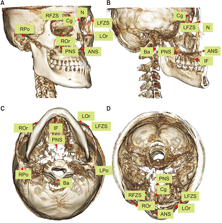
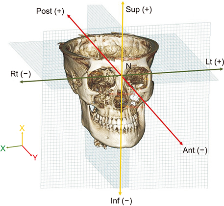
Forty-three patients (mean age, 23.0 ± 8.20 years) were grouped as follows: group 1 consisted of 10 patients with skeletal Class I and a menton (Me) deviation of < 2 mm; group 2, 11 patients with skeletal Class III and a Me deviation < 2 mm; group 3, nine patients with skeletal Class III and a Me deviation of 2 to less than 4 mm; and group 4, 13 patients with skeletal Class III and an Me deviation ≥ 4 mm. The candidate MSPs were established by three-dimensional (3D) cone beam computed tomography (CBCT) reorientation methods (RMs): (1) the MSP perpendicular to the Frankfort horizontal (FH) plane while passing through the crista galli and basion; (2) the MSP including the nasion, incisive foramen, and basion; (3) the MSP including the nasion, anterior nasal spine, and posterior nasal spine. The mean absolute distances (MADs) to the MSPs were calculated from the coordinates of 1,548 points on 129 CBCT images. The differences in the values of the 3D coordinates among RMs were compared.
RESULTS
The MADs of the three RMs showed significant differences (p < 0.05). Most of the differences in values of the coordinates were not significant among RMs.
CONCLUSIONS
Although the differences in distance among the three MSPs were minor, the MSP perpendicular to the FH plane while passing through the crista galli and basion best approximated the true symmetrical MSP.
Keyword
Figure
Reference
-
1. Kim SJ, Lee KJ, Yu HS, Jung YS, Baik HS. Three-dimensional effect of pitch, roll, and yaw rotations on maxillomandibular complex movement. J Craniomaxillofac Surg. 2015; 43:264–273.
Article2. Rohr R. Landmark-based image analysis: using geometric and intensity models. London: Kluwer Academic Publishers;2001.3. Cevidanes LH, Styner MA, Proffit WR. Image analysis and superimposition of 3-dimensional cone-beam computed tomography models. Am J Orthod Dentofacial Orthop. 2006; 129:611–618.
Article4. Andresen PR, Bookstein FL, Conradsen K, Ersbøll BK, Marsh JL, Kreiborg S. Surface-bounded growth modeling applied to human mandibles. IEEE Trans Med Imaging. 2000; 19:1053–1063.
Article5. da Motta AT, de Assis Ribeiro Carvalho F, Oliveira AE, Cevidanes LH, de Oliveira Almeida MA. Superimposition of 3D cone-beam CT models in orthognathic surgery. Dental Press J Orthod. 2010; 15:39–41.6. Ludlow JB, Gubler M, Cevidanes L, Mol A. Precision of cephalometric landmark identification: cone-beam computed tomography vs conventional cephalometric views. Am J Orthod Dentofacial Orthop. 2009; 136:312.e1–312.e10. discussion 312–3.
Article7. Gupta A, Kharbanda OP, Balachandran R, Sardana V, Kalra S, Chaurasia S, et al. Precision of manual landmark identification between as-received and oriented volume-rendered cone-beam computed tomography images. Am J Orthod Dentofacial Orthop. 2017; 151:118–131.
Article8. Damstra J, Fourie Z, De Wit M, Ren Y. A three-dimensional comparison of a morphometric and conventional cephalometric midsagittal planes for craniofacial asymmetry. Clin Oral Investig. 2012; 16:285–294.
Article9. Green MN, Bloom JM, Kulbersh R. A simple and accurate craniofacial midsagittal plane definition. Am J Orthod Dentofacial Orthop. 2017; 152:355–363.
Article10. Shin SM, Kim YM, Kim NR, Choi YS, Park SB, Kim YI. Statistical shape analysis-based determination of optimal midsagittal reference plane for evaluation of facial asymmetry. Am J Orthod Dentofacial Orthop. 2016; 150:252–260.
Article11. Moyers RE, Bookstein FL. The inappropriateness of conventional cephalometrics. Am J Orthod. 1979; 75:599–671.
Article12. Koh EH, Lee KH, Hwang HS. Effects of vertical head rotation on the posteroanterior cephalometric measurements. Korean J Orthod. 2003; 33:73–84.13. Solow B, Tallgren A. Natural head position in standing subjects. Acta Odontol Scand. 1971; 29:591–607.
Article14. Cooke MS, Wei SH. The reproducibility of natural head posture: a methodological study. Am J Orthod Dentofacial Orthop. 1988; 93:280–288.
Article15. Weber DW, Fallis DW, Packer MD. Three-dimensional reproducibility of natural head position. Am J Orthod Dentofacial Orthop. 2013; 143:738–744.
Article16. Park JU, Kook YA, Kim Y. Assessment of asymmetry in a normal occlusion sample and asymmetric patients with three-dimensional cone beam computed tomography: a study for a transverse reference plane. Angle Orthod. 2012; 82:860–867.
Article17. Miyashita K, Dixon AD. Contemporary cephalometric radiography. Tokyo: Quintessence;1996.18. Jung PK, Lee GC, Moon CH. Comparison of cone-beam computed tomography cephalometric measurements using a midsagittal projection and conventional two-dimensional cephalometric measurements. Korean J Orthod. 2015; 45:282–288.
Article19. Naji P, Alsufyani NA, Lagravère MO. Reliability of anatomic structures as landmarks in three-dimensional cephalometric analysis using CBCT. Angle Orthod. 2014; 84:762–772.
Article20. Gribel BF, Gribel MN, Frazäo DC, McNamara JA Jr, Manzi FR. Accuracy and reliability of craniometric measurements on lateral cephalometry and 3D measurements on CBCT scans. Angle Orthod. 2011; 81:26–35.
Article21. Hwang HS, Hwang CH, Lee KH, Kang BC. Maxillofacial 3-dimensional image analysis for the diagnosis of facial asymmetry. Am J Orthod Dentofacial Orthop. 2006; 130:779–785.
Article22. Kim HJ, Kim BC, Kim JG, Zhengguo P, Kang SH, Lee SH. Construction and validation of the midsagittal reference plane based on the skull base symmetry for three-dimensional cephalometric craniofacial analysis. J Craniofac Surg. 2014; 25:338–342.
Article23. An S, Lee JY, Chung CJ, Kim KH. Comparison of different midsagittal plane configurations for evaluating craniofacial asymmetry by expert preference. Am J Orthod Dentofacial Orthop. 2017; 152:788–797.
Article24. Lagravère MO, Low C, Flores-Mir C, Chung R, Carey JP, Heo G, et al. Intraexaminer and interexaminer reliabilities of landmark identification on digitized lateral cephalograms and formatted 3-dimensional cone-beam computerized tomography images. Am J Orthod Dentofacial Orthop. 2010; 137:598–604.
Article25. Lou L, Lagravere MO, Compton S, Major PW, Flores-Mir C. Accuracy of measurements and reliability of landmark identification with computed tomography (CT) techniques in the maxillofacial area: a systematic review. Oral Surg Oral Med Oral Pathol Oral Radiol Endod. 2007; 104:402–411.
Article26. Bjehin R. A comparison between the Frankfort horizontal and the sella turcica-nasion as reference planes in cephalometric analysis. Acta Odontol Scand. 1957; 15:1–12.
Article27. Ricketts RM, Schulhof RJ, Bagha L. Orientation-sella-nasion or Frankfort horizontal. Am J Orthod. 1976; 69:648–654.
Article28. Severt TR, Proffit WR. The prevalence of facial asymmetry in the dentofacial deformities population at the University of North Carolina. Int J Adult Orthodon Orthognath Surg. 1997; 12:171–176.29. Kwon TG, Park HS, Ryoo HM, Lee SH. A comparison of craniofacial morphology in patients with and without facial asymmetry--a three-dimensional analysis with computed tomography. Int J Oral Maxillofac Surg. 2006; 35:43–48.
Article30. Ruellas AC, Tonello C, Gomes LR, Yatabe MS, Macron L, Lopinto J, et al. Common 3-dimensional coordinate system for assessment of directional changes. Am J Orthod Dentofacial Orthop. 2016; 149:645–656.
Article
- Full Text Links
- Actions
-
Cited
- CITED
-
- Close
- Share
- Similar articles
-
- Comparison of cone-beam computed tomography cephalometric measurements using a midsagittal projection and conventional two-dimensional cephalometric measurements
- The impact of reorienting cone-beam computed tomographic images in varied head positions on the coordinates of anatomical landmarks
- Differences in positions of cone-beam computed tomography landmarks in patients with skeletal Class III facial asymmetry according to midsagittal planes
- Three-dimensional imaging modalities in endodontics
- Formulation of a reference coordinate system of three-dimensional head and neck images: Part II. Reproducibility of the horizontal reference plane and midsagittal plane


