Coronary Artery Dose-Volume Parameters Predict Risk of Calcification After Radiation Therapy
- Milgrom SA

- Varghese B
- Gladish GW
- Choi AD
- Dong W
- Patel ZS
- Chung CC
- Rao A
- Pinnix CC
- Gunther JR
- Dabaja BS
- Lin SH
- Hoffman KE
- Huff JL
- Slagowski
- Abe JI
- Iliescu CA
- Banchs J
- Yusuf SW
- Lopez-Mattei JC
- Affiliations
-
- 1Division of Radiation Oncology, Department of Radiation Oncology, MD Anderson Cancer Center, Houston, TX, USA.
- 2Department of Diagnostic Imaging, MD Anderson Cancer Center, Houston, TX, USA. jlopez9@mdanderson.org
- 3Division of Cardiology and Department of Radiology, George Washington University School of Medicine, Washington, DC, USA.
- 4Department of Biostatistics, University of Texas MD Anderson Cancer Center, Houston, TX, USA.
- 5KBR, Houston, TX, USA.
- 6Department of Computational Medicine and Bioinformatics, University of Michigan, Ann Arbor, MI, USA.
- 7Johnson Space Center, National Aeronautics and Space Administration, Houston, TX, USA.
- 8MEI Technologies, Houston, TX, USA.
- 9Division of Radiation Oncology, Department of Radiation Physics, MD Anderson Cancer Center, Houston, TX, USA.
- 10Department of Cardiology, University of Texas MD Anderson Cancer Center, Houston, TX, USA.
- KMID: 2468354
- DOI: http://doi.org/10.4250/jcvi.2019.27.e38
Abstract
- BACKGROUND
Radiation exposure increases the risk of coronary artery disease (CAD). We explored the association of CAD with coronary artery dose-volume parameters in patients treated with 3D-planned radiation therapy (RT).
METHODS
Patients who received thoracic RT and were evaluated by cardiac computed tomography ≥ 1 year later were included. Demographic data and cardiac risk factors were retrospectively collected. Dosimetric data (mean heart dose, d(max), d(mean), V₅₀ - V₅) were collected for the whole heart and for each coronary artery. A coronary artery calcium (CAC) Agatston score was calculated on a per-coronary basis and as a total score. Multivariable generalized linear mixed models were generated. The predicted probabilities were used for receiver operating characteristic analyses.
RESULTS
Twenty patients with a median age of 53 years at the time of RT were included. Nine patients (45%) had ≥ 3/6 conventional cardiac risk factors. Patients received RT for breast cancer (10, 50%), lung cancer (6, 30%), or lymphoma/myeloma (4, 20%) with a median dose of 60 Gy. CAC scans were performed a median of 32 months after RT. CAC score was significantly associated with radiation dose and presence of diabetes. In a multivariable model adjusted for diabetes, segmental coronary artery dosimetric parameters (d(max), d(mean), V5â‚€, Vâ‚„â‚€ V₃₀, Vâ‚‚â‚€, Vâ‚â‚€, and Vâ‚…) were significantly associated with CAC score > 0. Vâ‚…â‚€ had the highest area under the ROC curve (0.89, 95% confidence interval, 0.80-0.97).
CONCLUSIONS
Coronary artery radiation exposure is strongly correlated with subsequent segmental CAC score. Coronary calcification may occur soon after RT and in individuals with conventional cardiac risk factors.
MeSH Terms
Figure
-

Figure 1 Comparison of 3D-RT plan CT with post-RT CAC score using CAC-DRS method. Cardiac risk factors and coronary radiation dose volumes measurements might play a role in development of atherosclerosis. 3D: 3-dimensional, CAC: coronary artery calcium, CT: computed tomography, DRS: data and reporting system, RT: radiation therapy.
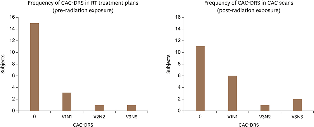
Figure 2 Changes in CAC-DRS categories in subjects (n = 20) before and after RT. Side to side comparison of changes in CAC-DRS in treatment plan (before RT) with calcium score scans (after RT). CAC: coronary artery calcium, DRS: data and reporting system, RT: radiation therapy.

Figure 3 Axial slice from CCT scans and 3-dimensional RT plans demonstrate regional association between radiation dose and coronary calcification. (A) RT to left breast and regional lymph nodes was delivered 1 year before CCT in (B). Prescription dose was 50 Gy. This patient had history of hypertension and dyslipidemia. (B) CCT demonstrating calcification of LAD. LAD CAC score was 46.55. (C) Chemoradiation therapy was administered for non-small cell lung cancer 10 years prior to CCT in (D). Prescription dose was 70 Gy. This patient had history of hypertension, dyslipidemia, and cigarette smoking. (D) CCT demonstrating calcification of LAD and left circumflex artery. LAD CAC score was 318.05, and left circumflex CAC score was 255.43. CAC: coronary artery calcium, CCT: cardiac computed tomography, LAD: left anterior descending artery, RT: radiation therapy.
Reference
-
1. Aleman BM, van den Belt-Dusebout AW, De Bruin ML, et al. Late cardiotoxicity after treatment for Hodgkin lymphoma. Blood. 2007; 109:1878–1886.
Article2. Clarke M, Collins R, Darby S, et al. Effects of radiotherapy and of differences in the extent of surgery for early breast cancer on local recurrence and 15-year survival: an overview of the randomised trials. Lancet. 2005; 366:2087–2106.3. Hancock SL, Donaldson SS, Hoppe RT. Cardiac disease following treatment of Hodgkin's disease in children and adolescents. J Clin Oncol. 1993; 11:1208–1215.
Article4. Hancock SL, Tucker MA, Hoppe RT. Factors affecting late mortality from heart disease after treatment of Hodgkin's disease. JAMA. 1993; 270:1949–1955.
Article5. Rademaker J, Schöder H, Ariaratnam NS, et al. Coronary artery disease after radiation therapy for Hodgkin's lymphoma: coronary CT angiography findings and calcium scores in nine asymptomatic patients. AJR Am J Roentgenol. 2008; 191:32–37.
Article6. Darby SC, Ewertz M, McGale P, et al. Risk of ischemic heart disease in women after radiotherapy for breast cancer. N Engl J Med. 2013; 368:987–998.
Article7. van Nimwegen FA, Schaapveld M, Cutter DJ, et al. Radiation dose-response relationship for risk of coronary heart disease in survivors of Hodgkin lymphoma. J Clin Oncol. 2016; 34:235–243.
Article8. Feng M, Moran JM, Koelling T, et al. Development and validation of a heart atlas to study cardiac exposure to radiation following treatment for breast cancer. Int J Radiat Oncol Biol Phys. 2011; 79:10–18.
Article9. Moignier A, Broggio D, Derreumaux S, et al. Coronary stenosis risk analysis following Hodgkin lymphoma radiotherapy: A study based on patient specific artery segments dose calculation. Radiother Oncol. 2015; 117:467–472.
Article10. Hahn E, Jiang H, Ng A, et al. Late cardiac toxicity after mediastinal radiation therapy for Hodgkin lymphoma: contributions of coronary artery and whole heart dose-volume variables to risk prediction. Int J Radiat Oncol Biol Phys. 2017; 98:1116–1123.
Article11. Polonsky TS, McClelland RL, Jorgensen NW, et al. Coronary artery calcium score and risk classification for coronary heart disease prediction. JAMA. 2010; 303:1610–1616.
Article12. Greenland P, Bonow RO, Brundage BH, et al. ACCF/AHA 2007 clinical expert consensus document on coronary artery calcium scoring by computed tomography in global cardiovascular risk assessment and in evaluation of patients with chest pain: a report of the American College of Cardiology Foundation Clinical Expert Consensus Task Force (ACCF/AHA Writing Committee to Update the 2000 Expert Consensus Document on Electron Beam Computed Tomography) developed in collaboration with the Society of Atherosclerosis Imaging and Prevention and the Society of Cardiovascular Computed Tomography. J Am Coll Cardiol. 2007; 49:378–402.13. Budoff MJ, Shaw LJ, Liu ST, et al. Long-term prognosis associated with coronary calcification: observations from a registry of 25,253 patients. J Am Coll Cardiol. 2007; 49:1860–1870.14. Budoff MJ, Nasir K, McClelland RL, et al. Coronary calcium predicts events better with absolute calcium scores than age-sex-race/ethnicity percentiles: MESA (Multi-Ethnic Study of Atherosclerosis). J Am Coll Cardiol. 2009; 53:345–352.15. Detrano R, Guerci AD, Carr JJ, et al. Coronary calcium as a predictor of coronary events in four racial or ethnic groups. N Engl J Med. 2008; 358:1336–1345.
Article16. Azour L, Kadoch MA, Ward TJ, Eber CD, Jacobi AH. Estimation of cardiovascular risk on routine chest CT: Ordinal coronary artery calcium scoring as an accurate predictor of Agatston score ranges. J Cardiovasc Comput Tomogr. 2017; 11:8–15.
Article17. Agatston AS, Janowitz WR, Hildner FJ, Zusmer NR, Viamonte M Jr, Detrano R. Quantification of coronary artery calcium using ultrafast computed tomography. J Am Coll Cardiol. 1990; 15:827–832.
Article18. Hecht HS, Blaha MJ, Kazerooni EA, et al. CAC-DRS: coronary artery calcium data and reporting system. An expert consensus document of the Society of Cardiovascular Computed Tomography (SCCT). J Cardiovasc Comput Tomogr. 2018; 12:185–191.
Article19. Li J, Lu D, Dou H, et al. Publisher correction: Desumoylase SENP6 maintains osteochondroprogenitor homeostasis by suppressing the p53 pathway. Nat Commun. 2018; 9:646.
Article20. Schabenberger O. SAS Institute Inc.Introducing the GLIMMIX procedure for generalized linear mixed models. In : Proceeding of the 30th annual SAS user group international conference; Cary, NC: SAS Institute Inc.;2005.21. Kirsch J, Buitrago I, Mohammed TL, Gao T, Asher CR, Novaro GM. Detection of coronary calcium during standard chest computed tomography correlates with multi-detector computed tomography coronary artery calcium score. Int J Cardiovasc Imaging. 2012; 28:1249–1256.
Article22. Kim YK, Sung YM, Cho SH, Park YN, Choi HY. Reliability analysis of visual ranking of coronary artery calcification on low-dose CT of the thorax for lung cancer screening: comparison with ECG-gated calcium scoring CT. Int J Cardiovasc Imaging. 2014; 30:Suppl 2. 81–87.
Article23. Hecht HS, Cronin P, Blaha MJ, et al. 2016 SCCT/STR guidelines for coronary artery calcium scoring of noncontrast noncardiac chest CT scans: A report of the Society of Cardiovascular Computed Tomography and Society of Thoracic Radiology. J Cardiovasc Comput Tomogr. 2017; 11:74–84.
Article24. Apter S, Shemesh J, Raanani P, et al. Cardiovascular calcifications after radiation therapy for Hodgkin lymphoma: computed tomography detection and clinical correlation. Coron Artery Dis. 2006; 17:145–151.
Article
- Full Text Links
-
- Actions
-
Cited
- CITED
-
- Close
- Share
-
- Similar articles
-
- The Relationship between Aortic Calcification Volume and Obstructive Coronary Artery Disease: Comparison with Coronary Calcification Volume
- Coronary Artery Calcification Its Incidence and Significance in Patients Detected by Cineangiography
- Anger and Coronary Calcification in Individuals with and without Risk Factors of Coronary Artery Disease
- Utility of Quantification of Coronary Artery Calcification Using Spiral CT
- Technical Aspect of Coronary CT Angiography: Imaging Tips and Safety Issues
