Transfusional Iron Overload and Choroid Plexus Hemosiderosis in a Pediatric Patient: Brain Magnetic Resonance Imaging Findings
- Affiliations
-
- 1Department of Radiology, Inha University School of Medici ne, Inha University Hospital, Incheon, Korea. pengoon@gmail.com
- KMID: 2468058
- DOI: http://doi.org/10.13104/imri.2019.23.4.390
Abstract
- Hemosiderosis is characterized by the deposition of excess iron in body tissues. The choroid plexus is an important part of the central nervous system that can be the primary site of iron overload. T2*-weighted gradient echo (GRE) sequence provides high sensitivity for demonstrating cerebral microhemorrhagic foci and iron deposition. In the present study, we describe the case of a 15-year-old boy with acute lymphoblastic leukemia, in whom repeated transfusion led to iron accumulation in the brain. GRE sequence effectively demonstrated hemosiderin deposition in the choroid plexus.
MeSH Terms
Figure
-
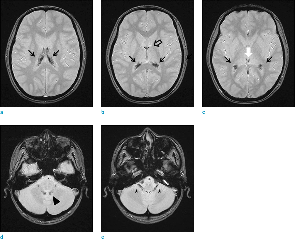
Fig. 1 MR images of choroid plexus hemosiderosis in a 15-year-old male with acute lymphoblastic leukemia in whom repeated blood transfusions led to iron overload. (a-e) Axial T2*-weighted gradient echo images demonstrate the presence of hypointense hemosiderin deposits in the choroid plexus of both the lateral ventricles (black arrows), both the foramina of Monro (open arrow), 3rd ventricle (white arrow), 4th ventricle (arrowhead), and both the foramina of Luschka (asterisks).
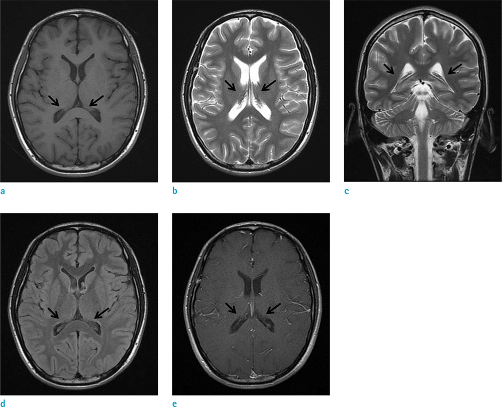
Fig. 2 T1-weighted image (WI) (a), axial (b) and coronal (c) T2WI, and fluid-attenuated inversion recovery (d) and postcontrast T1WI (e) show no remarkable abnormality in the ventricular system (arrows).

Fig. 3 CT images of choroid plexus. (a, b) No calcification or hemorrhage is observed in the choroid plexus (arrows) on precontrast CT images.
Reference
-
1. Fleming RE, Ponka P. Iron overload in human disease. N Engl J Med. 2012; 366:348–359.2. Morris CM, Keith AB, Edwardson JA, Pullen RG. Uptake and distribution of iron and transferrin in the adult rat brain. J Neurochem. 1992; 59:300–306.3. Rouault TA, Zhang DL, Jeong SY. Brain iron homeostasis, the choroid plexus, and localization of iron transport proteins. Metab Brain Dis. 2009; 24:673–684.4. Duprez T, Maiter D, Cosnard G. Transfusional hemochromatosis of the choroid plexus in beta-thalassemia major. J Comput Assist Tomogr. 2001; 25:487–488.5. Sossa DE, Chiang F, Verde AR, Sossa DG, Castillo M. Transfusional iron overload presenting as choroid plexus hemosiderosis. JBR-BTR. 2013; 96:39.6. Kira R, Ohga S, Takada H, Gondo K, Mihara F, Hara T. MR choroid plexus sign of iron overload. Neurology. 2000; 55:1340.7. Fearnley JM, Stevens JM, Rudge P. Superficial siderosis of the central nervous system. Brain. 1995; 118(Pt 4):1051–1066.8. Eng J, Fish JD. Insidious iron burden in pediatric patients with acute lymphoblastic leukemia. Pediatr Blood Cancer. 2011; 56:368–371.9. Halonen P, Mattila J, Suominen P, Ruuska T, Salo MK, Makipernaa A. Iron overload in children who are treated for acute lymphoblastic leukemia estimated by liver siderosis and serum iron parameters. Pediatrics. 2003; 111:91–96.10. Haacke EM, DelProposto ZS, Chaturvedi S, et al. Imaging cerebral amyloid angiopathy with susceptibility-weighted imaging. AJNR Am J Neuroradiol. 2007; 28:316–317.
- Full Text Links
-
- Actions
-
Cited
- CITED
-
- Close
- Share
-
- Similar articles
-
- MR Imaging Findings of the Pituitary Gland in Patients with Transfusional Hemochromatosis: Two Case Reports
- MR Findings of Choroid Plexus Papilloma: Case Report
- Effect of Desferrioxamine Therapy in Patients with Transfusional Hemosiderosis Due to Severe Aplastic Anemia
- Transfusional Hemosiderosis; Correlation of MR Findings with Clinical Findings
- Atypical Choroid Plexus Papilloma in an Adult
