J Adv Prosthodont.
2019 Dec;11(6):324-330. 10.4047/jap.2019.11.6.324.
Influence of preparation design on fracture resistance of different monolithic zirconia crowns: A comparative study
- Affiliations
-
- 1Department of Conservative Dentistry, College of Dentistry, Mustansiriyah University, Baghdad, Iraq. meelad_06@yahoo.com
- KMID: 2466247
- DOI: http://doi.org/10.4047/jap.2019.11.6.324
Abstract
- PURPOSE
The aim of the study was to evaluate and compare the fracture resistance and modes of fracture of monolithic zirconia crowns with two preparation designs.
MATERIALS AND METHODS
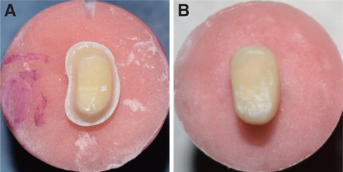
Forty human maxillary first premolar teeth were extracted for orthodontic purposes and divided into two main groups (n=20): Group A: monolithic traditional zirconia; Group B: monolithic translucent zirconia. The groups were further subdivided into two subgroups (n=10): (A1, B1) shoulder margin design; (A2, B2) feather-edge margin design. Teeth were prepared with either a 1 mm shoulder margin design or a feather-edge margin design. The prepared teeth were scanned using a digital intraoral scanner. The crowns were cemented using self-adhesive resin cement. All cemented teeth were stored in water for 7 days and thermocycling was done before testing. All samples were subjected to compressive axial loading until fracture. The fractographic analysis was done to assess the modes of fracture of the tested samples.
RESULTS
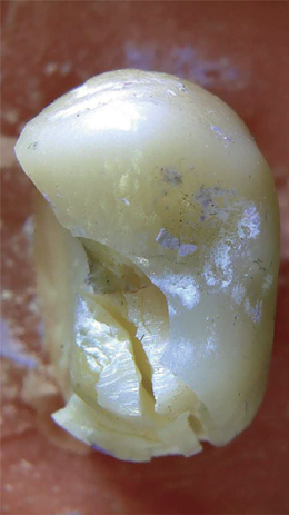
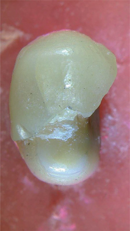
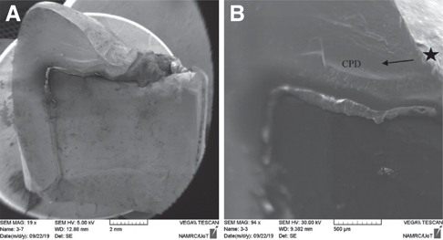
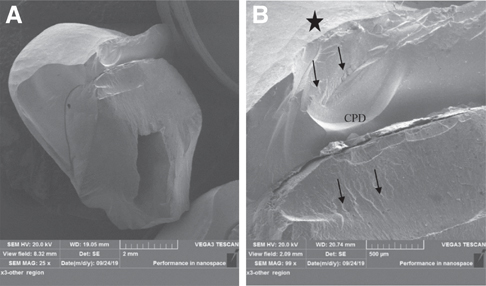
The highest mean values of fracture resistance were recorded in kilo-newton and were in the order of subgroup A1 (2.903); subgroup A2 (2.3); subgroup B1 (1.854) and subgroup B2 (1.523). One-way ANOVA showed a statistically significant difference among the 4 subgroups. Concerning modes of fracture, the majority of samples in subgroups A1 and B1 were fracture of restoration and/or tooth, while in subgroups A2 and B2, the majority of samples fractured through the central fossa.
CONCLUSION
Even though all the tested crowns fractured at a higher level than the maximum occlusal forces, the shoulder margin design was better than the feather-edge margin design and the monolithic traditional zirconia was better than the monolithic translucent zirconia in terms of fracture strength.
Keyword
Figure
Reference
-
1. Beuer F, Stimmelmayr M, Gueth JF, Edelhoff D, Naumann M. In vitro performance of full-contour zirconia single crowns. Dent Mater. 2012; 28:449–456.2. Zhang Y, Kelly JR. Dental Ceramics for Restoration and Metal Veneering. Dent Clin North Am. 2017; 61:797–819.3. Monaco C, Caldari M, Scotti R. AIOP Clinical Research Group. Clinical evaluation of 1,132 zirconia-based single crowns: a retrospective cohort study from the AIOP clinical research group. Int J Prosthodont. 2013; 26:435–442.4. Zhang Y. Making yttria-stabilized tetragonal zirconia translucent. Dent Mater. 2014; 30:1195–1203.5. de Lima E, Meira JBC, Özcan M, Cesar PF. Chipping of veneering ceramics in zirconium dioxide fixed dental prosthesis. Curr Oral Health Rep. 2015; 2:169–173.6. Mitov G, Anastassova-Yoshida Y, Nothdurft FP, von See C, Pospiech P. Influence of the preparation design and artificial aging on the fracture resistance of monolithic zirconia crowns. J Adv Prosthodont. 2016; 8:30–36.7. Johansson C, Kmet G, Rivera J, Larsson C, Vult Von Steyern P. Fracture strength of monolithic all-ceramic crowns made of high translucent yttrium oxide-stabilized zirconium dioxide compared to porcelain-veneered crowns and lithium disilicate crowns. Acta Odontol Scand. 2014; 72:145–153.8. Vichi A, Sedda M, Fabian Fonzar R, Carrabba M, Ferrari M. Comparison of contrast ratio, translucency parameter, and flexural strength of traditional and “Augmented translucency” zirconia for CEREC CAD/CAM system. J Esthet Restor Dent. 2016; 28:S32–S39.9. Schmitt J, Wichmann M, Holst S, Reich S. Restoring severely compromised anterior teeth with zirconia crowns and feather-edged margin preparations: a 3-year follow-up of a prospective clinical trial. Int J Prosthodont. 2010; 23:107–109.10. Poggio CE, Dosoli R, Ercoli C. A retrospective analysis of 102 zirconia single crowns with knife-edge margins. J Prosthet Dent. 2012; 107:316–321.11. Schmitz JH, Cortellini D, Granata S, Valenti M. Monolithic lithium disilicate complete single crowns with feather-edge preparation design in the posterior region: A multicentric retrospective study up to 12 years. Quintessence Int. 2017; 601–608.12. Patroni S, Chiodera G, Caliceti C, Ferrari P. CAD/CAM technology and zirconium oxide with feather-edge marginal preparation. Eur J Esthet Dent. 2010; 5:78–100.13. Reich S, Petschelt A, Lohbauer U. The effect of finish line preparation and layer thickness on the failure load and fractography of ZrO2 copings. J Prosthet Dent. 2008; 99:369–376.14. Dentsply Sirona. In fire HTC speed. Operating instructions. 2017. https://manuals.sirona.com/home.HomeDmsDocument.download.html?id=20054.15. Burke FJ. Maximising the fracture resistance of dentine-bonded all-ceramic crowns. J Dent. 1999; 27:169–173.16. Ramos RM, Clark D, Mazza M, Venuti P, Maiolino M, Kopanja S, Cirimpei V, Tawfik AA, Bordonali D, Acatrinei B, Sutradhar JC, Czerwinski M, Sienkiewicz A, Khademi J. The shoulderless approach a new rationale in prosthetic dentistry. Tomorrow Tooth J. 2017; 1:1–29.17. Church TD, Jessup JP, Guillory VL, Vandewalle KS. Translucency and strength of high-translucency monolithic zirconium oxide materials. Gen Dent. 2017; 65:48–52.18. Lawson NC, Burgess JO. Dental ceramics: a current review. Compend Contin Educ Dent. 2014; 35:161–166.19. Jang GW, Kim HS, Choe HC, Son MK. Fracture strength and mechanism of dental ceramic crown with zirconia thickness. Procedia Eng. 2011; 10:1556–1560.20. Wu XP, Li DS, Zhang J, Ma X, Zhao K, Zhang XP. Effects of different die materials on fracture and failure behaviors of all-ceramic crowns. Zhonghua Kou Qiang Yi Xue Za Zhi. 2009; 44:483–487.21. Yucel MT, Yondem I, Aykent F, Eraslan O. Influence of the supporting die structures on the fracture strength of all-ceramic materials. Clin Oral Investig. 2012; 16:1105–1110.22. Chitmongkolsuk S, Heydecke G, Stappert C, Strub JR. Fracture strength of all-ceramic lithium disilicate and porcelain-fused-to-metal bridges for molar replacement after dynamic loading. Eur J Prosthodont Restor Dent. 2002; 10:15–22.23. Dolidze T, Bitarova I. Advantages and dısadvantages of E-max and Zırconıa crowns. Eur Sci J. 2017; 12:465–468.24. Skjold A, Schriwer C, Øilo M. Effect of margin design on fracture load of zirconia crowns. Eur J Oral Sci. 2019; 127:89–96.25. ISO/TR 11405. Dental materials - Guidance on testing of adhesion to tooth structure. Geneva; Switzerland: International Standards Organization (ISO);1994. Available at: https://www.iso.org/standard/19347.html.26. Beuer F, Aggstaller H, Edelhoff D, Gernet W. Effect of preparation design on the fracture resistance of zirconia crown copings. Dent Mater J. 2008; 27:362–367.27. Miura S, Kasahara S, Yamauchi S, Egusa H. Effect of finish line design on stress distribution in bilayer and monolithic zirconia crowns: a three-dimensional finite element analysis study. Eur J Oral Sci. 2018; 126:159–165.28. Jalalian E, Rostami R, Atashkar B. Comparison of chamfer and deep chamfer preparation designs on the fracture resistance of zirconia core restorations. J Dent Res Dent Clin Dent Prospects. 2011; 5:41–45.29. Sun T, Zhou S, Lai R, Liu R, Ma S, Zhou Z, Longquan S. Load-bearing capacity and the recommended thickness of dental monolithic zirconia single crowns. J Mech Behav Biomed Mater. 2014; 35:93–101.30. Lang NP, Kiel RA, Anderhalden K. Clinical and microbiological effects of subgingival restorations with overhanging or clinically perfect margins. J Clin Periodontol. 1983; 10:563–578.31. Rekow ED, Harsono M, Janal M, Thompson VP, Zhang G. Factorial analysis of variables influencing stress in all-ceramic crowns. Dent Mater. 2006; 22:125–132.32. Shahrbaf S, van Noort R, Mirzakouchaki B, Ghassemieh E, Martin N. Fracture strength of machined ceramic crowns as a function of tooth preparation design and the elastic modulus of the cement. Dent Mater. 2014; 30:234–241.33. Sornsuwan T, Ellakwa A, Swain MV. Occlusal geometrical considerations in all-ceramic pre-molar crown failure testing. Dent Mater. 2011; 27:1127–1134.34. Nakamura K, Harada A, Inagaki R, Kanno T, Niwano Y, Milleding P, Örtengren U. Fracture resistance of monolithic zirconia molar crowns with reduced thickness. Acta Odontol Scand. 2015; 73:602–608.
- Full Text Links
- Actions
-
Cited
- CITED
-
- Close
- Share
- Similar articles
-
- Influence of the preparation design and artificial aging on the fracture resistance of monolithic zirconia crowns
- Fracture resistance and marginal fit of the zirconia crowns with varied occlusal thickness
- Fracture strength of zirconia monolithic crowns and metal-ceramic crowns after cyclic loading and thermocycling
- Fracture strength of zirconia monolithic crowns
- Fracture resistance of implant- supported monolithic crowns cemented to zirconia hybrid-abutments: zirconia-based crowns vs. lithium disilicate crowns

