J Endocr Surg.
2019 Dec;19(4):95-105. 10.16956/jes.2019.19.4.95.
Which Clinicopathological Factors Are Related to Tumor Size in Papillary Thyroid Cancer?
- Affiliations
-
- 1Department of Surgery, Ajou University School of Medicine, Suwon, Korea. servent-lee@hanmail.net
- KMID: 2466235
- DOI: http://doi.org/10.16956/jes.2019.19.4.95
Abstract
- PURPOSE
Papillary thyroid cancer (PTC) is the most common endocrine cancer worldwide. Tumor size on observation, together with lymph node metastasis, serves as a determinant of surgery. However, not all patients with PTC experience an increase in the size of tumor. We investigated various clinicopathological factors associated with the size of tumor to discern the group that can be observed without the need for early surgery.
METHODS
The records of 1,401 patients diagnosed with PTC (excluding the follicular variant) between 2015 and 2017, were reviewed. Clinicopathological features suspected to have a link to tumor growth were included, such as diabetes mellitus (DM), use of metformin, a surgical history of breast, ovary, uterine, or familial non-medullary thyroid cancer, B-type Raf kinase (BRAF) V600E mutation, and lymphovascular invasion (LVI).
RESULTS
Size of tumor was found to be related to sex, age, hypertension, ovary-uterine surgery, LVI and psammoma bodies. However, after adjusting for the effects of other factors on tumor size, both age and LVI were found to be significantly related to tumor size, with age also being significantly related to LVI.
CONCLUSION
Age and LVI are significant factors in the enlargement of tumors, unlike several other features, including DM, breast cancer history, familial cancer history, Graves's disease, Hashimoto's thyroiditis, and BRAF mutation.
MeSH Terms
Figure
-

Fig. 1 The distribution of mean tumor size according to age.
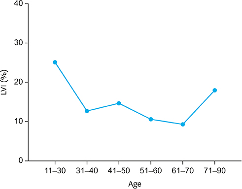
Fig. 2 The distribution of LVI according to age. LVI = lymphovascular invasion.
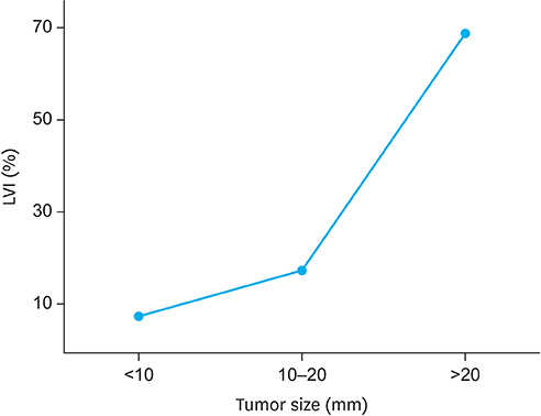
Fig. 3 The distribution of LVI according to tumor size. LVI = lymphovascular invasion.
Reference
-
1. Grebe SK, Hay ID. Follicular cell-derived thyroid carcinomas. Cancer Treat Res. 1997; 89:91–140.
Article2. Ahn HS, Kim HJ, Welch HG. Korea's thyroid-cancer “epidemic”--screening and overdiagnosis. N Engl J Med. 2014; 371:1765–1767.
Article3. Pellegriti G, Frasca F, Regalbuto C, Squatrito S, Vigneri R. Worldwide increasing incidence of thyroid cancer: update on epidemiology and risk factors. J Cancer Epidemiol. 2013; 2013:965212.
Article4. Miyauchi A. Clinical trials of active surveillance of papillary microcarcinoma of the thyroid. World J Surg. 2016; 40:516–522.
Article5. Ito Y, Miyauchi A, Inoue H, Fukushima M, Kihara M, Higashiyama T, et al. An observational trial for papillary thyroid microcarcinoma in Japanese patients. World J Surg. 2010; 34:28–35.
Article6. Ito Y, Miyauchi A. Appropriate treatment for asymptomatic papillary microcarcinoma of the thyroid. Expert Opin Pharmacother. 2007; 8:3205–3215.
Article7. Ito Y, Miyauchi A, Kihara M, Higashiyama T, Kobayashi K, Miya A. Patient age is significantly related to the progression of papillary microcarcinoma of the thyroid under observation. Thyroid. 2014; 24:27–34.
Article8. Zelenko Z, Gallagher EJ. Diabetes and cancer. Endocrinol Metab Clin North Am. 2014; 43:167–185.
Article9. Algire C, Amrein L, Zakikhani M, Panasci L, Pollak M. Metformin blocks the stimulative effect of a high-energy diet on colon carcinoma growth in vivo and is associated with reduced expression of fatty acid synthase. Endocr Relat Cancer. 2010; 17:351–360.
Article10. Zhou G, Myers R, Li Y, Chen Y, Shen X, Fenyk-Melody J, et al. Role of AMP-activated protein kinase in mechanism of metformin action. J Clin Invest. 2001; 108:1167–1174.
Article11. Rajoria S, Suriano R, George A, Shanmugam A, Schantz SP, Geliebter J, et al. Estrogen induced metastatic modulators MMP-2 and MMP-9 are targets of 3,3′-diindolylmethane in thyroid cancer. PLoS One. 2011; 6:e15879.
Article12. Seethala RR, Baloch ZW, Barletta JA, Khanafshar E, Mete O, Sadow PM, et al. Noninvasive follicular thyroid neoplasm with papillary-like nuclear features: a review for pathologists. Mod Pathol. 2018; 31:39–55.
Article13. Hayashida N, Namba H, Kumagai A, Hayashi T, Ohtsuru A, Ito M, et al. A rapid and simple detection method for the BRAF(T1796A) mutation in fine-needle aspirated thyroid carcinoma cells. Thyroid. 2004; 14:910–915.
Article14. Mai KT, Truong LD, Ball CG, Olberg B, Lai CK, Purgina B. Lymphatic endothelial cancerization in papillary thyroid carcinoma: hidden evidence of lymphatic invasion. Pathol Int. 2015; 65:220–230.
Article15. Ito Y, Fukushima M, Higashiyama T, Kihara M, Takamura Y, Kobayashi K, et al. Tumor size is the strongest predictor of microscopic lymph node metastasis and lymph node recurrence of N0 papillary thyroid carcinoma. Endocr J. 2013; 60:113–117.
Article16. Rosário PW, de Faria S, Bicalho L, Alves MF, Borges MA, Purisch S, et al. Ultrasonographic differentiation between metastatic and benign lymph nodes in patients with papillary thyroid carcinoma. J Ultrasound Med. 2005; 24:1385–1389.
Article17. Shimamoto K, Satake H, Sawaki A, Ishigaki T, Funahashi H, Imai T. Preoperative staging of thyroid papillary carcinoma with ultrasonography. Eur J Radiol. 1998; 29:4–10.
Article18. Noguchi S, Noguchi A, Murakami N. Papillary carcinoma of the thyroid. I. Developing pattern of metastasis. Cancer. 1970; 26:1053–1060.
Article19. Wise LA, Palmer JR, Stewart EA, Rosenberg L. Age-specific incidence rates for self-reported uterine leiomyomata in the Black Women's Health Study. Obstet Gynecol. 2005; 105:563–568.
Article20. Spitz IM. Clinical utility of progesterone receptor modulators and their effect on the endometrium. Curr Opin Obstet Gynecol. 2009; 21:318–324.
Article21. Donnez J, Donnez O, Courtoy GE, Dolmans MM. The place of selective progesterone receptor modulators in myoma therapy. Minerva Ginecol. 2016; 68:313–320.22. Onitilo AA, Engel JM, Glurich I, Stankowski RV, Williams GM, Doi SA. Diabetes and cancer I: risk, survival, and implications for screening. Cancer Causes Control. 2012; 23:967–981.
Article23. Park YJ, Ahn HY, Choi HS, Kim KW, Park DJ, Cho BY. The long-term outcomes of the second generation of familial nonmedullary thyroid carcinoma are more aggressive than sporadic cases. Thyroid. 2012; 22:356–362.
Article24. Wang X, Cheng W, Li J, Su A, Wei T, Liu F, et al. Endocrine tumours: familial nonmedullary thyroid carcinoma is a more aggressive disease: a systematic review and meta-analysis. Eur J Endocrinol. 2015; 172:R253–62.
Article25. Zhou YM, Luo H, Gou JX, Zhao WJ, Dai WY, Zhu J, et al. Second generation of familial nonmedullary thyroid carcinoma: a meta-analysis on the clinicopathologic features and prognosis. Eur J Surg Oncol. 2017; 43:2248–2256.
Article26. Xing M. BRAF mutation in papillary thyroid cancer: pathogenic role, molecular bases, and clinical implications. Endocr Rev. 2007; 28:742–762.
Article27. Elisei R, Ugolini C, Viola D, Lupi C, Biagini A, Giannini R, et al. BRAF(V600E) mutation and outcome of patients with papillary thyroid carcinoma: a 15-year median follow-up study. J Clin Endocrinol Metab. 2008; 93:3943–3949.
Article28. Betge J, Pollheimer MJ, Lindtner RA, Kornprat P, Schlemmer A, Rehak P, et al. Intramural and extramural vascular invasion in colorectal cancer: prognostic significance and quality of pathology reporting. Cancer. 2012; 118:628–638.
Article29. Huh JW, Kim HR, Kim YJ. Lymphovascular or perineural invasion may predict lymph node metastasis in patients with T1 and T2 colorectal cancer. J Gastrointest Surg. 2010; 14:1074–1080.
Article30. Sun W, Lan X, Zhang H, Dong W, Wang Z, He L, et al. Risk factors for central lymph node metastasis in CN0 papillary thyroid carcinoma: a systematic review and meta-analysis. PLoS One. 2015; 10:e0139021.
Article31. Karkkainen MJ, Haiko P, Sainio K, Partanen J, Taipale J, Petrova TV, et al. Vascular endothelial growth factor C is required for sprouting of the first lymphatic vessels from embryonic veins. Nat Immunol. 2004; 5:74–80.
Article32. Dias S, Hattori K, Zhu Z, Heissig B, Choy M, Lane W, et al. Autocrine stimulation of VEGFR-2 activates human leukemic cell growth and migration. J Clin Invest. 2000; 106:511–521.
Article33. Kriehuber E, Breiteneder-Geleff S, Groeger M, Soleiman A, Schoppmann SF, Stingl G, et al. Isolation and characterization of dermal lymphatic and blood endothelial cells reveal stable and functionally specialized cell lineages. J Exp Med. 2001; 194:797–808.
Article34. Streit M, Detmar M. Angiogenesis, lymphangiogenesis, and melanoma metastasis. Oncogene. 2003; 22:3172–3179.
Article35. Yamamoto S, Kawakami S, Yonese J, Fujii Y, Ohkubo Y, Suyama T, et al. Lymphovascular invasion is an independent predictor of prostate-specific antigen failure after radical prostatectomy in patients with pT3aN0 prostate cancer. Int J Urol. 2008; 15:895–899.
Article36. Compton C, Fenoglio-Preiser CM, Pettigrew N, Fielding LP. American Joint Committee on Cancer Prognostic Factors Consensus Conference: Colorectal Working Group. Cancer. 2000; 88:1739–1757.
Article
- Full Text Links
-
- Actions
-
Cited
- CITED
-
- Close
- Share
-
- Similar articles
-
- Expression of Vascular Endothelial Growth Factor (VEGF) in Papillary Thyroid Carcinoma
- Current Guidelines for Fine Needle Aspiration of Thyroid Nodules
- Risk factors for central and lateral lymph node metastasis in papillary thyroid carcinoma
- Clinico-pathologic Characteristics of the Primary Thyroid Cancer in Patients with Breast Cancer
- Proto-oncogene Pokemon in thyroid cancer: a potential promoter of tumorigenesis in papillary thyroid carcinoma
