A Case of Eosinophilic Esophagitis Associated with Herpes Esophagitis in a Pediatric Patient
- Affiliations
-
- 1Department of Pediatrics, Dankook University College of Medicine, Cheonan, Korea. pdlks@dankook.ac.kr
- 2Department of Pathology, Dankook University College of Medicine, Cheonan, Korea.
- KMID: 2465804
- DOI: http://doi.org/10.5946/ce.2019.021
Abstract
- Eosinophilic esophagitis is a rare disease in Asian countries, but its incidence is growing rapidly in Western countries. The main pathophysiology of eosinophilic esophagitis is esophageal epithelial barrier dysfunction; disruption of the esophageal epithelial barrier easily induces antigen sensitization to foods and aeroallergens, which leads to subsequent esophageal inflammation as a result of eosinophil recruitment. Here we report a case of an 11-year-old Korean boy who suffered from fever, odynophagia, dysphagia, and chest pain. His upper endoscopic findings showed longitudinal ulcers with a volcano-like appearance at the distal esophagus. Polymerase chain reaction test results and biopsy specimens were positive for herpes simplex virus type 1. He was treated with acyclovir and a proton pump inhibitor, but his follow-up endoscopy showed typical patterns of eosinophilic esophagitis, and the biopsy specimens were compatible with the diagnostic criteria for eosinophilic esophagitis. Therefore, we report a very rare case of eosinophilic esophagitis after herpes esophagitis in a Korean child with normal immunity.
MeSH Terms
-
Acyclovir
Asian Continental Ancestry Group
Biopsy
Chest Pain
Child
Deglutition Disorders
Endoscopy
Eosinophilic Esophagitis*
Eosinophils*
Esophagitis*
Esophagus
Fever
Follow-Up Studies
Herpesvirus 1, Human
Humans
Incidence
Inflammation
Male
Polymerase Chain Reaction
Proton Pumps
Rare Diseases
Simplexvirus
Ulcer
Acyclovir
Proton Pumps
Figure
-

Fig. 1. (A) Longitudinal ulcers with raised margins, yellow bases with a volcano-like appearance on the distal esophagus (black ellipses), and ulcers on the lip are shown. (B) The initial esophageal biopsy specimen shows nonspecific inflammatory and ulcerative findings without cytopathic changes or intranuclear inclusions (hematoxylin and eosin stain, ×200). The polymerase chain reaction test is positive for herpes simplex virus (HSV) 1; patient (P), positive control (PC), and negative control (NC).

Fig. 2. (A) After acyclovir treatment, although the middle and distal esophagus shows improvement in mucosal edema and ulcers, the distal esophagus still shows ulceration with scarring. (B) The post-treatment biopsy specimen shows an improvement in inflammation, and the herpes simplex virus (HSV) type 1 polymerase chain reaction test shows a negative change. P, patient; PC, positive control; NC, negative control.
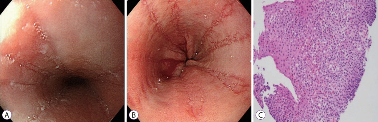
Fig. 3. (A) The follow-up endoscopy after acyclovir and proton pump inhibitor treatment shows multiple white exudates and moderate mucosal edema with furrows in the proximal esophagus. (B) The distal esophagus shows multiple longitudinal furrows with loss of mucosal vascularity. (C) The distal esophageal biopsy specimen from the follow-up endoscopy shows massive intraepithelial eosinophilic infiltration (up to 200 per high power field) and subepithelial fibrosis (hematoxylin and eosin stain, ×200).
Reference
-
1. Furuta GT, Katzka DA. Eosinophilic esophagitis. N Engl J Med. 2015; 373:1640–1648.
Article2. Warners MJ, de Rooij W, van Rhijn BD, et al. Incidence of eosinophilic esophagitis in the Netherlands continues to rise: 20-year results from a nationwide pathology database. Neurogastroenterol Motil. 2018; 30:e13165.
Article3. Moawad FJ. Eosinophilic esophagitis: incidence and prevalence. Gastrointest Endosc Clin N Am. 2018; 28:15–25.4. Clayton F, Peterson K. Eosinophilic esophagitis: pathophysiology and definition. Gastrointest Endosc Clin N Am. 2018; 28:1–14.5. Fritz J, Lerner D, Suchi M. Herpes simplex virus esophagitis in immunocompetent children: a harbinger of eosinophilic esophagitis? J Pediatr Gastroenterol Nutr. 2018; 66:609–613.
Article6. Žaja Franulović O, Lesar T, Busic N, Tešović G. Herpes simplex primo-infection in an immunocompetent host with eosinophilic esophagitis. Pediatr Int. 2013; 55:e38–e41.
Article7. Sehgal S, Darbari A, Bader A. Herpes simplex virus and eosinophilic esophagitis. J Pediatr Gastroenterol Nutr. 2013; 56:e1.
Article8. Rodrigues F, Brandão N, Duque V, Ribeiro C, António AM. Herpes simplex virus esophagitis in immunocompetent children. J Pediatr Gastroenterol Nutr. 2004; 39:560–563.
Article9. Iriarte Rodriguez A, Frago Marquínez I, de Lima Piña GP. A case report: asymptomatic esophageal eosinophilia after herpes simplex esophagitis. Controversies in the therapeutic approach. Rev Esp Enferm Dig. 2018; 110:471–472.
Article10. Zimmermann D, Criblez DH, Dellon ES, et al. Acute herpes simplex viral esophagitis occurring in 5 immunocompetent individuals with eosinophilic esophagitis. ACG Case Rep J. 2016; 3:165–168.
Article11. Monsanto P, Almeida N, Cipriano MA, Gouveia H, Sofia C. Concomitant herpetic and eosinophilic esophagitis--a causality dilemma. Acta Gastroenterol Belg. 2012; 75:361–363.12. Squires KA, Cameron DJ, Oliver M, da Fonseca Junqueira JC. Herpes simplex and eosinophilic oesophagitis: the chicken or the egg? J Pediatr Gastroenterol Nutr. 2009; 49:246–250.
Article13. Lee K, Furuta GT, Nguyen N. Eosinophilic esophagitis is an underlying cause for gastrointestinal concerns in children. Front Pediatr. 2018; 6:113.
Article14. Kim GH, Jung KW, Jung HY, et al. Diagnostic trends and clinical characteristics of eosinophilic esophagitis: a Korean, single-center database study. J Neurogastroenterol Motil. 2018; 24:248–254.
Article15. Al-Hussaini AA, Fagih MA. Herpes simplex ulcerative esophagitis in healthy children. Saudi J Gastroenterol. 2011; 17:353–356.
Article16. Cristoforo TA, Rietsma K, Wilsey A, Swan EK, Wilsey M. Herpes esophagitis with concomitant eosinophilic esophagitis in a child: a case report. Clin Pediatr (Phila). 2018; 57:618–620.
Article17. Machicado JD, Younes M, Wolf DS. An unusual cause of odynophagia in a patient with eosinophilic esophagitis. Gastroenterology. 2014; 147:37–38.
Article18. Lindberg GM, Van Eldik R, Saboorian MH. A case of herpes esophagitis after fluticasone propionate for eosinophilic esophagitis. Nat Clin Pract Gastroenterol Hepatol. 2008; 5:527–530.
Article
- Full Text Links
-
- Actions
-
Cited
- CITED
-
- Close
- Share
-
- Similar articles
-
- CT Findings of Eosinophilic Esophagitis: Case Report
- Herpes Simplex Esophagitis: A report of two cases
- A case of herpes simplex virus esophagitis with candidal esophagitis in the immunocompetent patient
- A Case of Herpes simplex Esophagitis in an Immunocompetent Boy
- Clinical Review of Eosinophilic Esophagitis
