Effect of additional firing process after sintering of monolithic zirconia crown on marginal and internal fitness
- Affiliations
-
- 1Central Dental Laboratory, Kyung Hee University Dental Hospital, Seoul, Republic of Korea.
- 2Department of Prosthodontics, School of Dentistry, Pusan National University, Pusan, Republic of Korea. prostho.hjlee@pusan.ac.kr
- KMID: 2461137
- DOI: http://doi.org/10.4047/jkap.2019.57.4.321
Abstract
- PURPOSE
To evaluate an effect of additional firing process after sintering of monolithic zirconia crown on marginal and internal fit through three-dimensional analysis.
MATERIALS AND METHODS
Ten monolithic zirconia crowns were fabricated using titanium abutment model. Monolithic zirconia crowns were designed, milled, and sintered as a control group, and additional firing with coloring was performed as a test group. Three dimensional analysis were performed by using triple-scan protocol, and cross-section analysis on mesio-distal and disto-lingual section was evaluated to measure marginal and internal fitness. Then, three-dimensional surface difference on between two groups was evaluated (α=.05).
RESULTS
There was statistically significant difference between the control group (32.0 ± 24.3 µm) and the test group (17.0 ± 10.8 µm) in the mesial axial wall (P < .02) and the control group (60.2 ± 24.3 µm) and the test group (71.8 ± 21.5 µm) in the distal axial wall (P < .01). There was no statistically significant difference at the remaining point.
CONCLUSION
There was no statistical significance on the deviation of inner surface of crown according to firing number, and the results of both group were considered clinically acceptable.
Keyword
Figure
-
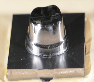
Fig. 1 Master model with titanium block made by using CAD/CAM.

Fig. 2 The constant seating force.

Fig. 3 Scanned three-dimensional surface data. (A) Titanium master die, (B) Monolithic zirconia crown, (C) Superimposed crown on the master die.
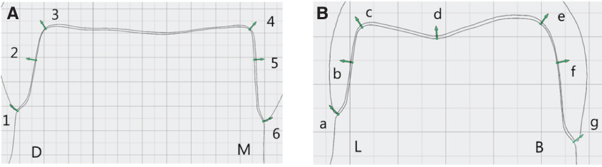
Fig. 4 Measurement points for marginal gap (1, 6, a, g) and internal gap (2, 3, 4, 5, b, c, d, e, f). (A) Mesio-distal section, (B) Bucco-lingual section.

Fig. 5 Mean of marginal and internal gap of monolithic zirconia crown: Mesio-distal section (*Significant at P < .05).

Fig. 6 Mean of marginal and internal gap of monolithic zirconia crown: Bucco-lingual section.
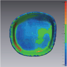
Fig. 7 Three-dimensional inner surface comparison of monolithic zirconia crown.
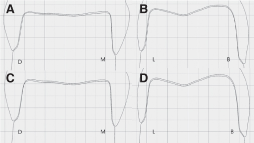
Fig. 8 Monolithic zirconia crown superimposed on the master die. (A, B) control group, (C, D) test group.
Reference
-
1. Suárez MJ, Lozano JF, Paz Salido M, Martínez F. Three-year clinical evaluation of In-Ceram Zirconia posterior FPDs. Int J Prosthodont. 2004; 17:35–38.2. Conrad HJ, Seong WJ, Pesun IJ. Current ceramic materials and systems with clinical recommendations: a systematic review. J Prosthet Dent. 2007; 98:389–404.
Article3. Lebon N, Tapie L, Duret F, Attal JP. Understanding dental CAD/CAM for restorations-dental milling machines from a mechanical engineering viewpoint. Part B: labside milling machines. Int J Comput Dent. 2016; 19:115–134.4. Sturdevant JR, Bayne SC, Heymann HO. Margin gap size of ceramic inlays using second-generation CAD/CAM equipment. J Esthet Dent. 1999; 11:206–214.
Article5. Gardner FM. Margins of complete crowns-literature review. J Prosthet Dent. 1982; 48:396–400.
Article6. Jacobs MS, Windeler AS. An investigation of dental luting cement solubility as a function of the marginal gap. J Prosthet Dent. 1991; 65:436–442.
Article7. Abduo J, Lyons K, Swain M. Fit of zirconia fixed partial denture: a systematic review. J Oral Rehabil. 2010; 37:866–876.
Article8. Vojdani M, Safari A, Mohaghegh M, Pardis S, Mahdavi F. The effect of porcelain firing and type of finish line on the marginal fit of zirconia copings. J Dent (Shiraz). 2015; 16:113–120.9. Kim JH, Kim KB. Influence of high temperature of the porcelain firing process on the marginal fit of zirconia core. J Dent Hyg Sci. 2013; 13:135–141.10. Balkaya MC, Cinar A, Pamuk S. Influence of firing cycles on the margin distortion of 3 all-ceramic crown systems. J Prosthet Dent. 2005; 93:346–355.
Article11. Holmes JR, Bayne SC, Holland GA, Sulik WD. Considerations in measurement of marginal fit. J Prosthet Dent. 1989; 62:405–408.
Article12. Moon BH, Yang JH, Lee SH, Chung HY. A study on the marginal fit of all ceramic using CCD camera. J Korean Acad Prosthodont. 1998; 36:273–292.13. McLean JW, von Fraunhofer JA. The estimation of cement film thickness by an in vivo technique. Br Dent J. 1971; 131:107–111.
Article14. Molin MK, Karlsson SL, Kristiansen MS. Influence of film thickness on joint bend strength of a ceramic/resin composite joint. Dent Mater. 1996; 12:245–249.
Article15. Holst S, Karl M, Wichmann M, Matta RE. A new triple-scan protocol for 3D fit assessment of dental restorations. Quintessence Int. 2011; 42:651–657.16. Matta RE, Schmitt J, Wichmann M, Holst S. Circumferential fit assessment of CAD/CAM single crowns-a pilot investigation on a new virtual analytical protocol. Quintessence Int. 2012; 43:801–809.17. Faucher RR, Nicholls JI. Distortion related to margin design in porcelain-fused-to-metal restorations. J Prosthet Dent. 1980; 43:149–155.
Article
- Full Text Links
-
- Actions
-
Cited
- CITED
-
- Close
- Share
-
- Similar articles
-
- Color stability of fully- and pre-crystalized chair-side CAD-CAM lithium disilicate restorations after required and additional sintering processes
- Effect of milling and sintering process on integrity of zirconia prosthesis: a literature review
- Fracture strength of zirconia monolithic crowns
- Color stability of fully- and pre-crystalized chair-side CAD-CAM lithium disilicate restorations after required and additional sintering processes
- Effect of sintering programs and surface treatments on monolithic zirconia
